Case Presentation Thoracic and Cardiovascular surgery department SMC


 Anorexia/Nausea/Vomiting(-/-/-) General Weakness(-) Constipation/Diarrhea(-/-) Fever/Chill/Night sweat(-/-/-)")

 Large PMOE VSD (9. 6 mm) with left-to-right")


 Page 10 YOUR LOGO")


 50%")

 : 25% - Diagnostic difficulties: preop. echo,")
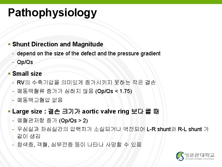

 Pulmonary vascular disease : as")


- Slides: 30
Case Presentation Thoracic and Cardiovascular surgery department, SMC 2007313075 Son Eui Young YOUR LOGO
Chief Complaint M / 2개월 11일 Abnormal echocardiography result - Onset : 2012 -04 -03 Page 2 YOUR LOGO
Present illness 2개월 남아, 31+0 wk, 1. 69 kg, syphilis mother's baby, C/sec d/t PPROM and breech presentation, Apgar score 2/2으로 출생 출산 후 시행한 TTE 상 VSD 및 ASD 소견 관찰 이에 대해 further evaluation 및 management 위하여 본원 NICU 전원 Page 3 YOUR LOGO
Other Histories Development: normal Surgical history: none Current medication: none Page 4 YOUR LOGO
Review of System 특이사항 없음 Abdominal pain (-) Anorexia/Nausea/Vomiting(-/-/-) General Weakness(-) Constipation/Diarrhea(-/-) Fever/Chill/Night sweat(-/-/-) Melena/hematochezia/ hematemesis (-/-/-) Headache/Dizziness(-/-) Dysuria/Frequent voiding(-/-) Visual disturbance/ocular pain(-/-) Red urine/Foamy urine(-/-) Sore throat/Rhinorrhea/Sneezing(-/-/-) Arthralgia/Myalgia (-/-) Cough/Sputum (-/-/-) Morning stiffness(-/-/-) Dyspnea/Hemoptysis (-/-) Easily bruisilibility (-) Chest pain (-) Palpitation (-) Page 5 YOUR LOGO
Physical Exam Vital sign - BP 110/70, HR 94, RR 16, BT 36. 3℃ - Sp. O 2 94% General appearance: Not so ill-looking Mental state: alert & well oriented - GCS (E 4, V 5, M 6) Head and Neck - Pinkish Conjunctiva/ unicteric Sclera Isocoric Pupil size, prompt light reflex at both eyes Chest - Symmetric expansion chest wall retraction (+) : subcostal Vesicular Breath sound s wheezing Murmur (+) Lower Left sternal border, pansystolic grade 3 Subclavicular node / Axillary node ( - / - ) Abdomen - Liver palpation( - ) - Spleen palpation ( - ) Nystagmus ( - ) Otoscopy (정상) oral ulcer ( - ), tongue dehydration ( - ) Tonsilar hypertrophy( - ), Pharyngeal injection( - ) Skin - Rash/Purpura/erythema (- / - ) Cervical LNE( -); Thyroid enlargement ( - ) Carotid bruit Rt/Lt ( - / - ) Paranasal tenderness ( - ) Page 6 YOUR LOGO
Echocardiogram 2012 -04 -30, #18 1) Large PMOE VSD (9. 6 mm) with left-to-right dominant bidirectional shung 2) Large secundum ASD (8. 8 x 7. 4 mm) with left-to-right shunt 3) dilated RA & RV 4) no PDA, no Co. A Page 7 YOUR LOGO
Initial Lab CBC 8180 - 9. 1 - 278 k CRP 0. 03 T-bil/AST/ALP 0. 8/23/11/▲ 399 BUN/Cr 8. 5/0. 19 e’ 141 - ▲ 5. 3 - 103 Page 8 YOUR LOGO
Problem List #1. Preterm AGA #2. Low birth weight infant #3. Syphilis mother's baby #4. Abnormal echocardiographic finding including VSD Page 9 YOUR LOGO
Assessment #1, #2, #4 - VSD (SA, small to moderate) Page 10 YOUR LOGO
Plan VSD closure with bovine pericardium ASD primary closure Page 11 YOUR LOGO
Operation - VSD closure with bovine pericardium - ASD primary closure 수술 후 진단명 - VSD (PMOE, large) - ASD (secundum, large) Page 12 YOUR LOGO
Hospital Course POD #1 - vital sign stable - pain tolerable - wound clear - no immediate complication POD #5 - extubation done POD #12 - C-line remove - 퇴원 고려 POD #14 (2012 -06 -12) - Vital sign stable Page 13 YOUR LOGO
Disease Review Ventricular Septal Defect 2007313075 Son Eui Young YOUR LOGO
Introduction 1 in 1000 live births 선천성 심질환 중 가장 흔하다. (단독으로는 25%) 50% associated with other congenital malformations First described by Roger in 1879, hence small VSDs are also known as the ‘maladie de Roger’. First VSD closed under direct vision by Lillehei in 1955 Page 15 YOUR LOGO
Anatomic Classification of VSD Perimembranous: 80% Subarterial: 14% Muscular VSD : 10% Figure 117– 4 Classification of ventricular septal defects (VSDs): YOUR LOGO
Commonly Associated Defects Patent ductus arteriosus (PDA) : 25% - Diagnostic difficulties: preop. echo, TEE etc - should be ligated or clipped Coarctation or Aorta : 10% - Augmented L-R shunt left ventricular outflow tract obstruction(Congenital valvar or subvalvar aortic stenosis): 4% large atrial septal defects (ASDs), right ventricular outflow tract obstruction, vascular ring, and persistent left superior vena cava. YOUR LOGO
YOUR LOGO
Complication Growth failure Congestive heart failure (left heart failure) Pulmonary vascular disease : as Eisenmenger syndrome or complex Severe illness with viral or bacterial pneumonia Infective endocarditis Acquired left ventricular outflow tract obstruction Aneurysm of the ventricular septum Paradoxical emboli Sudden death Heart block secondary to intracardiac repair Aortic regurgitation Impaired left ventricular function in some patients Stenosis in the right ventricular outflow tract Increase in weight following VSD closure Discrete fibrous subaortic stenosis YOUR LOGO
Diagnostic work-up Symptoms - tachypnea, growth failure, profuse sweating during feeding, a bulging precordium, a pansystolic murmur, an enlarged liver, and thready pulses the physical examination, chest radiograph, and electrocardiogram (ECG) YOUR LOGO
VSD Natural Course Spontaneous closure or decrease of size Perimembranous or muscular type Aggressive medical management cf. Malalignment type, SA, MO, Endocardial cushion type Usually within 6 -12 Mo of age YOUR LOGO
VSD Natural Course Eisenmenger or Severe PHT Usually after 1 yr of age Contraindications for op d/t PHT - PVR > 8 -10 Wood unit - No response to pulmonary vasodilators such as O 2 or nitric oxide - Mainly R-L shunt or no L-R shunt through VSD YOUR LOGO
VSD Natural Course AV Deformity Mainly subarterial type Some of PM type MO VSD Increased incidence of infective endocarditis if there is AR YOUR LOGO
Treatment of VSD : Indications Approximately 30% of infants - surgery within the first year of life Significant shunt - Medically uncontrolled CHF - PA pressure > 1/2 of systemic artery pressure - Shunt amount; Qp/Qs > 1. 5 or 1. 7 Other problems - DCRV, Subaortic stenosis, AV deformity Consider natural course, patient’s age YOUR LOGO
Treatment of VSD : depends on size Large VSD < 3 months – CHF, failure to thrive Elective repair at 6 -12 months (PVR < 8. 0 units) Small VSD (Qp: Qs<2: 1) Endocarditis Cardiac Enlargement Any Aortic Incompetence Subarterial (supracristal) VSD - any size, operate early YOUR LOGO
Surgical methods • Corrective surgery • RA, RV, PA, LV 등을 통해 접근 가능 • Dacron, Gore-tex patch, autopericardial patch • 크기가 작으면 primary closure • Pulmonary artery bending • 폐동맥혈류가 과한 환자에게 폐동맥을 좁혀줌으로써 합병증 예방 • 요즘엔 교정수술의 성적이 향상되어 잘 안 함 YOUR LOGO
THANK YOU FOR YOUR ATTENTION Page YOUR LOGO
Page 29 YOUR LOGO
Page 30 YOUR LOGO