
Case Presentation Staci Smith DO April 17 th


- Slides: 50
Case Presentation Staci Smith DO April 17 th, 2009
Differential Diagnosis l l l hematuria proteinuria red blood cell casts glomerulonephritis
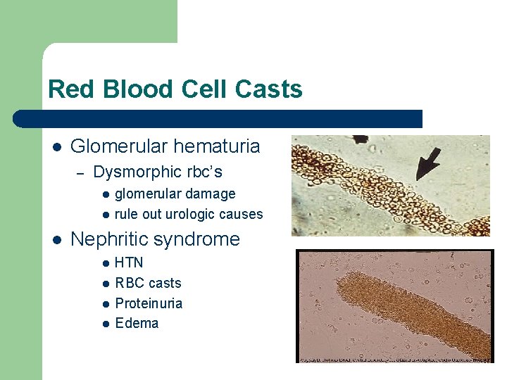
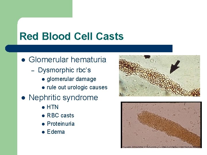
Red Blood Cell Casts l Glomerular hematuria – Dysmorphic rbc’s l l l glomerular damage rule out urologic causes Nephritic syndrome l l HTN RBC casts Proteinuria Edema
Dysmorphic rbc l l dysmorphic red cells acanthocytes – ring form
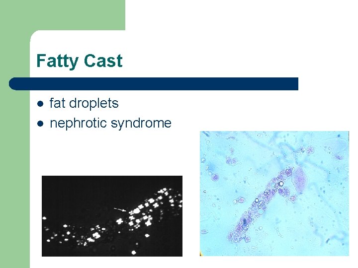
Fatty Cast l l fat droplets nephrotic syndrome
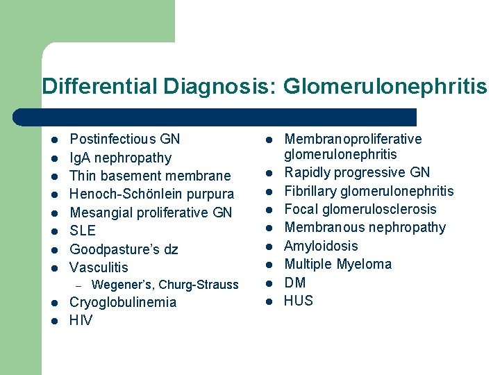
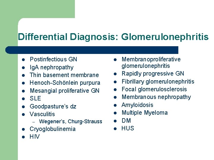
Differential Diagnosis: Glomerulonephritis l l l l Postinfectious GN Ig. A nephropathy Thin basement membrane Henoch-Schönlein purpura Mesangial proliferative GN SLE Goodpasture’s dz Vasculitis – l l Wegener’s, Churg-Strauss Cryoglobulinemia HIV l l l l l Membranoproliferative glomerulonephritis Rapidly progressive GN Fibrillary glomerulonephritis Focal glomerulosclerosis Membranous nephropathy Amyloidosis Multiple Myeloma DM HUS
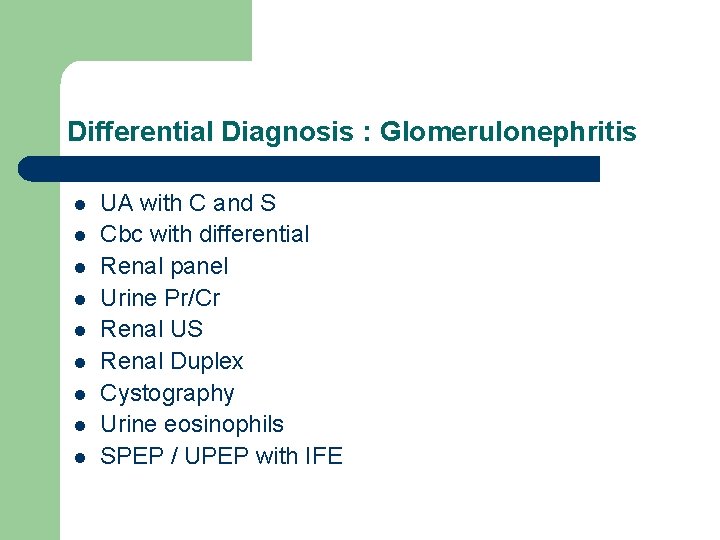
Differential Diagnosis : Glomerulonephritis l l l l l UA with C and S Cbc with differential Renal panel Urine Pr/Cr Renal US Renal Duplex Cystography Urine eosinophils SPEP / UPEP with IFE
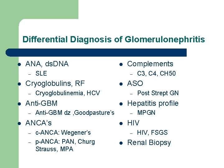
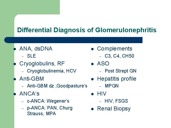
Differential Diagnosis of Glomerulonephritis l ANA, ds. DNA – l l – – l l c-ANCA: Wegener’s p-ANCA: PAN, Churg Strauss, MPA MPGN HIV – l Post Strept GN Hepatitis profile – l C 3, C 4, CH 50 ASO – Anti-GBM dz , Goodpasture’s ANCA’s Complements – Cryoglobulinemia, HCV Anti-GBM – l SLE Cryoglobulins, RF – l HIV, FSGS Renal Biopsy
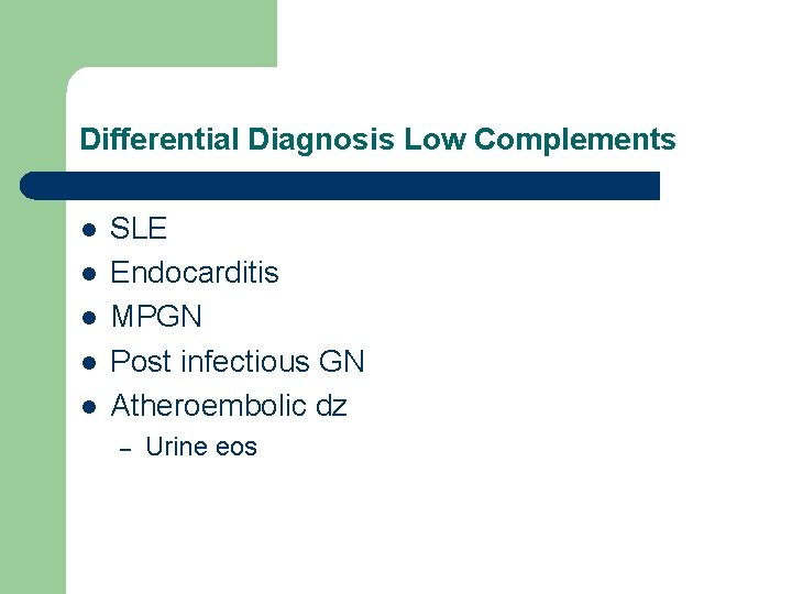
Differential Diagnosis Low Complements l l l SLE Endocarditis MPGN Post infectious GN Atheroembolic dz – Urine eos
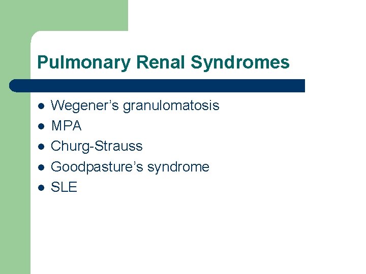
Pulmonary Renal Syndromes l l l Wegener’s granulomatosis MPA Churg-Strauss Goodpasture’s syndrome SLE
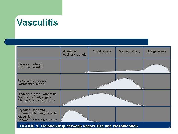
Vasculitis
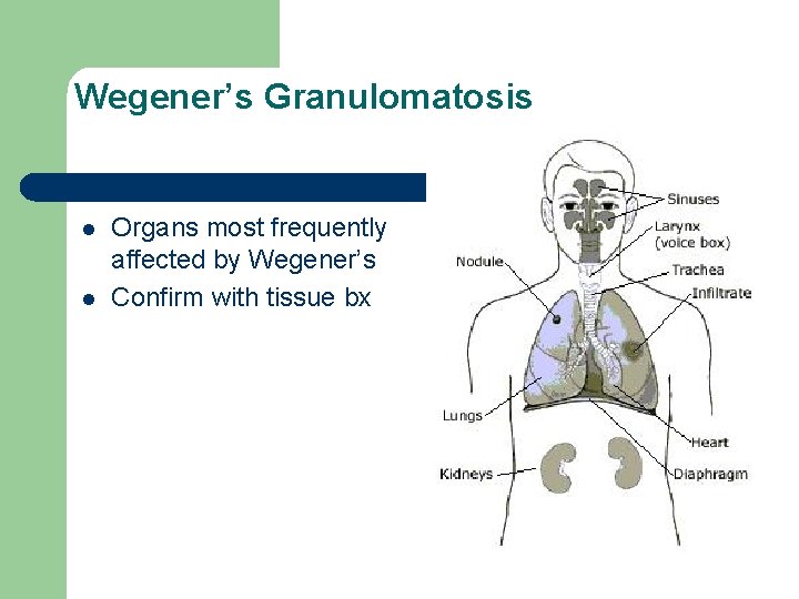
Wegener's Granulomatosis l l distinguished from other vasculitides by the pattern of organ involvement histologic features of granulomatosis and necrotizing inflammation – primary involvement l l upper and lower respiratory tracts kidneys - glomerulonephritis
Wegener's Granulomatosis l l l pathogenesis is unknown interplay of an initiating inflammatory event and a highly specific immune response is directed against neutrophil granule proteins – high titer autoantibodies known as anti-neutrophil cytoplasmic antibodies (ANCA)
Wegener’s Triad l l 1954 - Goodman and Churg triad of pathological features – – – systemic necrotizing angiitis necrotizing granulomatous inflammation of the respiratory tract necrotizing glomerulonephritis
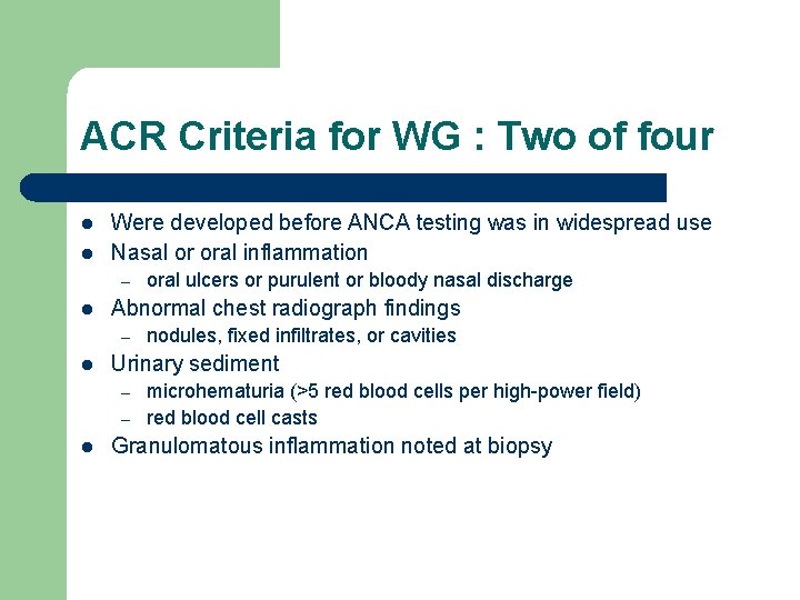
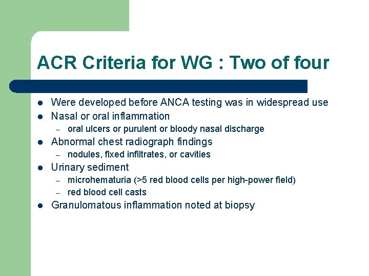
ACR Criteria for WG : Two of four l l Were developed before ANCA testing was in widespread use Nasal or oral inflammation – l Abnormal chest radiograph findings – l nodules, fixed infiltrates, or cavities Urinary sediment – – l oral ulcers or purulent or bloody nasal discharge microhematuria (>5 red blood cells per high-power field) red blood cell casts Granulomatous inflammation noted at biopsy
Wegener’s Granulomatosis l l Organs most frequently affected by Wegener’s Confirm with tissue bx
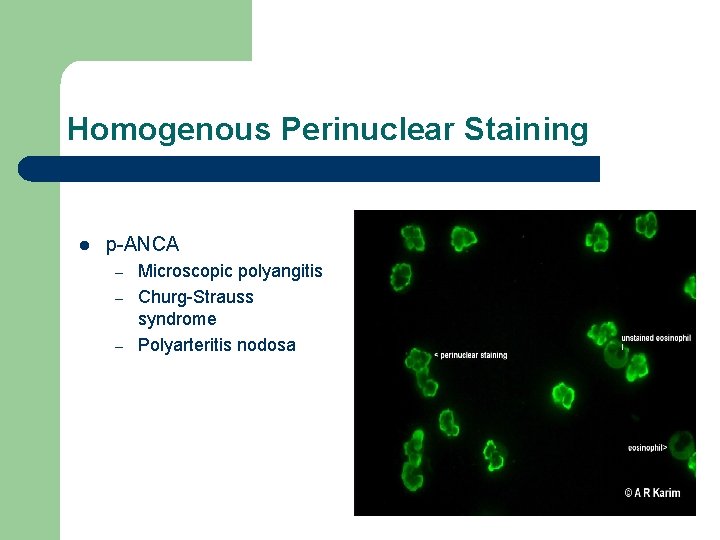
Homogenous Perinuclear Staining l p-ANCA – – – Microscopic polyangitis Churg-Strauss syndrome Polyarteritis nodosa
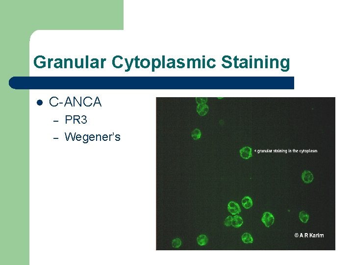
Granular Cytoplasmic Staining l C-ANCA – – PR 3 Wegener’s
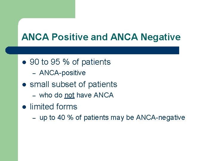
ANCA Positive and ANCA Negative l 90 to 95 % of patients – l small subset of patients – l ANCA-positive who do not have ANCA limited forms – up to 40 % of patients may be ANCA-negative
Clinical History l ELK classification – – broad spectrum of organ involvement ears, nose, and throat or upper respiratory tract (E); lungs (L); and kidneys (K)
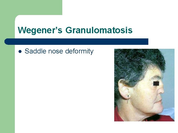
ELK Classification: Ears, nose, and throat l l l nasal obstruction with serosanguineous discharge chronic sinusitis deep central facial pain saddle nose deformity middle ear involvement -hearing loss subglottic stenosis-hoarseness and stridor
Wegener’s Granulomatosis l Saddle nose deformity
ELK Classification l Lungs – – – asymptomatic or may present with cough and occasional hemoptysis progressive dyspnea and respiratory failure massive pulmonary hemorrhage
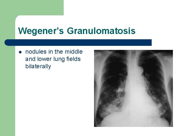
Wegener’s Granulomatosis l nodules in the middle and lower lung fields bilaterally
ELK Classification l l Patients with kidney involvement generally are asymptomatic, but some patients may notice mild hematuria. Edema secondary to nephrotic syndrome may be present
Wegener’s Granulomatosis l Neurologic involvement – – – approximately one third of patients peripheral neuropathy with mononeuritis multiplex cranial neuropathy involving the second, sixth, and seventh cranial nerves
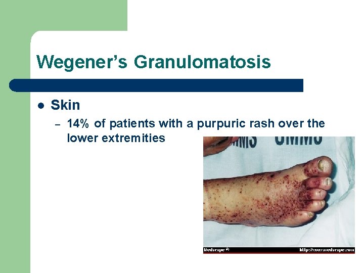
Wegener’s Granulomatosis l Skin – 14% of patients with a purpuric rash over the lower extremities
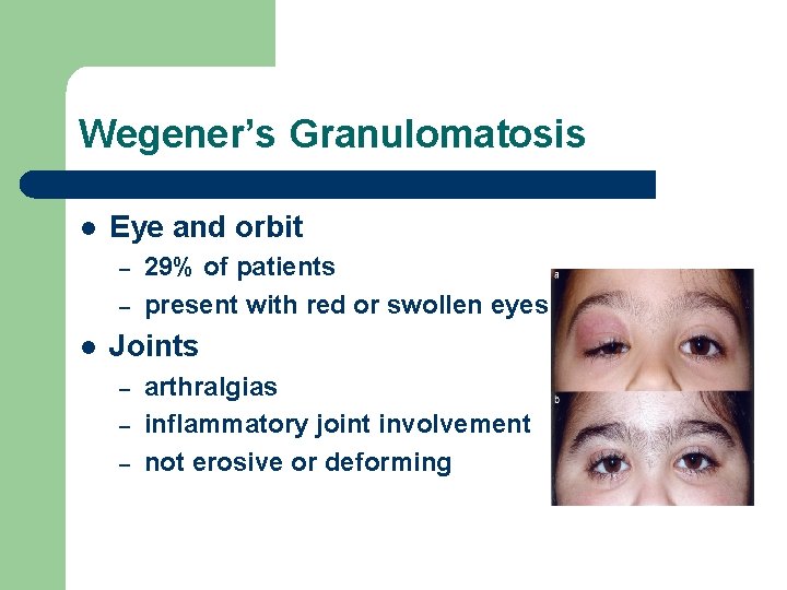
Wegener’s Granulomatosis l Eye and orbit – – l 29% of patients present with red or swollen eyes Joints – – – arthralgias inflammatory joint involvement not erosive or deforming
Limited form of Wegener’s l clinical findings isolated to the upper respiratory tract or the lungs – – – one-fourth of cases up to 80 percent eventually develop glomerulonephritis incompletely understood phenotypic differences younger at disease onset more likely to be women
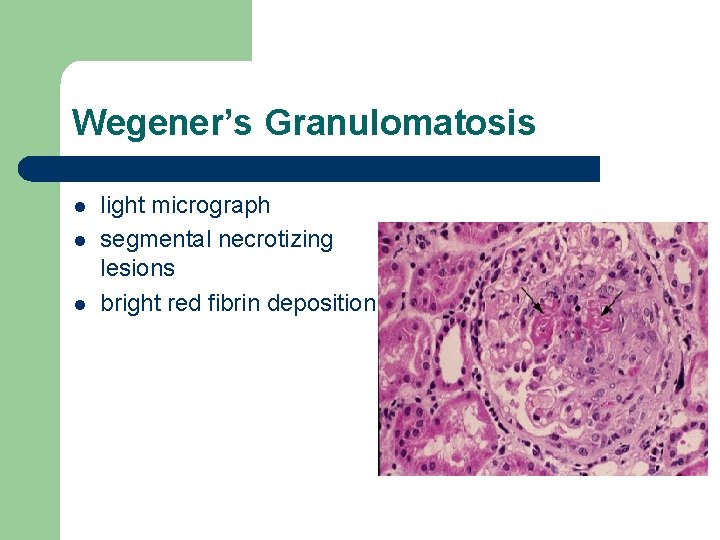
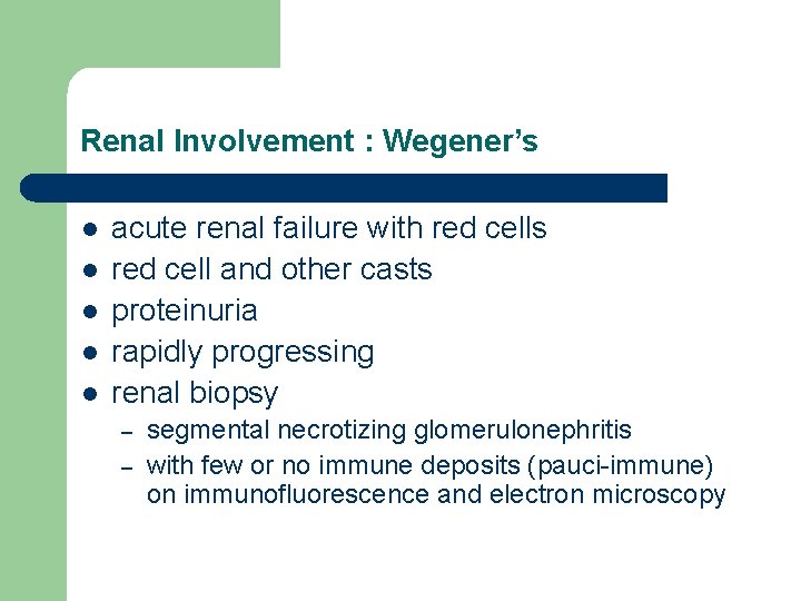
Renal Involvement : Wegener’s l l l acute renal failure with red cells red cell and other casts proteinuria rapidly progressing renal biopsy – – segmental necrotizing glomerulonephritis with few or no immune deposits (pauci-immune) on immunofluorescence and electron microscopy
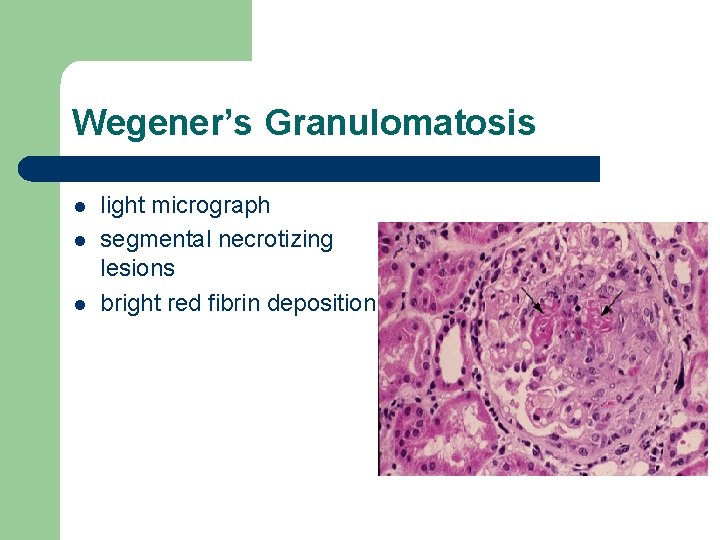
Wegener’s Granulomatosis l light micrograph segmental necrotizing lesions bright red fibrin deposition
Renal Involvement : Wegener’s l glomerular involvement – – often accompanied by mononuclear tubulointerstitial infiltrates small vessel involvement l l venules, capillaries, arterioles reoccurs in renal transplant
Renal Involvement : Wegener’s l l may be severe enough to require dialysis hypertension – – mediated by ischemia-induced activation of the renin-angiotensin system angiotensin converting enzyme inhibitors l l risk of worsening renal function removal of the angiotensin II effect on autoregulation and maintenance of the glomerular filtration rate
Treatment l untreated systemic WG – – poor prognosis 90% of patients dying within 2 years l l from respiratory or renal failure long-lasting remission – – cyclophosphamide in combination with corticosteroids 90% of patients respond to cyclophosphamide
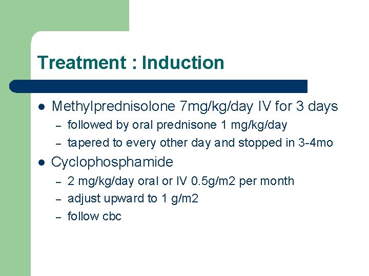
Treatment : Induction l Methylprednisolone 7 mg/kg/day IV for 3 days – – l followed by oral prednisone 1 mg/kg/day tapered to every other day and stopped in 3 -4 mo Cyclophosphamide – – – 2 mg/kg/day oral or IV 0. 5 g/m 2 per month adjust upward to 1 g/m 2 follow cbc
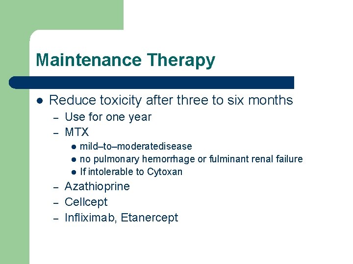
Maintenance Therapy l Reduce toxicity after three to six months – – Use for one year MTX l l l – – – mild–to–moderatedisease no pulmonary hemorrhage or fulminant renal failure If intolerable to Cytoxan Azathioprine Cellcept Infliximab, Etanercept
Predicting Relapse l l ANCA titers usually parallel the course of vasculitis, especially C-ANCA decreasing titers of ANCA predict a lower risk for clinical relapse
Plasma Exchange l l l role is controversial no real added benefits may be beneficial if life threatening pulmonary hemorrhage especially if dialysis dependent
Key Points : Decreased Complements l l l SLE Endocarditis MPGN Post infectious GN Atheroembolic dz – Urine eos
Differential Diagnosis l l l hematuria proteinuria red blood cell casts glomerulonephritis
Differential Diagnosis Low Complements l l l SLE Endocarditis MPGN Post infectious GN Atheroembolic dz – Urine eos
Red Blood Cell Casts l Glomerular hematuria – Dysmorphic rbc’s l l l glomerular damage rule out urologic causes Nephritic syndrome l l HTN RBC casts Proteinuria Edema
Differential Diagnosis: Glomerulonephritis l l l l Postinfectious GN Ig. A nephropathy Thin basement membrane Henoch-Schönlein purpura Mesangial proliferative GN SLE Goodpasture’s dz Vasculitis – l l Wegener’s, Churg-Strauss Cryoglobulinemia HIV l l l l l Membranoproliferative glomerulonephritis Rapidly progressive GN Fibrillary glomerulonephritis Focal glomerulosclerosis Membranous nephropathy Amyloidosis Multiple Myeloma DM HUS
Differential Diagnosis of Glomerulonephritis l ANA, ds. DNA – l l – – l l c-ANCA: Wegener’s p-ANCA: PAN, Churg Strauss, MPA MPGN HIV – l Post Strept GN Hepatitis profile – l C 3, C 4, CH 50 ASO – Anti-GBM dz , Goodpasture’s ANCA’s Complements – Cryoglobulinemia, HCV Anti-GBM – l SLE Cryoglobulins, RF – l HIV, FSGS Renal Biopsy
ACR Criteria for WG : Two of four l l Were developed before ANCA testing was in widespread use Nasal or oral inflammation – l Abnormal chest radiograph findings – l nodules, fixed infiltrates, or cavities Urinary sediment – – l oral ulcers or purulent or bloody nasal discharge microhematuria (>5 red blood cells per high-power field) red blood cell casts Granulomatous inflammation noted at biopsy
Renal Involvement : Wegener’s l l l acute renal failure with red cells red cell and other casts proteinuria rapidly progressing renal biopsy – – segmental necrotizing glomerulonephritis with few or no immune deposits (pauci-immune) on immunofluorescence and electron microscopy
Renal Involvement : Wegener’s l l l acute renal failure with red cells red cell and other casts proteinuria rapidly progressing renal biopsy – – segmental necrotizing glomerulonephritis with few or no immune deposits (pauci-immune) on immunofluorescence and electron microscopy
Wegener’s Granulomatosis l light micrograph segmental necrotizing lesions bright red fibrin deposition
Thank You
Reference l l Duna, GF, Cotch, MF, Galperin, C, et al. Wegener's granulomatosis: Role of environmental exposures. Clin Exp Rheumatol 1998; 16: 669. Hoffman, GS, Kerr, GS, Leavitt, RY, et al. Wegener's granulomatosis: An analysis of 158 patients. Ann Intern Med 1992; 116: 488. Banerjee, A, Mc. Kane, W, Thiru, S, Farrington, K. Wegener's granulomatosis presenting as acute suppurative interstitial nephritis. J Clin Pathol 2001; 54: 787. Wen, YK, Chen, ML. Transformation from tubulointerstitial nephritis to crescentic glomerulonephritis: an unusual presentation of ANCA-associated renal vasculitis. Ren Fail 2006; 28: 189