Case Presentation New England Society of Interventional Radiology


 Pulmonary AVM B) Pulmonary")
 Pulmonary AVM B) Pulmonary")

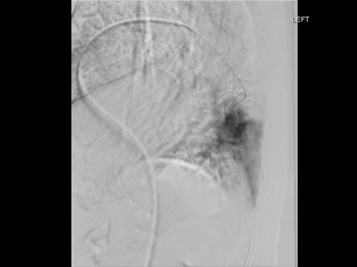
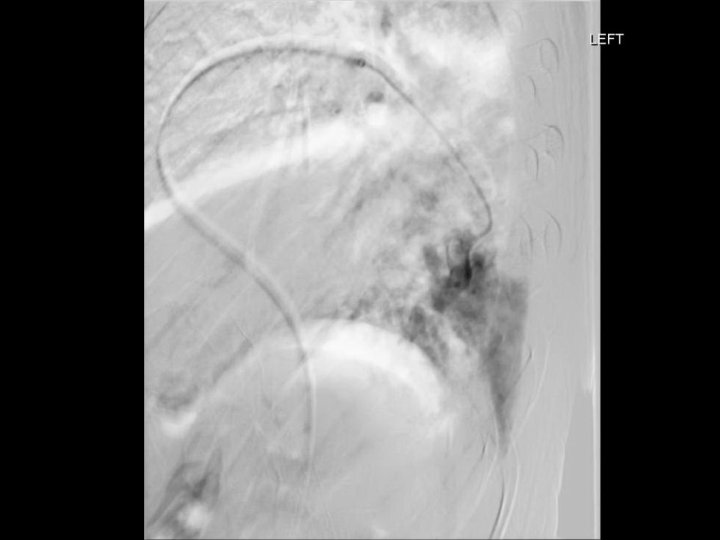
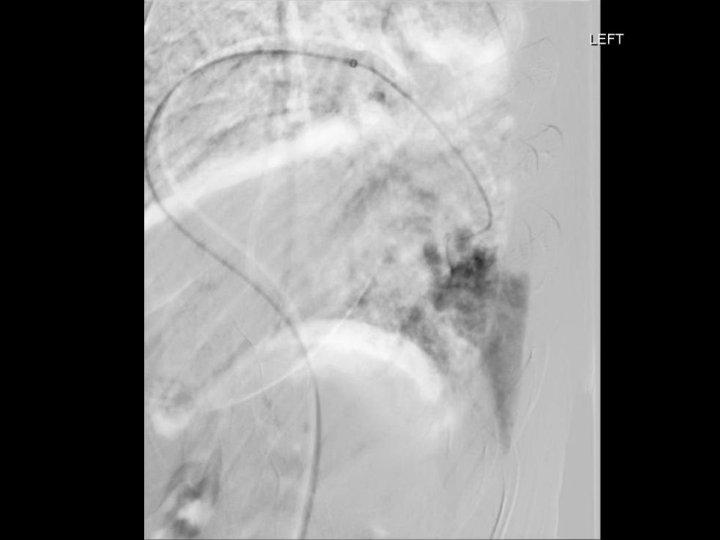
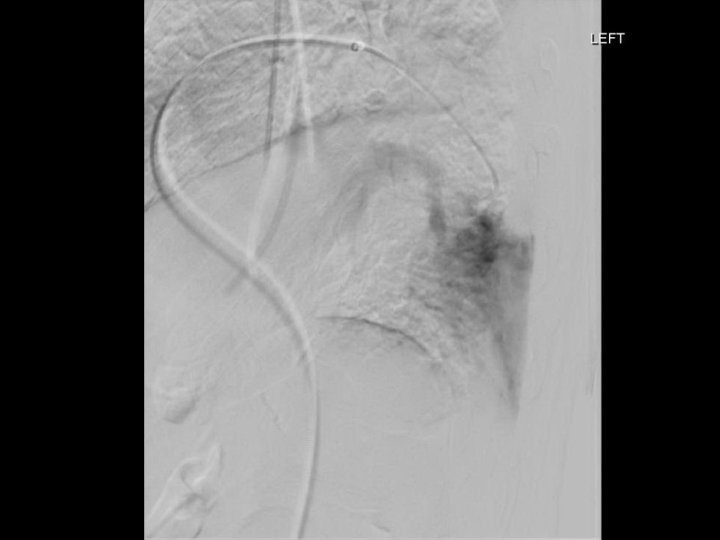
 Pulmonary artery normal in arterial phase 2) No pulmonary arteriovenous shunt")

- Slides: 57
Case Presentation New England Society of Interventional Radiology November 14, 2016 Aaron L. Harman, MD and Chieh-Min Fan, MD Interventional Radiology Brigham and Women’s Hospital
Disclosures • None
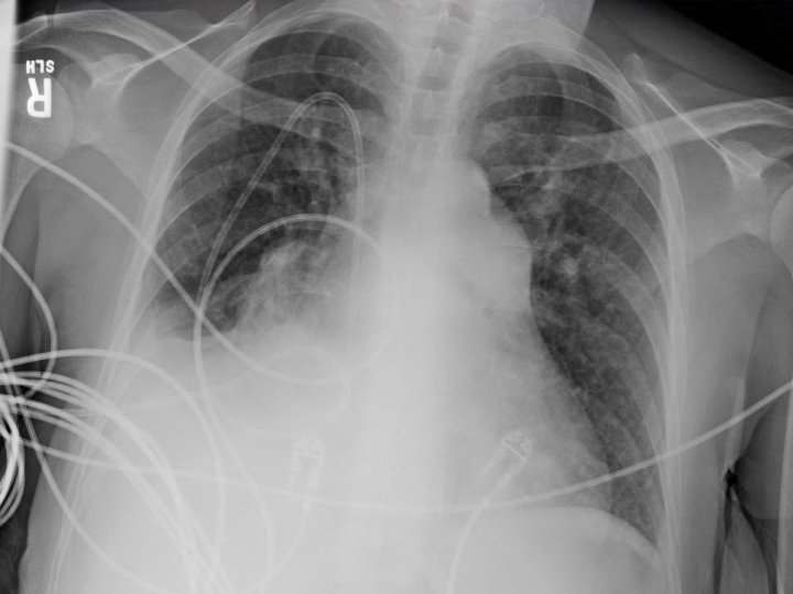
Case • 39 F with history of familial pulmonary hypertension on epoprostenol, on transplant list, p/w hemoptysis estimated at 300 -500 m. L • At current presentation, hypotensive and hypoxic • Prior episode of hemoptysis 1. 5 months prior treated with bronchial artery embolization
Case • 39 F with history of familial pulmonary hypertension on epoprostenol, on transplant list, p/w hemoptysis estimated at 300 -500 m. L • At current presentation, hypotensive and hypoxic • Prior episode of hemoptysis 1. 5 months prior treated with bronchial artery embolization
1. 5 months prior
Coronal
Coronal Sagittal
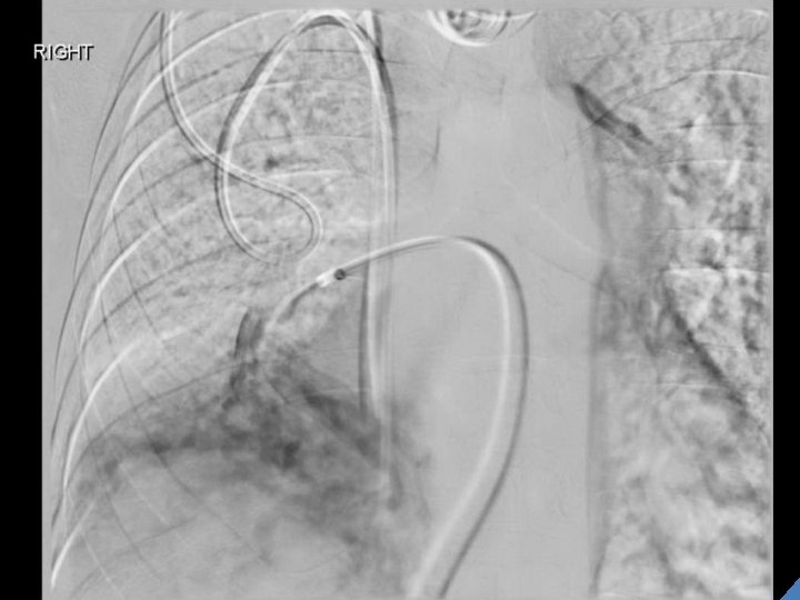
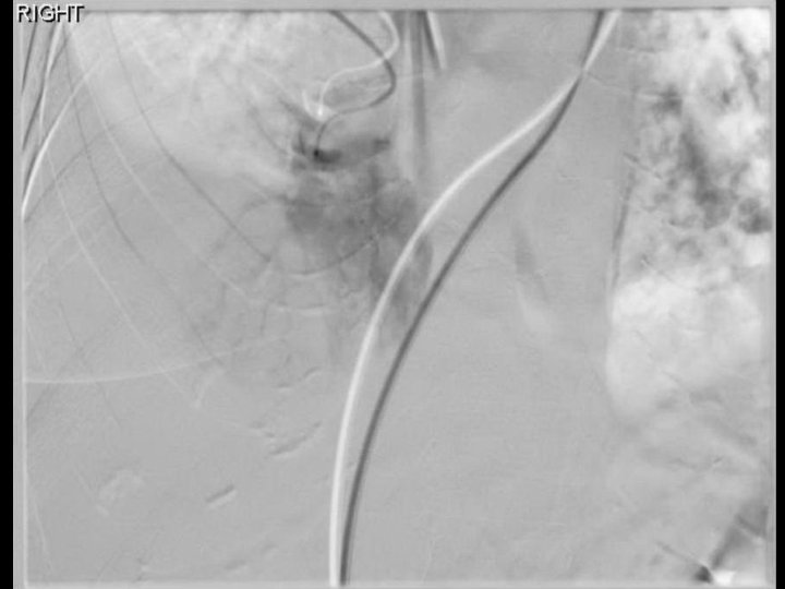
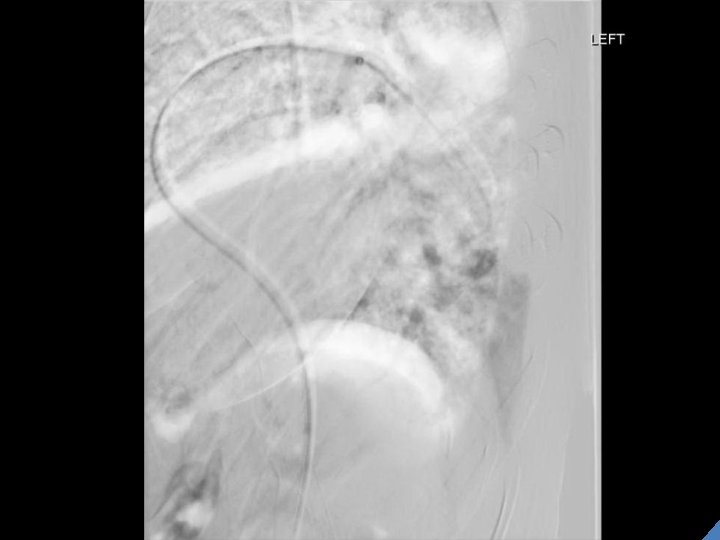
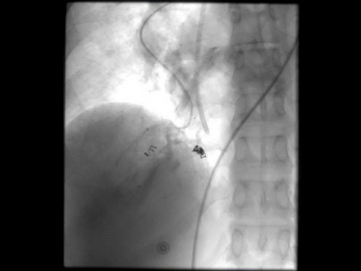
Attempt #1 • Systemic BP 90 s/50 s • Main PA systolic and diastolic pressures measured at 130 s and 50 s respectively.
Attempt #1 • Systemic BP 90 s/50 s • Main PA systolic and diastolic pressures measured at 130 s and 50 s respectively. • Is this normal?
Attempt #1 • Systemic BP 90 s/50 s • Main PA systolic and diastolic pressures measured at 130 s and 50 s respectively. • Is this normal? NO.
Attempt #1 • Systemic BP 90 s/50 s • Main PA systolic and diastolic pressures measured at 130 s and 50 s respectively. • Which is the normal PA systolic pressure range? A) 2 -6 mm Hg B) 15 -30 mm Hg C) 60 -90 mm Hg D) 90 -140 mm Hg
Attempt #1 • Systemic BP 90 s/50 s • Main PA systolic and diastolic pressures measured at 130 s and 50 s respectively. • Which is the normal PA systolic pressure range? A) 2 -6 mm Hg B) 15 -30 mm Hg C) 60 -90 mm Hg D) 90 -140 mm Hg
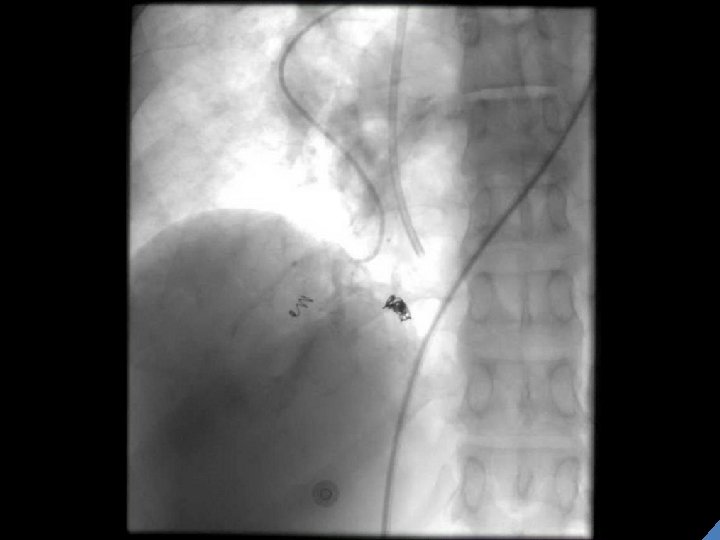
Attempt #1 • Systemic BP 90 s/50 s • Main PA systolic and diastolic pressures measured at 130 s and 50 s respectively. • With even small hand injections of contrast, the SBP dropped into 80 s despite pressors. • Therefore, the procedure was terminated.
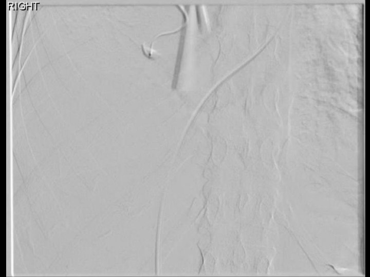
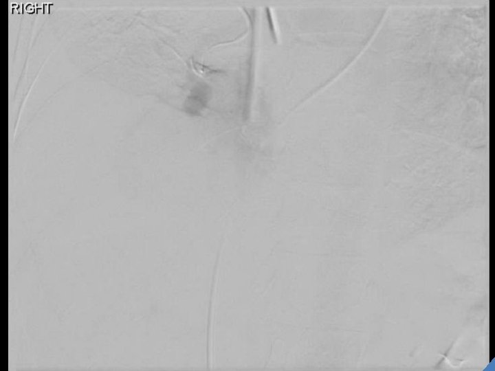
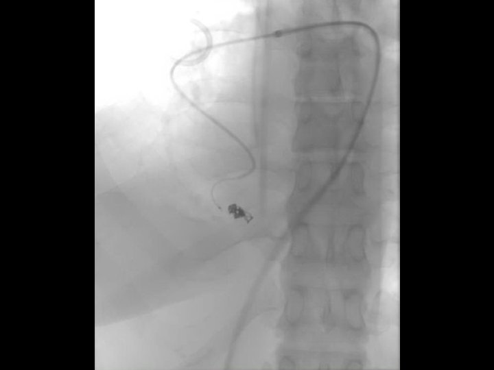
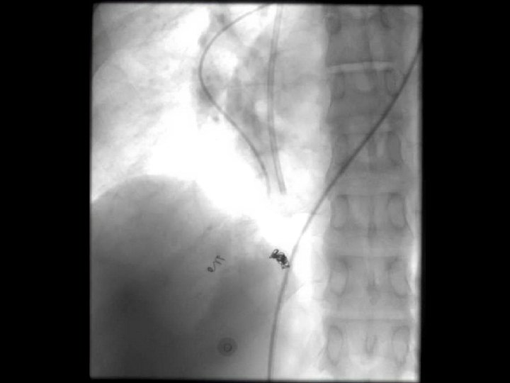
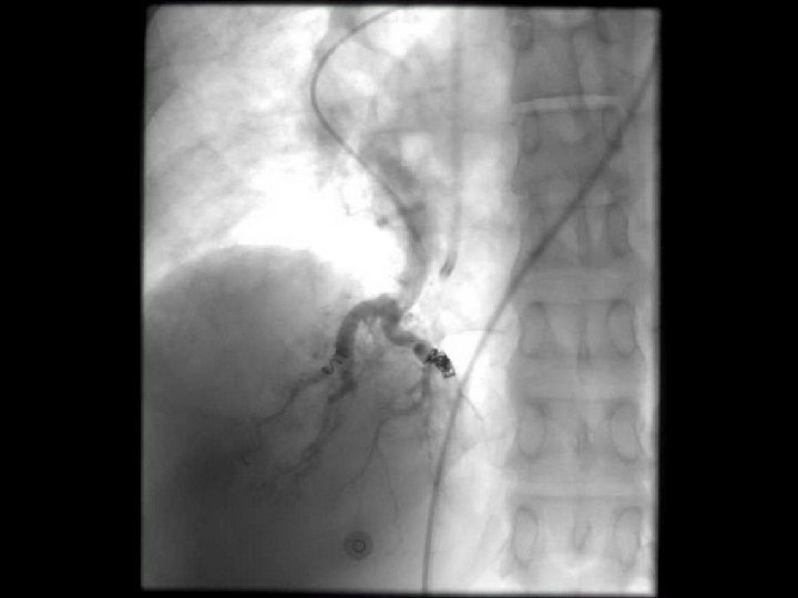
Attempt #2
What’s This?
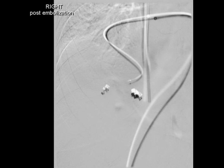
Case continued • Following the procedure, the patient clinically improved, decreased oxygen requirement. • The patient was discharged 6 days later. • 2 months later presented with hemoptysis.
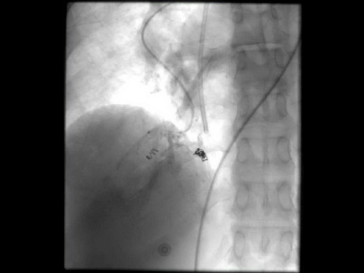
Sagittal
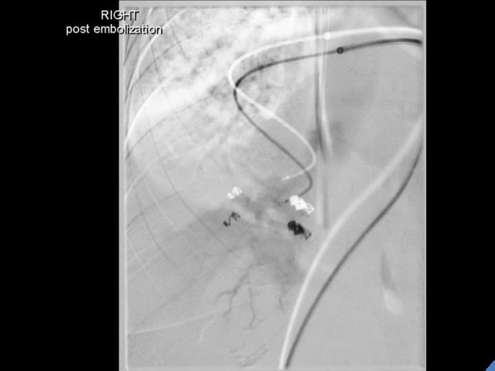
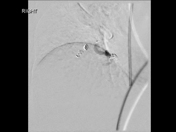
Post embolization • Following the procedure, the patient clinically improved, decreased oxygen requirement. • The patient was discharged 4 days later. Sagittal
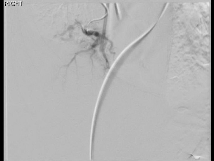
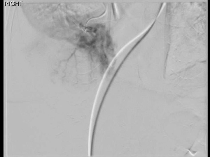
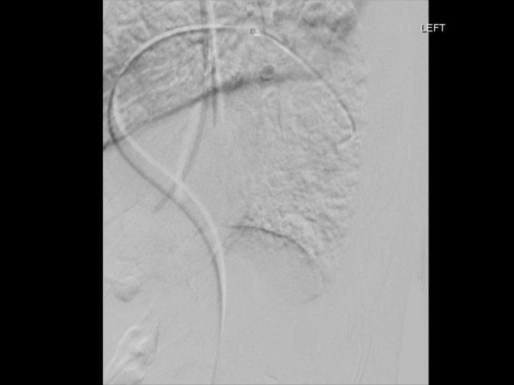
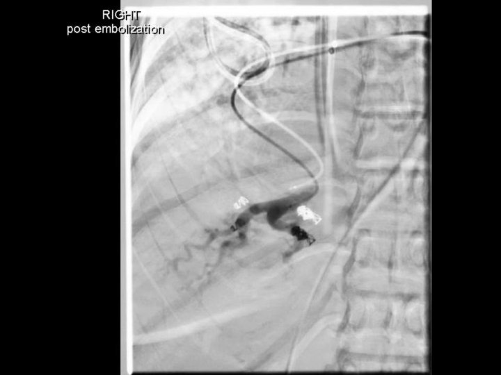
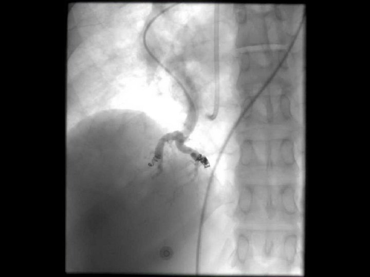
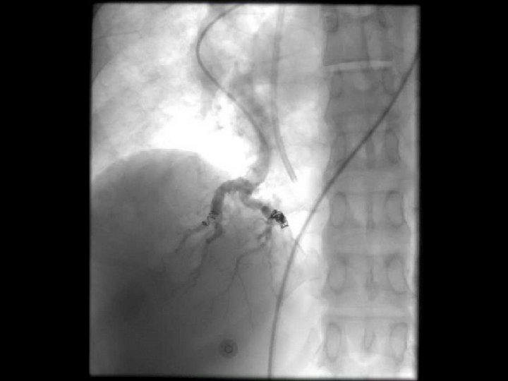
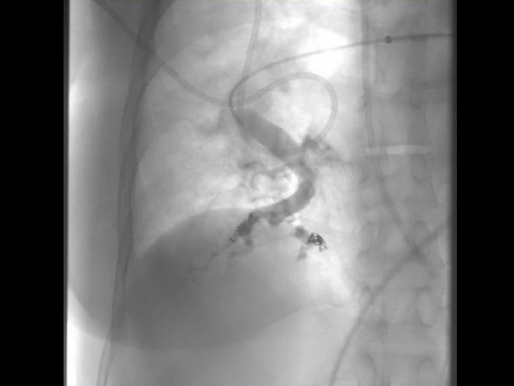
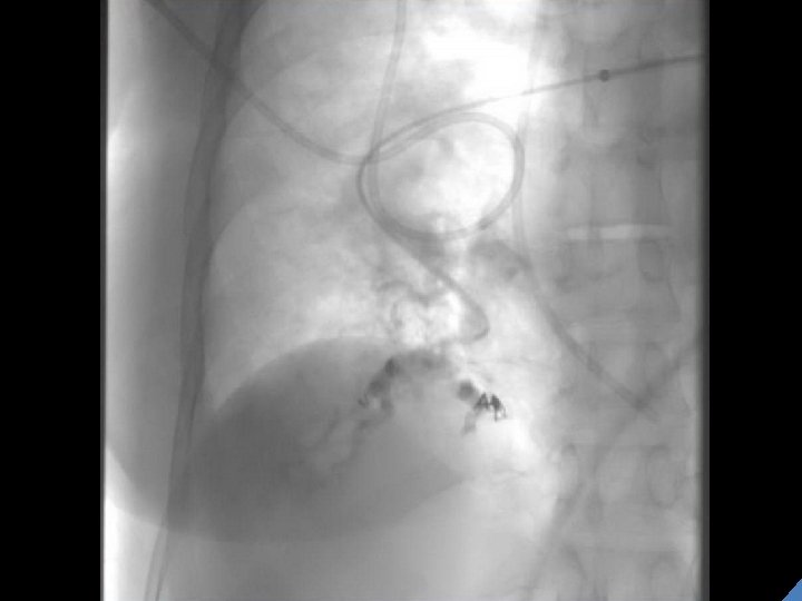
Which of the following is the most likely diagnosis? A) Pulmonary AVM B) Pulmonary AVF C) Pulmonary varix D) Pulmonary aneurysm
Which of the following is the most likely diagnosis? A) Pulmonary AVM B) Pulmonary AVF C) Pulmonary varix D) Pulmonary aneurysm
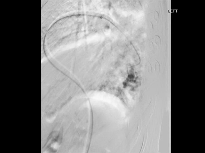
Pulmonary Varix Rare M=F May be congenital or acquired Acquired associated with mitral valve disease, hereditary hemorrhagic telangiectasia, or chronic pulmonary hypertension • Usually asymptomatic • May present with hemoptysis or thromboembolic disease • • Diego VP et al. Learning from the pulmonary veins. Radiographics 2013; 33: 999 -1022.
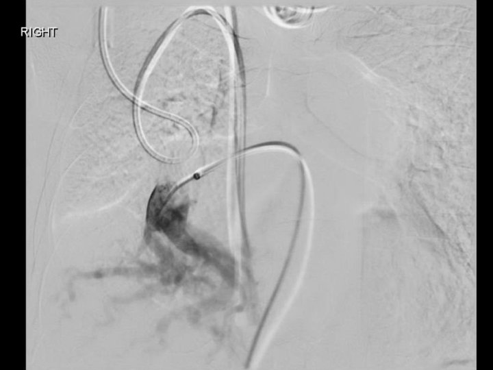
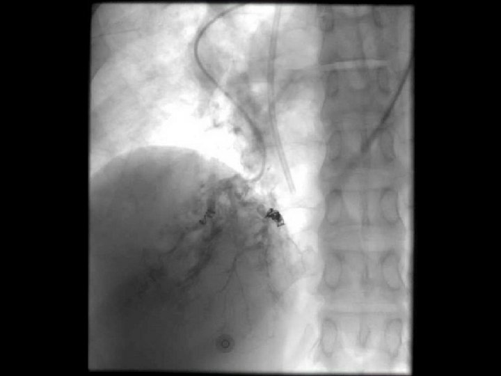
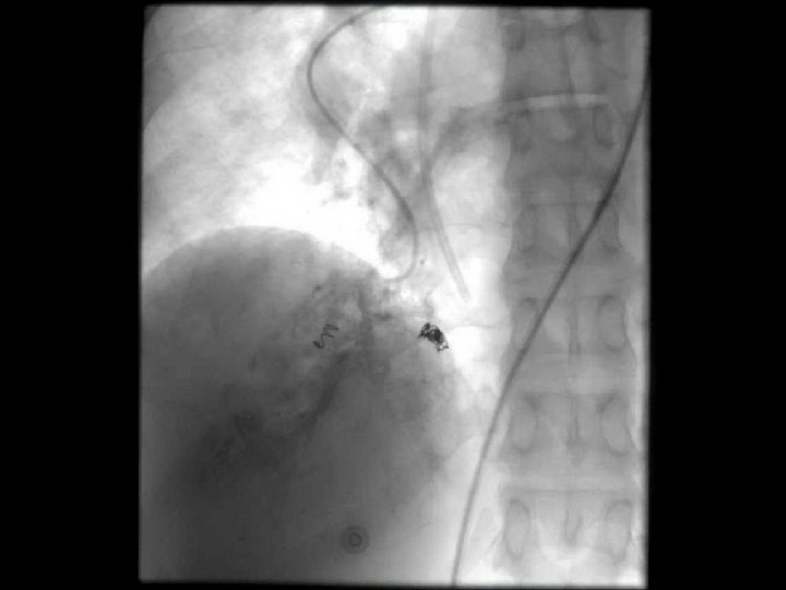
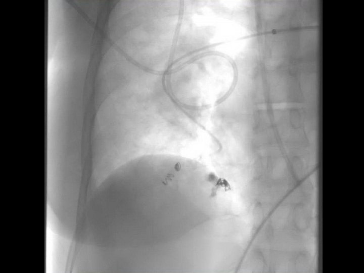
Angiographic findings 1) Pulmonary artery normal in arterial phase 2) No pulmonary arteriovenous shunt 3) A blood vessel flowed directly from the varix to the left atrium 4) Contrast remained in the varix, compared to retention in normal pulmonary vein 5) Peripheral region of the dilated, tortuous vein was normal Bartram C, Strickland B. Pulmonary varices. Br J Radiol 1971; 44: 927 -35.
Thank you