Case presentation DR AHMED KENSARAH History This is


 4")


- Slides: 51
Case presentation DR. AHMED KENSARAH
History This is a 60 year old Saudi lady. known case of • HTN • Hypothyroidism • RA C/O Left large ulcerating breast mass.
History of present illness • Lt breast lump 6 years ago • Ignored • in last 1 year. • • The mass increased in size Bloody nipple discharge Skin changes Fungating Ulcer
History • No Family history of breast cancer • No history of benign breast disease or biopsy • No history of smoking.
History • Age of menarche: 14 • Age of menopause : 50 • She had her 1 st child birth at 30 years of age • No History of OCP
• Systemic Review unremarkable • Past Surgical History Lateral anal sphincterotomy 2 years back • Allergies -ve • Medication – Thyroxin – Mobic – Capotin
Physical Examination Vital signs CVS Unremarkable Chest Abdomen
Breast Examination • Right breast Normal with free axilla • Left breast Enlarged Red Edematous Fungated ulcer Bloody discharge
• Left breast Firm Hot Tender Left arm swelling No axillary LN
Investigation Blood work: • • CBC U&E LFT Serum Calcium NORMAL
Investigation Radiological • Mammogram – Left breast : was not done. – Right breast : Benign prominent ducts.
Investigation Ultrasound : • Left : – Whole Left breast parenchyma was involved – single hypoechoic L. N in the Left axilla measuring 1. 8 cm. • Right: – Normal appreaing parenchyma. – Multiple small L. N in the Right axilla.
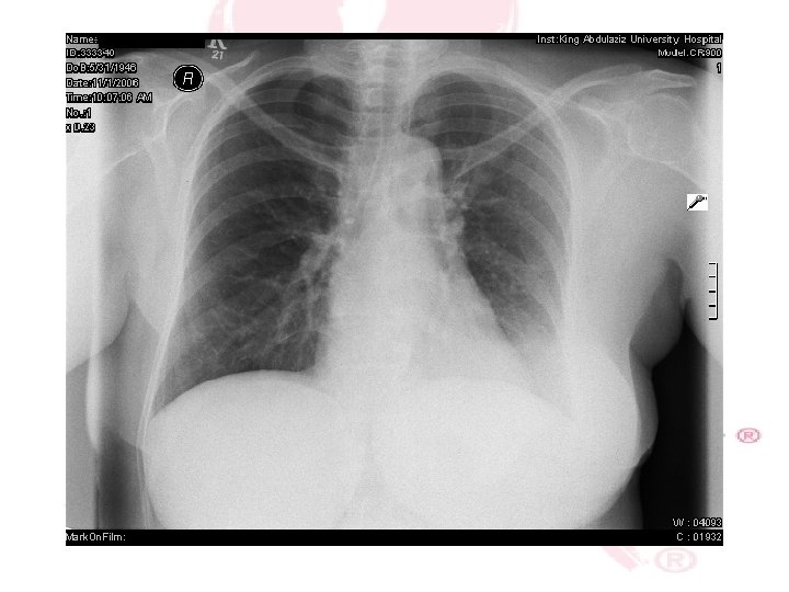
CHEST X-RAY
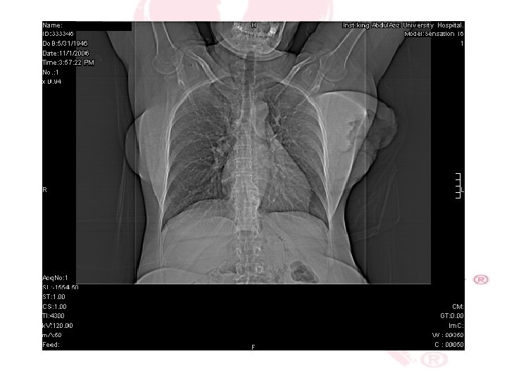
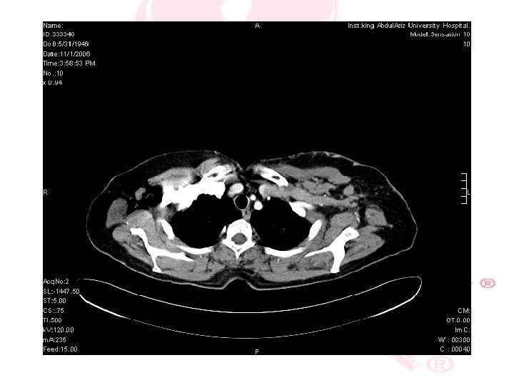
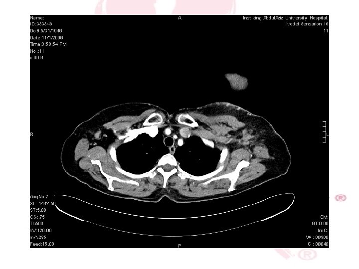
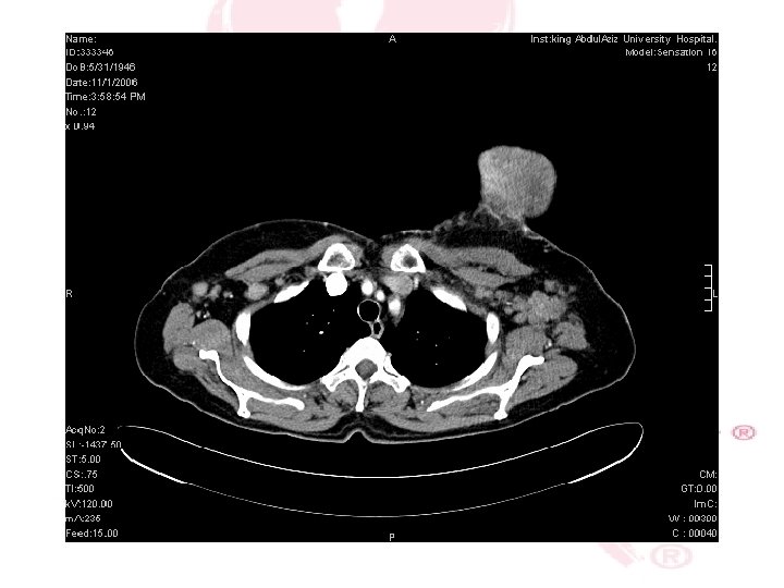
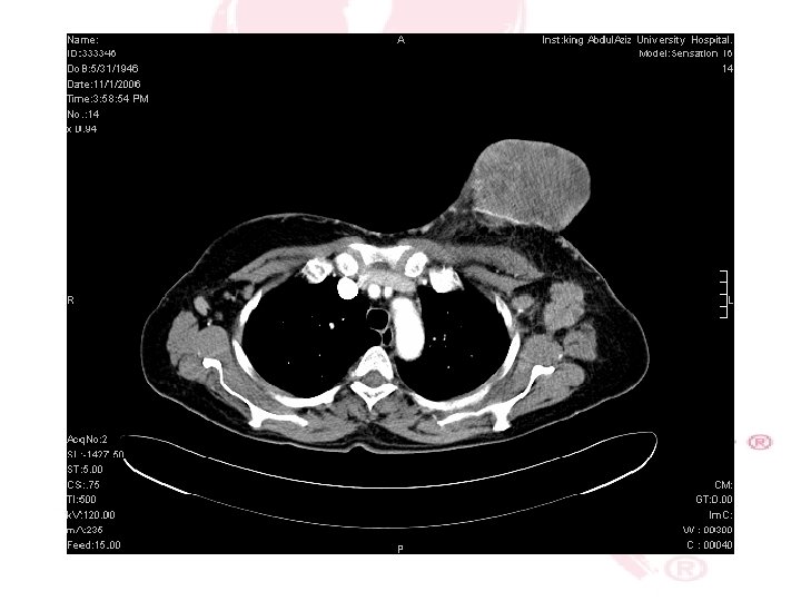
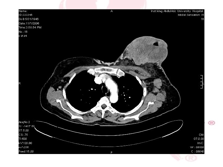
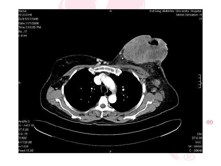
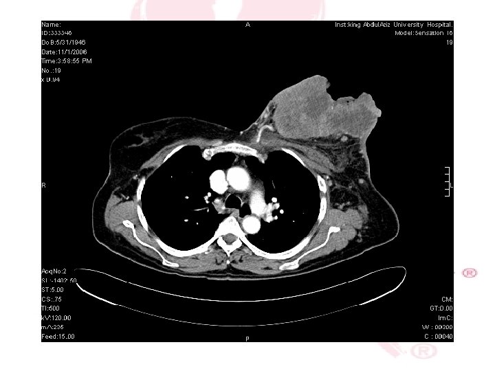
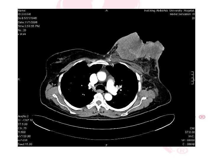
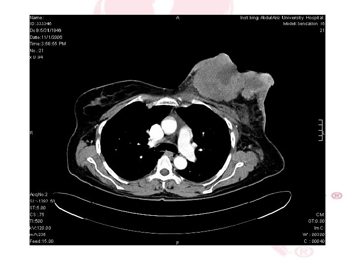
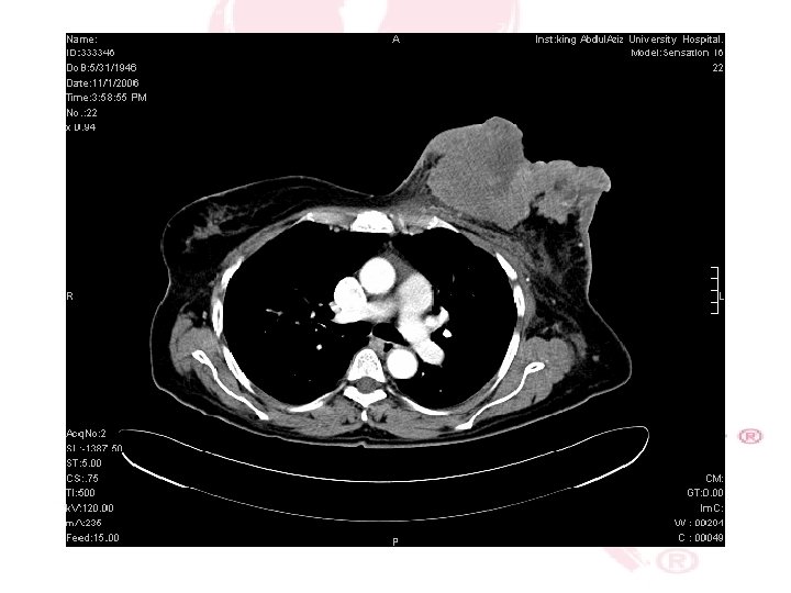
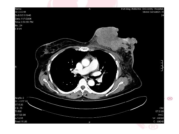
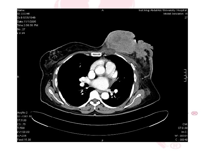
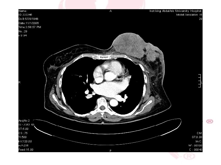
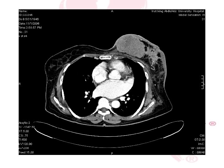
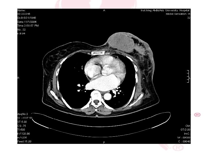
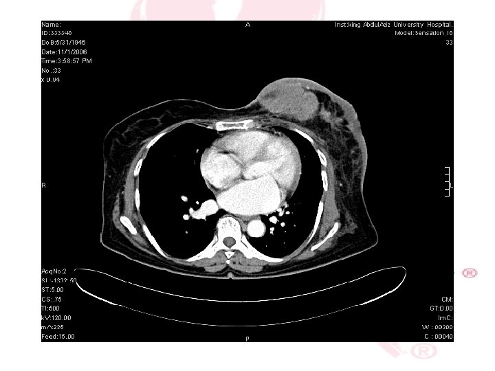
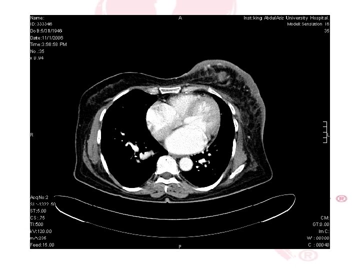
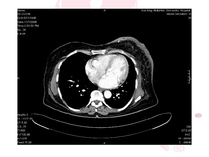
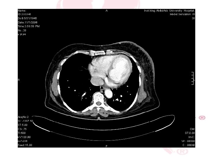
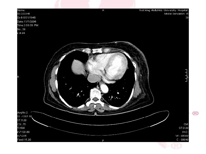
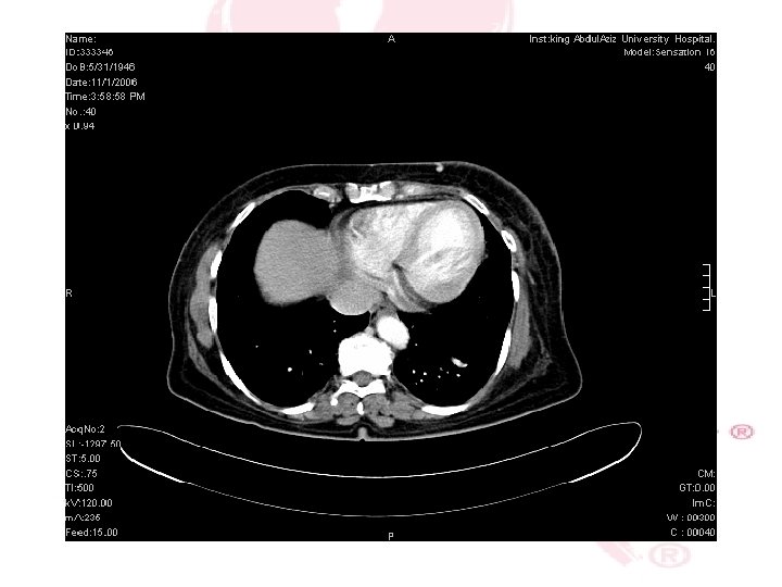
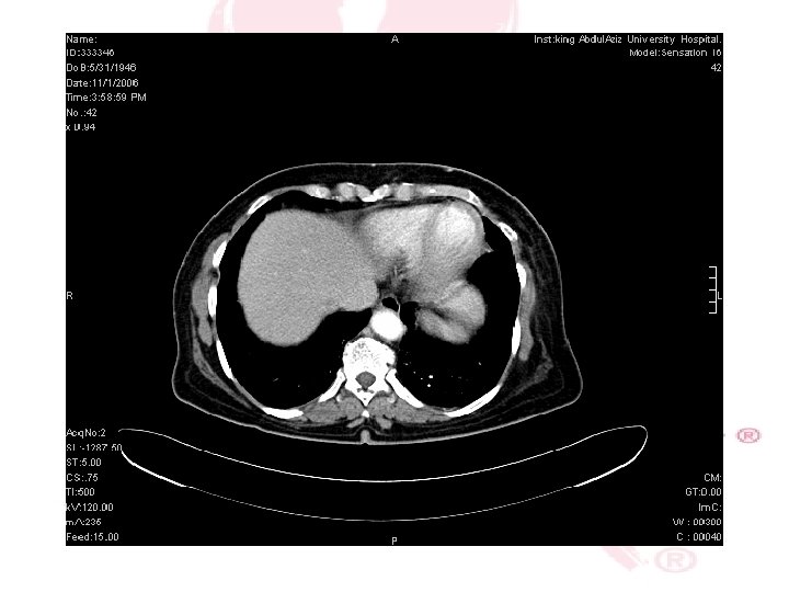
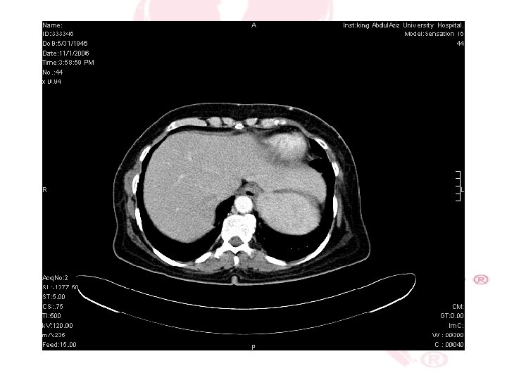
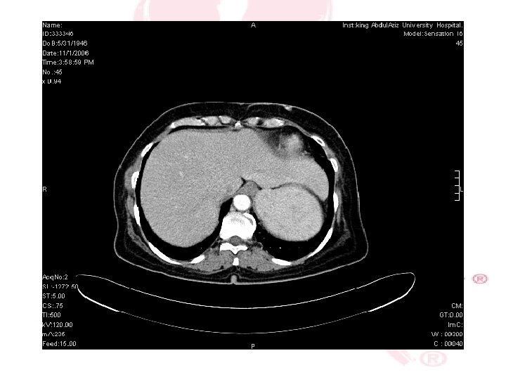
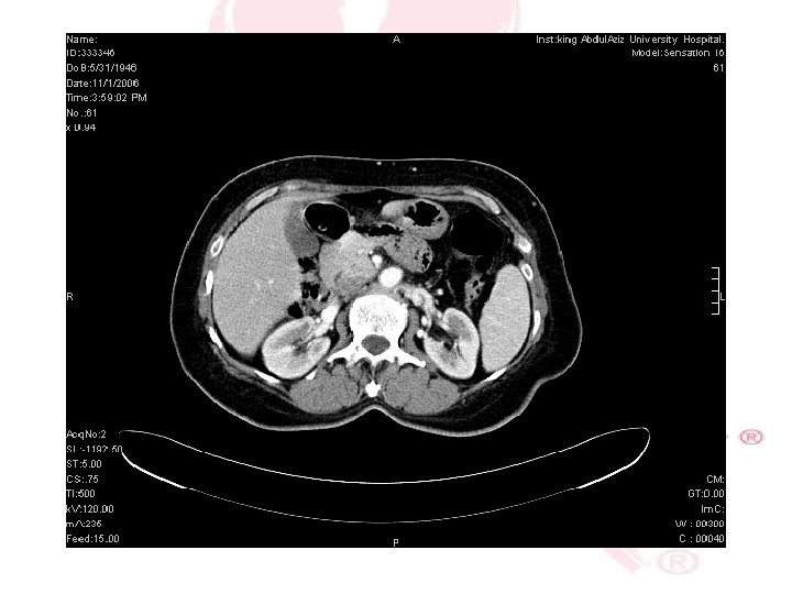
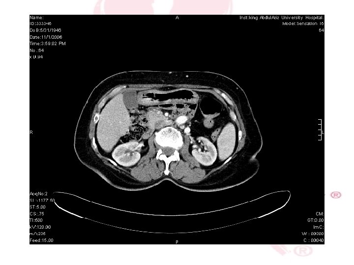
CT SCAN CHEST , ABDOMEN &PELVIS
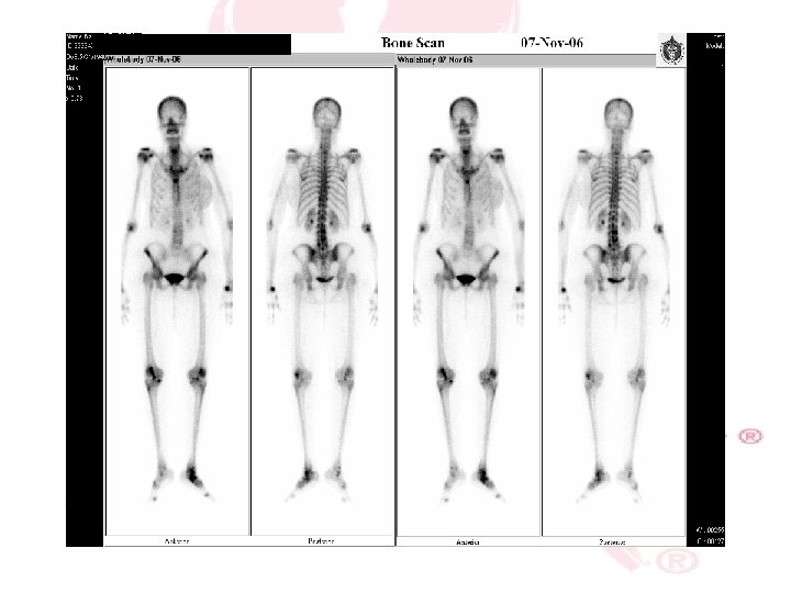
Bone scan
Histopathology True cut Biposy Invasive Ductal Carcinoma ER/PR Status : +ve HER 2 score : +1
Diagnosis Invasive Ductal Carcinoma. Stage 3 Locally advanced Breast Cancer.
mangement referred to the Oncology. Started on the AC Regimen ( Adriamicin, Cyclophosphamide) 4 cyles, once every 3 weeks. It will be followed by the taxanes for 4 cyles.
mangement Down staging of the tumor for operation. MRM with axillary L. N dissection. Plastic surgery Referral for possible Pectoralis Muscle flap.