Case Presentation CC Abdominal pain HPI 53 yo

Case Presentation CC Abdominal pain HPI 53 yo woman presents with acute epigastric pain radiates to her back associated with nausea & emesis No Et. OH, trauma, new meds PMH RA, HTN, Cholecystectomy, C/S

Case Presentation MEDS ALL Remicade, Lodine, Altace, Methotrexate Codeine SH Married, 20 ppd, occ Et. OH FH Negative ROS Negative

Case Presentation Vitals Gen HEENT Lungs Heart Abd Ext Neuro 226/110 84 20 98. 6 NAD Anicteric CTA RRR with flow murmur Epigastric pain w/o R/G No c/c/e A&Ox 3. Non-focal

Case Presentation Lipase LFTs WBC HGB Ca Alb BUN Cr Tri 1157 NL 16, 900 13. 1 9. 7 4. 4 20 0. 8 96

Case Presentation

Pancreatic Cystic Neoplasms Rajeev Jain, M. D.

Cystic Lesions of the Pancreas Congenital Cysts Acquired Cysts Dermoid VHL Polycystic Simple Pseudocyst Parasitic Retention Primary Cystic Nonpancreatic Neoplasms Lesions Adrenal cyst Biloma Choledochal Choledochocele Diverticulum Duplication Mesenteric Retroperitoneal Splenic artery aneurysm Levy MJ & Clain JE. Clin Gastroenterol Hepatol. 2(8): 639 -53. 2004.

Primary Pancreatic Cystic Neoplasms • Mucinous Tumors – Cystadenoma – Cystadenocarcinoma – Intraductal papillary mucinous tumor (IPMN) • Acinar cell cystadenocarcinoma • Angiomatous tumor – Angioma – Hemangioma – Lymphangioma • Nonmucin Tumors – Serous cystadenoma – Solid-pseudopapillary tumor • Cystic degeneration • • Islet cell tumors Lymphoepithelial cyst Pancreaticoblastoma Teratoma Levy MJ & Clain JE. Clin Gastroenterol Hepatol. 2(8): 639 -53. 2004.

Epidemiology TYPE GENDER AGE % Serous cystadenoma F>M 60 s 32 -39 Mucinous cystic neoplasm F>M 40 s 10 -45 IPMN M=F 50 s 21 -33 Solid pseudo-papillary F>M 30 s <10 Cystic endocrine neoplasm M=F 40 s <10 M>F 50 s <1 Ductal adenocarcinoma with cystic degeneration Acinar-cell cystadenocarcinoma Brugge WR. et al. N Engl J Med. 351: 1218 -26. 2004.

Serous Cystadenoma • Microcystic, serous, or glycogen-rich adenomas • Presentation – 50 -60% have abdominal pain (up to 25 cm) – 30% palpable mass – Occ. obstructive jaundice, pancreatitis, pancreatic insufficiency, or gastric outlet obst. – 25% small & asymptomatic found on CT Levy MJ & Clain JE. Clin Gastroenterol Hepatol. 2(8): 639 -53. 2004.
, small (<1 -2 cm), fluid-filled")
Serous Cystadenoma • Focal, welldemarcated lesions • Multiple (>6), small (<1 -2 cm), fluid-filled microcysts • Dense fibrous septations give honeycomb appearance 8 mm Levy MJ & Clain JE. Clin Gastroenterol Hepatol. 2(8): 639 -53. 2004.

Serous Cystadenoma Source: www. jichi. ac. jp

WHO Classification Mucin-producing cystic neoplasms of the pancreas Mucinous cystadenoma Intraductal Papillary Mucinous Neoplasm (IPMN) Mucinous cystadenocarcinoma

Mucinous Cystadenoma • Macrocystic adenoma • Premalignant – 25% contain malignancy at time of diagnosis • Presentation – – Pain (60 -80%) Diabetes (25%) Pancreatitis (10 -20%) Incidental (10 -30%) • Ovarian stroma Grogan JR et al. AJR 176: 921 -9. 2001. Levy MJ & Clain JE. Clin Gastroenterol Hepatol. 2(8): 639 -53. 2004.

Ovarian Stroma
 Main Duct Variant Side Branch Variant Grogan JR et")
Intraductal Papillary Mucinous Neoplasm (IPMN) Main Duct Variant Side Branch Variant Grogan JR et al. AJR 176: 921 -9. 2001.
 – Acute pancreatitis (22 -45%) – Asymptomatic")
IPMN • Presentation – Pain (50 -100%) – Acute pancreatitis (22 -45%) – Asymptomatic (up to 30%) • Variants – Main duct (47 -75%) – Side branch (25 -39%) – Both (14%) Levy MJ & Clain JE. Clin Gastroenterol Hepatol. 2(8): 639 -53. 2004.

Evaluation of PCNs • 10 -37% initially diagnosed erroneously as pseudocyst – Delay in diagnosis – Lost opportunity for curative resection • Type of PCN – Demographics – Radiology – EUS with FNA

Imaging of PCNs SCA MCN IPMN Even Body/tail Head Septae Yes No Locularity Multi Central sunburst Peripheral curvilinear None PD displaced PD dilated & mucin Location TUS/CT/EUS Calcifications ERCP Levy MJ & Clain JE. Clin Gastroenterol Hepatol. 2(8): 639 -53. 2004.

Serous Cystadenoma CT Findings Demos TC et al. AJR. 179: 1375 -1388. 2002.
: 701 -7. 2002.")
IPMN and ERCP Aithal GP et al. Gastrointest Endosc. 56(5): 701 -7. 2002.

Pancreatic Duct

IPMN Histology

Pancreatic Cyst Fluid Analysis Viscosity Amylase CA 19 -9 CEA Cytology Serous cystadenoma Low Variable Low Glycogen Mucinous cystadenoma High Variable High Mucinous cystadeno. CA High Variable High Mucin IPMN High Variable Mucin Pseudocyst Low High Variable Low Histiocytes Levy MJ & Clain JE. Clin Gastroenterol Hepatol. 2(8): 639 -53. 2004.

Pancreatic Cyst Fluid Analysis • 19 pancreatic cystic masses, 31 pseudocysts • CA 19 -9 > 50, 000 U/m. L – Sens 75%, Spec 90% – MCN > other cysts • CEA < 5 ng/m. L – Sens 100%, Spec 86% – SCN > other cysts • Amylase > 5, 000 U/m. L – Sens 94%, Spec 74% – Pseudocysts > other cysts Hammel P. et al. Gastroenterology. 108: 1230 -5. 1995.

Diagnosis of Pancreatic Cystic Neoplasms • • Multicenter trial 341 pts EUS-FNA 112 pts surgical resection Prospective evaluation: – EUS imaging, – Cyst fluid cytology, – Cyst fluid tumor markers • CEA, CA 72 -4, CA 125, CA 19 -9, and CA 153 Brugge WR. et al. Gastroenterology. 126: 1330 -6. 2004.
 –")
Diagnosis of Pancreatic Cystic Neoplasms Accuracy – CEA • (88 of 111, 79%) – EUS morphology • (57 of 112, 51%) – Cytology • (64 of 109, 59%) Brugge WR. et al. Gastroenterology. 126: 1330 -6. 2004.

Proposed Management Algorithm for Symptomatic PCNs Scheiman JM. Gastroenterology. 128: 463 -9. 2005.

Proposed Management Algorithm for Asymptomatic PCNs Castillo, C. F. -d. et al. Arch Surg. 138: 427 -34. 2003.

Pancreatic Cystic Neoplasm
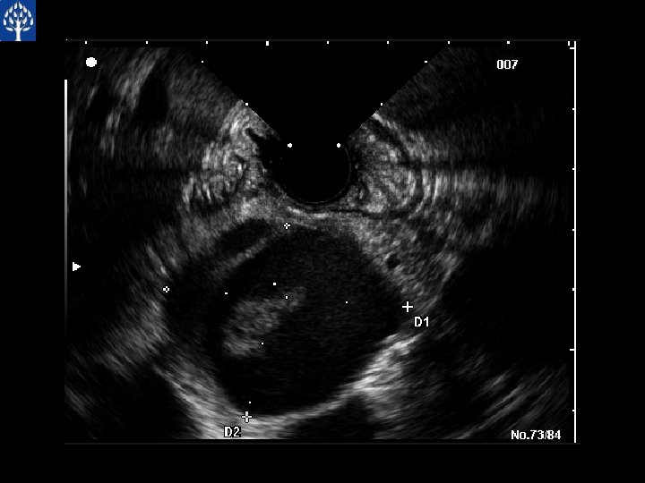

EUS Guided Cyst Aspiration Amylase CEA CA 19 -9 93 906 1, 890, 000

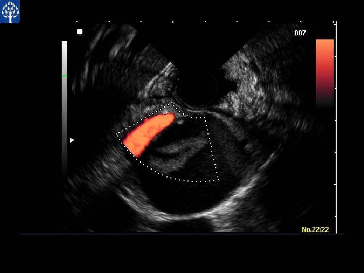
Pancreatic Cystic Neoplasm

Pancreatic Cyst Amylase 502 CEA 2. 5 CA 19 -9 ----

Case Presentation
- Slides: 36