Carpal Instability Weiling Chang Carpal Instability Definition Inability

Carpal Instability Weiling Chang

Carpal Instability - Definition • Inability to maintain normal alignment and distribute load under physiologic conditions. – Results from ligamentous and osseous injuries. – “Static” pattern: abnormal carpal alignment at rest – “Dynamic” pattern: normal carpal alignment at rest but abnormal alignment with movement or stress.

Carpal Instability - Caveats • Little consensus in literature regarding classification. • Biomechanics of the many extrinsic and intrinsic ligaments still being investigated.

Diagnosis of carpal disorders • Start with plain radiographs to assess alignment. • Articular bones have opposing surfaces 2 mm or less. • Check Gilulas lines. • Three smooth arcs. Disruption in the continuity suggests abnormality at site of broken arc.

Overview of Patterns of Carpal Instability • Dissociative – Scapholunate dissociation – Lunotriquetral dissociation • Non-dissociative – Radiocarpal – Midcarpal

Normal DISI VISI MR imaging of the major carpal stabilizing ligaments: normal anatomy and clinical examples. . Radiographics. 1995 May; 15(3): 575 -87

Normal

DISI: SCAPHOLUNATE ANGLE > 60

VISI SCAPHOLUNATE ANGLE < 30

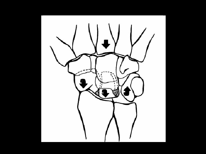
Normal Carpal Kinematics • At the carpus, OPPOSING torques are always acting: – Under axial load or radial deviation, the scaphoid flexes and the triquetrum extends. – With ulnar deviation, the scaphoid extends and the triquetrum flexes. • Lunate is the intercalated segment b/n these opposing forces of the scaphoid and triquetrum. • Forces are balanced by a ligamentous ring.

DISI PATTERN VISI PATTERN LUNATE is the intercalated element

Overview of Patterns of Carpal Instability • Dissociative – Scapholunate dissociation – Lunotriquetral dissociation • Non-dissociative – Radiocarpal – Midcarpal

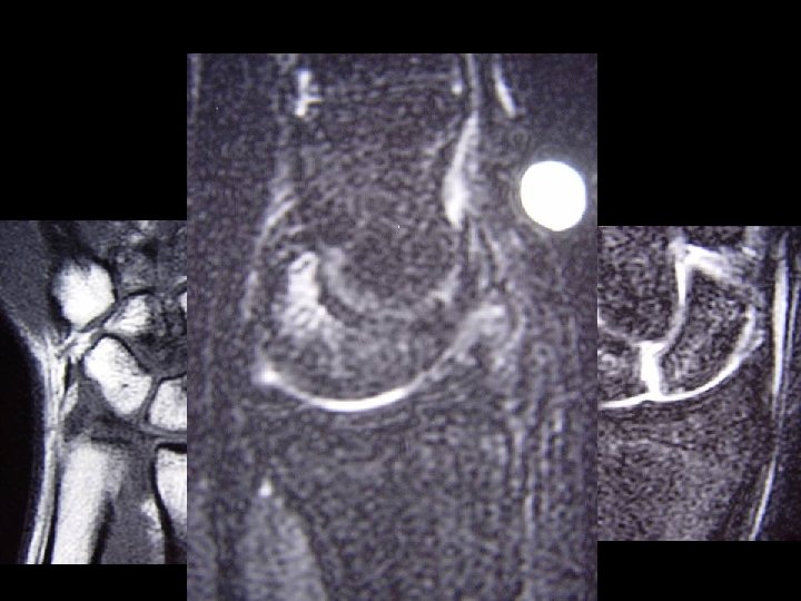
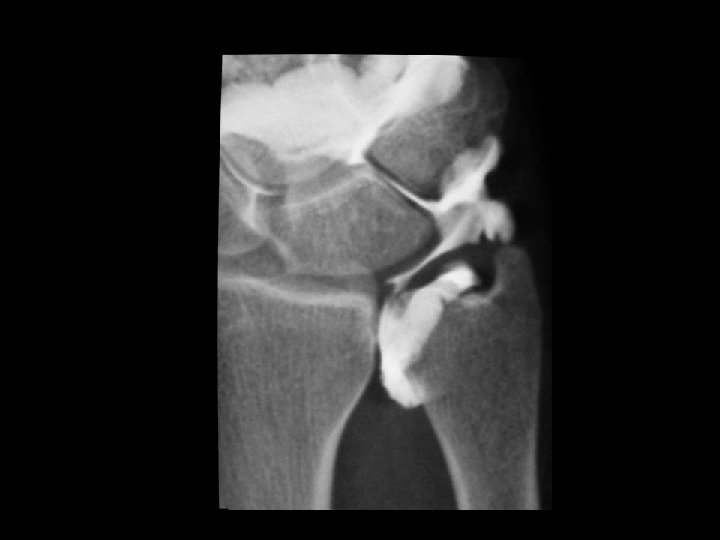
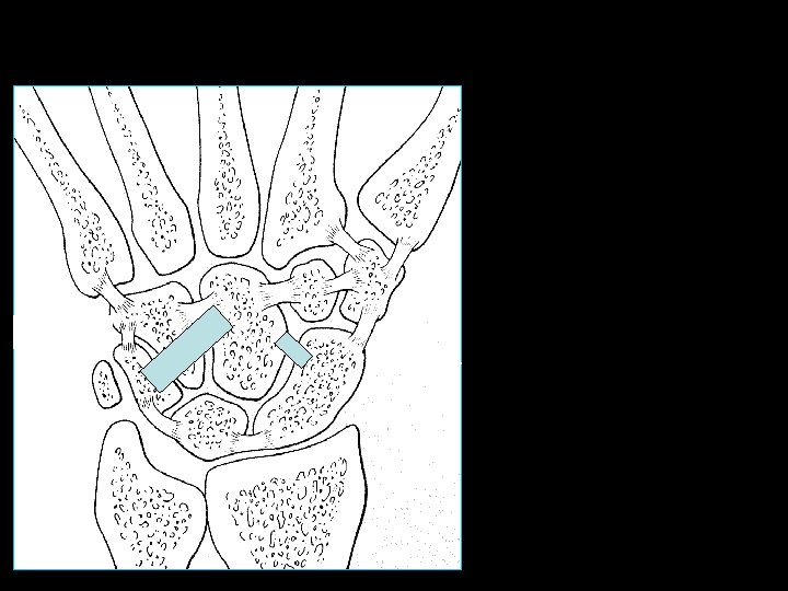
Scapholunate dissociation • Scapholunate interosseous ligament is the strongest and stiffest of the interosseous ligaments. • Occurs as an isolated injury or with distal radius or scaphoid fractures. • Tenderness in the anatomic snuffbox. • Rupture site most often at scaphoid attachment sites because fibers less dense. • Tears traumatic and degenerative.

Scapholunate ligament

Skeletal Radiol. 2006 Apr 12

Classification of Scapholunate Dissociation Stage I identified with MRI. Stage II diagnosed with stress views with a clenched hand. Stage III and IV demonstrates DISI pattern. Eur Radiol. 2006 Mar 1

Scapholunate dissociation DISI PATTERN LUNATE is the intercalated element


S/L Dissociation, Scaphoid Rotary Subluxation DISI

Eur Radiol. 2006 Mar 1
: 575 -87.")
Radiographics. 1995 May; 15(3): 575 -87.

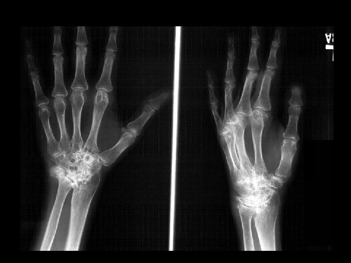
Endstage: SLAC Wrist

Scaphoid Fracture DISI PATTERN LUNATE is the intercalated element
: 575")
Scaphoid Fracture DISI PATTERN LUNATE is the intercalated element Radiographics. 1995 May; 15(3): 575 -87.
:")
Humpback Deformity Wrist fractures: what the clinician wants to know. Radiology. 2001 Apr; 219(1): 11 -28
: 575 -87.")
Radiographics. 1995 May; 15(3): 575 -87.

Overview of Patterns of Carpal Instability • Dissociative – Scapholunate dissociation – Lunotriquetral dissociation • Non-dissociative – Radiocarpal – Midcarpal

Lunotriquetral Dissociation • Like the scapholunate interosseous ligament, disruption may be either traumatic or degenerative. • Many tears associated with Palmer II TFCC tears. • Studies and literature regarding this ligament is scarce and controversial. • Occur both in isolation or part of the perilunate instability.
: 114 -20")
J Am Acad Orthop Surg. 1998 Mar-Apr; 6(2): 114 -20

Lunotriquetral Dissociation • Heterogeneity of clinical symptoms from asymptomatic tears to collapse of the carpus with a fork-like deformity of the wrist. • Generally pain aggavated with ulnar deviation. • Sensation of weakness or instability.
: 701 -7.")
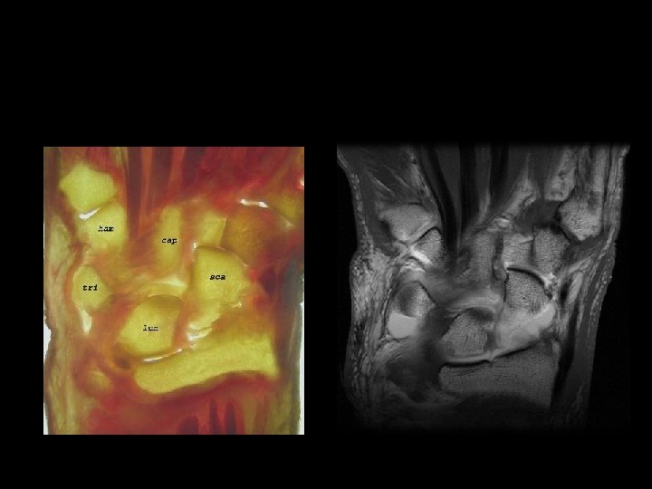
Normal Lunotriquetral Ligament Radiology. 2003 Jun; 227(3): 701 -7.

Normal Lunotriquetral Ligament Skeletal Radiol. 2006 Apr 12

VISI PATTERN LUNATE is the intercalated element

Eur Radiol. 2006 Mar 1

Eur Radiol. 2006 Mar

Overview of Patterns of Carpal Instability • Dissociative – Scapholunate dissociation – Lunotriquetral dissociation • Non-dissociative – Radiocarpal – Midcarpal

Radiocarpal Instability • Results in shift of the entire carpus. • Palmar, dorsal, radial, or ulnar translocation. • Dorsal and volar translocations with Barton or reverse Barton fractures. • Ulnocarpal translocations more frequently occur with RA, CPPD.

Normal. Inclination of the radial articulating surface

Eur Radiol. 2006 Mar 1

TRAUMA Eur Radiol. 2006 Mar CPPD ARTHROPATHY

Overview of Patterns of Carpal Instability • Dissociative – Scapholunate dissociation – Lunotriquetral dissociation • Non-dissociative – Radiocarpal – Midcarpal

Midcarpal Instability • Disruption of normal smooth motion of the proximal carpal row. Normal carpal kinematics

Midcarpal Instability • Occurs from repetitive stress in young patients. • Grip strength can be reduced by 50% • Painful and audible “snapping” caused by en bloc extension of the proximal carpal row during ulnar deviation.

Dynamic Midcarpal instability Eur Radiol. 2006 Mar

Static Midcarpal Instability • Static MCI results in flexion of the proximal carpal row and VISI

Eur Radiol. 2006 Mar

Conclusion • Dissociative – Scapholunate dissociation – Lunotriquetral dissociation • Non-dissociative – Radiocarpal – Midcarpal
- Slides: 53