Cardiovascular Testing A careful patient history and physical


 and changes in")


- Slides: 18
Cardiovascular Testing
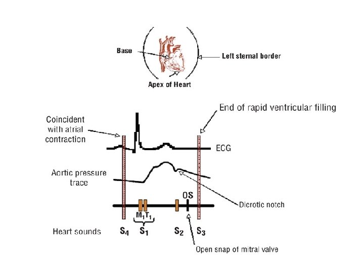
• A careful patient history and physical examination are extremely important in diagnosing cardiovascular disease and should be done prior to any test. • Heart sounds and heart murmurs are important in identifying heart valve abnormalities and other structural cardiac defects. • Elevated jugular venous pressure is an important sign of heart failure and may be used to assess severity and response to therapy.
• Electrocardiography is useful for determining rhythm disturbances (tachyor bradyarrhythmias) and changes in ventricular and atrial size. • Exercise stress testing provides important information concerning the likelihood and severity of coronary artery disease; changes in the electrocardiogram, blood pressure, and heart rate are used to assess the response to exercise. • Cardiac catheterization and angiography are used to assess coronary anatomy and ventricular performance.
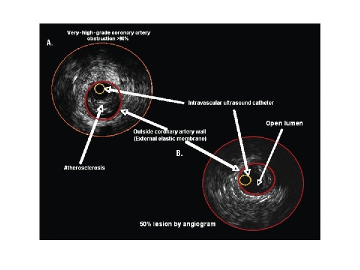
• Every 36 seconds 1 person dies from cardiovascular disease and each day about 2, 500 people die in the United States. • Cardiovascular disease exceeds the next four leading causes of death combined (cancer, lung disease, accidents, and diabetes). • Another important factor in cardiovascular disease is that greater than 60% of unexpected cardiac deaths occur without prior history of heart disease and 70% of patients having a myocardial infarction have coronary artery blockages of about 40% to 60%.
• A comprehensive history is the cornerstone of a cardiovascular workup. • The value of the history depends on the clinician’s ability to elicit relevant information. • Family history is very important because of the genetic links involved in many cardiovascular diseases from early myocardial infarction, strokes, diabetes, valvular heart disease, hypertension and familial hypercholesterolemia.
• The elements of a comprehensive history include the chief complaint, present problems, past medical history, review of systems, and social history. • Ischemic heart disease is the most common cardiovascular disease seen in clinical practice. • A focus on chest pain history is very important. The clinical syndrome of angina is frequently described by patients as a discomfort from chest ache, pain, or pressure, to dull pain in the jaw, back, shoulder, or either arm.
• Symptoms of angina can occur in patients with noncardiac conditions such as gastrointestinal (esophageal), chest wall, or pulmonary disease. • Based on the history, classify the patient’s symptoms. • Three characteristics to consider are (a) whether the substernal chest discomfort has a classic quality and duration that is (b) provoked by exertion or emotional stress and (c) relieved by rest or nitroglycerin.
• The cardiovascular physical examination is divided into four categories: 1. Global examination of the patient for signs of cardiovascular disease (CVD) and a review of all body systems. 2. Observation and assessment of physical findings (e. g. , jugular venous pressure). 3. Measurement of parameters of CVD function (pulse, blood pressure). 4. Auscultation, percussion, and palpation of the chest and related cardiac structures.
• Heart rate is described by both rate and rhythm. • In healthy individuals, the heart rate is usually assessed by counting the pulse for 15 seconds and multiplying by 4. • In patients with irregular rhythms, the pulse should be taken over an extended period, approximately 1 to 2 minutes, to try to determine the patient’s average pulse and rhythm.
• Arterial pulses are an accurate measure of the ventricular rate in healthy persons with good ventricular function. • In patients with a rapid ventricular rate—because of supraventricular tachyarrhythmias such as atrial flutter or fibrillation or rapid ventricular rates (e. g. , ventricular tachycardia or premature ventricular beats)— extremity pulses (e. g. , radial pulse) may be considerably slower than the true ventricular rate. • A more accurate ventricular rate is determined by listening to the ventricles with the stethoscope (usually at the apex) or counting from an electrocardiogram (ECG).
TESTING MODALITIES: 1 - CHEST RADIOGRAPHY: • It gives global information about position and size of the heart and chambers and surrounding anatomy. • The standard chest radiographs for evaluation of lungs and heart are standing posteroanterior and lateral views taken at maximal inspiration.
• The posteroanterior view chest radiograph outlines the superior vena cava, right atrium on the right and left sides, aortic knob, main pulmonary artery, left atrial appendage (especially if enlarged), and left ventricle. • In the lateral view, the chest radiograph visualizes the right ventricle, inferior vena cava, and left ventricle. • These structures are visualized as shadows of differing density rather than discrete structures.
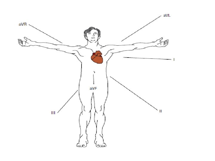
2 - ELECTROCARDIOGRAM: • Measurement of electrical activity in the heart, now known as the ECG, was introduced about 75 years ago by Willem Einthoven. • In its simplest interpretation, the ECG characterizes rhythms and conduction abnormalities. • However, the ECG also provides, by inference, information about the anatomy and structures of the heart, pathophysiologic changes, and hemodynamics of the CVD system. • ECG abnormalities are often the earliest sign of adverse drug effects, ischemia, and electrolyte abnormalities.
• The ECG can be used to evaluate ischemia following angioplasty or other surgical interventions and to monitor responses to antiarrhythmic agents or in patients receiving drugs with potential cardiac effects.
Drugs That May Affect the Electrocardiogram • Digoxin • Pentamidine • Antiarrhythmics—classes I–IV • Lithium • Tricyclic antidepressants • Catecholamines (e. g. , dopamine, albuterol) • H 1 antagonists • Diuretics (electrolyte abnormalities) • Methylxanthines • Doxorubicin