Cardiovascular System The Human Heart Human heart valves


, it pumps blood")


 and two")

")


 , the valve does ___________, causing")
 is a heart _______ in which there")
 node which is")

 of arteries contain _________________ that contract")
 of veins consist of _____ of")

- Slides: 25
Cardiovascular System The Human Heart
• Human heart valves are remarkable structures. These ________ membranes attached to the heart wall constantly _______ to _________ (causing the sound of a heartbeat). This flexing of the tissue occurs day after day, year after year. • In fact, the tissue withstands about _____ beats a year, or ___________________. Each beat is an amazing display of strength and flexibility.
Human Heart valves
• When the heart muscle _______ or beats (called _______), it pumps blood ____________. The heart contracts in two stages. • In the first stage, the _______ contract ___________, pumping blood to the _______ (through the ______ and _____ valves). • Then the ____________ to propel blood out of the heart (through the _________ valves).
• From the pulmonary valve the blood goes to the __________ then to the _____ where _______ occurs (________ circulation) • From the ______ the blood goes directly into the _______ to begin ___________ to the _____ of the body. • Then the heart muscle _____ (called diastole) before the next heartbeat. This allows blood to fill up the heart again
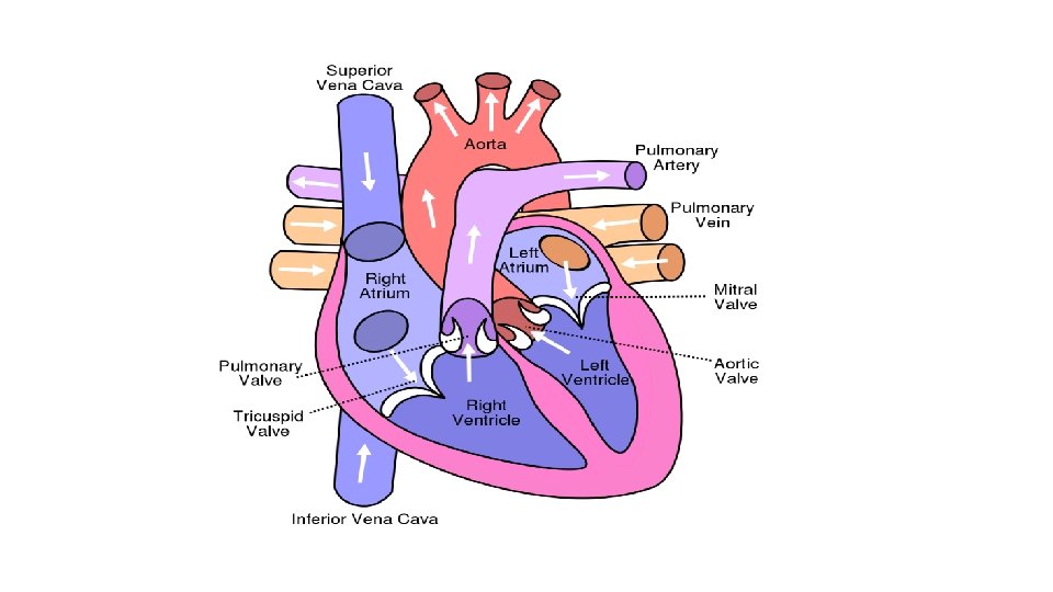
• The right and left sides of the heart have separate functions. • The ____ side of the heart collects _______ blood from the body and pumps it to the ____ where it picks up oxygen and releases carbon dioxide. • The _______ side of the heart then collects _______ blood from the lungs and pumps it to the body so that ______ throughout your body has the oxygen it needs to function properly. • The two sides of the heart are _____ by a very ________ called the ____, so that the O 2 blood and de-O 2 blood _______.
• The heart consists of four chambers, two ______ (_______ chambers) and two ______ (_____ chambers). • The atria are the _______ chambers of the heart, receiving blood flowing back to the heart. • The ventricles are the _______ chambers of the heart that pump the blood out of the heart. •
• Blood passes through a valve before leaving each chamber of the heart. • The valves prevent the _____________. • There are two _____ valves (atrioventricular) and 2 _________ valves • Valves are actually flaps (leaflets) that act as _____ inlets for blood coming ____ a ventricle and ______ outlets for blood ____ a ventricle. • Each of the 4 main valves has three flaps (leaflets), except the mitral (bicuspid) valve, which only has two flaps.
• The four heart valves include the following: • Tricuspid valve – (_____) located between the _______ atrium and the right ventricle. • Pulmonary valve – (_______ valve) located between the right ventricle and the _________. • Bicuspid (Mitral) valve – (____ valve) located between the _____ atrium and the left ventricle. • Aortic valve – (__________) located between the left ventricle and the aorta.
• The bicuspid/mitral valve and tricuspid valve control blood flow from the atria into the ventricles, this is blood flow ______ • • The aortic valve and pulmonary valve control blood flow ______ of the ventricles and _______ the aorta and pulmonary arteries, respectively. This is blood that flows ________ to the lungs and body systems
• Heart valves function this way. • As the heart muscle contracts and relaxes, the ______, letting blood flow into the ventricles and atria at _______ times. • The following is a step-by-step description of how the valves of the left ventricle function normally:
1. When the left ventricle _____, the __________ and the ______/mitral valve ______, to allow blood to flow from the left atrium into the left ventricle. 2. The left atrium ______, allowing even ______ to flow into the left ventricle. 3. When the left ______ contracts, the ____/mitral valve _____ and the _________, so blood flows into the aorta. Crazy cool huh? ? ?
• The normal heart sound is typically described as ”_____. " • The "lub" sound is usually ________ than the "dub", and it is associated with the ___________ at the beginning of systole. The sounds are also amplified by the chest, making them more audible. • The sounds of the heart are only from the ______, not opening. • The ”____" is the ______ heart sound. The ”____" sound is _____________ than the "lub" sound. It is associated with the ______________ (aortic and pulmonary). • The sound is shorter and louder because the ______ of the valves are _____ than the bicuspid and tricuspid valves.
Heart valve disease • When heart valves _____ open and close properly, the implications for the heart _______, possibly _______ the heart's _______ to pump blood adequately through the body. • Heart valves can malfunction in several ways, including the following: • .
• _______ – (also known as “prolapse”) , the valve does ___________, causing the blood to ________ instead of forward through the valve. • ______ - the valve opening is ________ or does not form properly, inhibiting the ability of the heart to pump blood properly. • _______- the valve opening ________, preventing blood from passing from an atria to a ventricle, or from a ventricle to the pulmonary artery or aorta. Blood must find an alternate route
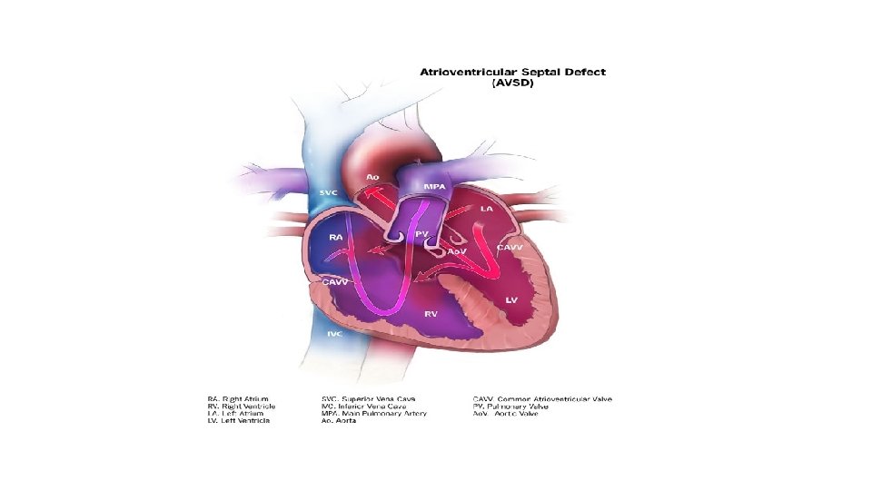
• An atrioventricular septal defect (AVSD) is a heart _______ in which there are ________ of the right and left sides of the heart, and the _____ that control the flow of blood between these chambers may ________
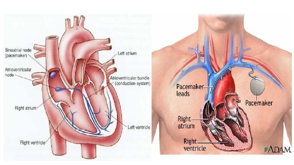
• The ____________ of the heart is the _______ (SA) node which is located in the ________. • The SA node generates an __________ which then gets sent around the heart, stimulating the cardiac muscle to contract and ___________.
• An "artificial pacemaker" is a small, ________ device that helps the heart beat in a _______. Most are permanent (_______) and inserted surgically (superficially) _______, and others are temporary (external). They can replace a ______ natural pacemaker or __________. • A pacemaker uses batteries to ___________ to the heart to help it pump properly. An _______ is placed next to the heart wall and small electrical _____________ to the right atrium of the heart. • Most pacemakers are _____ pacemakers. They have a ________. It turns the signal _____ when the heartbeat is above a certain level. It turns the signal ______ when the heartbeat is _______.
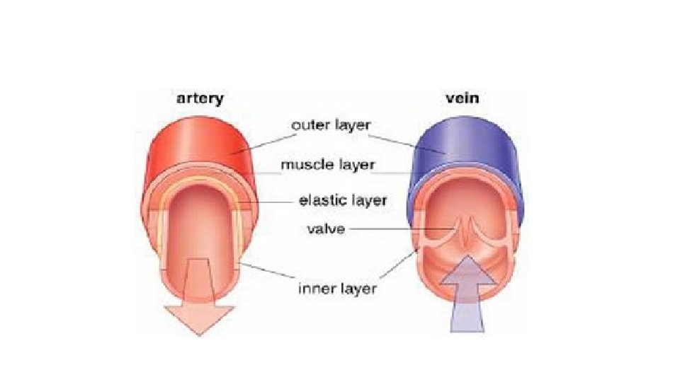
• Arteries • The _____ (outer structure) of arteries contain _________________ that contract and relax under the instructions of the sympathetic nervous system. • See your coloring plate 103 for detailed illustration of the anatomy of each of the blood vessels. • Arterioles – _____ arteries • Arteries - ______ have valves (except for the semi -lunar valves of the pulmonary artery and the aorta).
• Veins • The walls (outer structure) of veins consist of _____ of tissues that are ___________ than the corresponding layers of arteries. • Veins - ______ throughout the main veins of the body. These are to ____ blood flowing in the ____________________, as this could return waste materials back to the tissues. • Venules – branches of _____ veins
Arteries Veins - carry blood ______ from the heart - carry blood _____ the heart - ______ blood to various parts of body - ____ blood from various parts of body - _____, elastic muscle that can handle - _______, elastic muscle layer with ________ of blood flowing semilunar valves preventing backflow through them of blood - ________ in body - ______ to the skin - arterial walls are _______ - veins have _______ walls - there are ________ - ____ present, especially in limbs - thickest layer is ________________ - diseases – artherogenesis, myocardial - diseases – deep vein thrombosis ischemia