CARDIOVASCULAR SYSTEM heart macrocirculation microcirculation lymph vascular system


, which obeys ‘all or")


 >1 cm II. Distributing or Muscular")


 Tunica intima smaller with endothelium and incomplete or absent internal elastic")

 • direct connections between arterioles and venules allow blood to bypass")
.")

 • irregular vessels with large diameters • endothelium with gaps and")
 In comparison with arteries • Subjected to")


- Slides: 25
CARDIOVASCULAR SYSTEM • heart • macrocirculation, • microcirculation • lymph vascular system.
THE HEART Pericardium 1. Parietal pericardium outer membrane - fibrous layer tough, white fibrous, anchorage -serous layer a thin fibrous layer on top of a simple squamous mesothelium 2. Visceral pericardium= epicardium = serous layer Pericardial cavity filled with pericardial fluid preventing friction Pericarditis
The Heart Wall - three layers: A. The Epicardium
B. The Myocardium 1 - cardiac muscle 2 - fibrous connective tissue "skeleton of the heart" • • anchorage for the cardiac muscle and the atrioventricular valves. coronary trigone. The elastic component for the recoil that assists in filling the chambers Cardiac muscle ■ Striated. ■ Shorter than skeletal muscle ■ Rich in mitochondria (up to 40% of cell volume) ■ Branched. ■ Has one nucleus in the center of the cell. ■ No nerves are involved in the spread of contraction through the muscle. ■ Adjacent cells are interconnected end–toend by intercalated discs.
Intercalated discs where fibres interdigitate, function as a syncytium (unit), which obeys ‘all or none law’. Transverse Part: Transverse zonula (fasciae) adherents; A *hold cardiac muscle cells *anchor thin filaments Desmosomes (macula adherentes) rivets to prevent the cells from pulling apart B Lateral Part: Gap junctions (nexus) - for impulse transfer providing C (electrical communication between cardiac muscle cells) Lateral
C. The Endocardium • simple squamous endothelium • loose connective tissue • continuous with the endothelium of the blood vessels • Endocarditis. Cardiac Valves • fibrous trigone • lamina fibrosa, collagen fibers with elastic fibers) • endothelium
♥ Conduction pathway: Specialized muscle fibres without any direct stimulus from the nervous system, consisting of • Sinoatrial (SA) node located near the junction of the superior vena cava and the right atrium which initiates the beating action (called the pacemaker). -Internodal pathways (tracts) -Atrioventricular (AV) node -Bundle of His (AV bundle) -Rt & Lt bundle branches. -Purkinje fibers • generate electrical impulses • conduct electrical impulses through the myocardium • synchronize the heartbeat • are found along the innermost layer of the myocardium
BLOOD VESSELS vasoconstriction /vasodilatation • Regulate arterial blood pressure • Adjust blood flow within organs • Regulate capillary blood pressure • Distribute blood volume within the body arteries, arterioles, capillaries, venules and veins. General Structure of Blood Vessels three major layers or tunics • Tunica intima simple squamous endothelial, subendothelial connective tissue, internal elastic lamina • Tunica media circumferential smooth muscle, the external elastic lamina • Tunica adventitia connective tissue, nerves, vasa vasorum
ARTERIES I. Conducting or Elastic Arteries (large arteries) >1 cm II. Distributing or Muscular Arteries (medium arteries) 0. 1 -10 mm III. Arterioles (small arteries) <0. 1 mm Elastic Arteries (large arteries) e. g. aorta, pulmonary, branches of the aorta tunica intima thick, Endothelium with Weibel-Palade bodies/ von Willebrand factor (platelets coagulation), less prominent Internal elastic lamina tunica media most of the wall, concentric fenestrated lamellae of elastic fibers ( 50 elastic lamellae). Smooth muscle and collagen fibres (type III) Indistinct external elastic lamina tunica adventitia elastic, collagen fibres and is vasa vasorum
Weibel-Palade bodies Vasa vasorum
Muscular Arteries Tunica intima • thinner than in elastic arteries endothelium and Weibel-Palade bodies • well defined internal elastic lamina wavy pink line Tunica media • concentric layers of smooth muscle cells. • elastic and collagen fibres • the external elastic lamina can be clearly distinguished Tunica adventitia • variable thickness and appearance
Arterioles (small arteries) Tunica intima smaller with endothelium and incomplete or absent internal elastic lamina Tunica media circular smooth muscles i. e. single to few layers Tunica adventitia autonomic nerve fibres peripheral resistance blood pressure lumen size control arterial Arterioles are the major resistance vessels they branches into several arterioles with diameters small enough to offer considerable resistance to flow.
Microvascular bed Metarterioles communication between arterioles and venules and are important in bypassing the blood flow through the capillaries. True capillaries branch mainly from metarterioles Precapillary sphincters smooth muscles rings at the origin of true capillaries to regulate blood flow into true capillaries and tissues Thoroughfare channel NO Precapillary sphincters
Arteriovenous anastomosis (arterialvenous-shunt) • direct connections between arterioles and venules allow blood to bypass the capillary bed very common in the dermis of the skin, lips, nose and GIT • Functions as backup routes in case of blockage and assist in temprature regulation
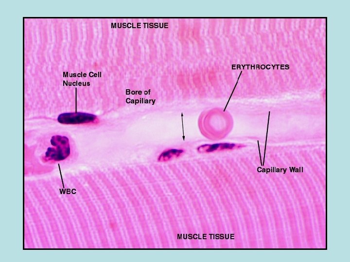
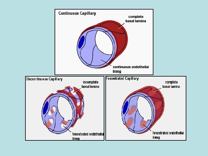
CAPILLARIES -Only the tunica intima -endothelium, its basal lamina and the pericytes (perivascular cells). -contractile -blood flow -repairing -phagocytic -Pinocytotic vesicles for transporting materials across endothelium in both directions -Ultrastructurally three different types of capillaries -Continuous capillaries -Fenestrated capillaries -Sinusoids (discontinuous capillaries) Pericytes
Continuous capillaries and fenestrated capillaries continuous endothelium lining continuous basal lamina fenestrated capillaries there a tiny pores (fenestrae) <100 nm in diameter control access of substances Continuous capillaries strict control on access of the substances e. g. blood-brain-barrier“, the "blood-thymus barrier, muscle and connective tissues Fenestrated capillaries e. g. the endocrine glands, renal glomeruli intestinal villi
Discontinuous capillaries (Sinusoid) • irregular vessels with large diameters • endothelium with gaps and discontinuous basal lamina found where a very free exchange of substances e. g. -liver -hematopoietic organs (bone marrow, spleen) -endocrine glands
VEINS large, medium or small veins (venules) In comparison with arteries • Subjected to structural variation • more numerous • diameter larger than that of adjacent arteries • walls are thinner and less elastic i. e. little elastic recoil • veins are highly stretchable i. e more compliant (less resistance) • the vasa vasorum are greater in the veins (necessary as the vessels have much less oxygenated blood) • valves are found in veins. • Veins have less smooth muscles than arteries
Venules Postcapillary venules and muscular venules • Postcapillary venules receive blood from capillaries and possess an endothelial lining with its basal lamina and Pericytes. • They are larger than capillaries. • Muscular venules are distinguished from postcapillary venules by the presence of a tunica media (which is present in muscular venules)
Medium veins The three tunics of the wall are most evident in medium sized vein Tunica intima consists of endothelium with its basal lamina Valves formed by loose, pocket-shaped folds of the tunica intima, endothelium /elastic fibres prevent the backflow of blood. varicose veins Tunica media thinner circularly arranged smooth muscles Tunica adventitia thicker than the tunica media and have longitudinal bundles of smooth muscles, collagen and elastic fibres
Large veins Tunica intima consists of endothelium/ basal lamina and subendothelial connective tissue. Often the boundaries with tunica media is not clear Tunica media thin and contains smooth muscle cells, collagen fibres Tunica adventitia is the thickest layer it has bundles of longitudinal smooth muscle cells, collagen and elastic fibres.
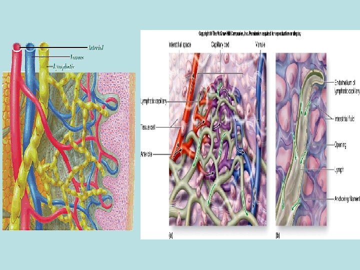
Lymphatic Vessels unidirectional flow, the lymph. Three types Lymph capillaries larger than blood capillaries and irregularly absent basal lamina no tight junctions entry of liquids into the lymph capillary temporary openings larger particles (lipid droplet) e. g. villi of the ileum and jejunum Lymph collecting vessels similar to lymph capillaries but larger and with valves empty into lymph nodes The lymph is moved by the compression of the lymph vessels by surrounding tissues Lymph ducts smooth muscle cells They also form valves which may give a beaded appearance to the lymph the right lymphatic duct and the thoracic duct subclavian veins