Cardiovascular System General Circulation Heart Anatomy Physiology II


 Depolarizations are positive in front and negative behind wave.")


")
")


")
 = End Diastolic Volume – End Systolic")


- Slides: 44
Cardiovascular System: General Circulation & Heart Anatomy & Physiology II Tony Serino, Ph. D. Biology Department Misericordia University
General Circulatory System 1. Cardiovascular – – Consists of a closed system of vessels which transport blood Two circuits: Systemic and Pulmonary Arteries move blood away from the heart Veins move blood toward the heart
General Circulatory System 2. Lymphvascular – moves lymph – – Consist of blind end tubes which collect interstitial fluid (now called lymph) and returns it to circulation The lymph is cleaned before returned to the blood vessels
Heart as a Dual Pump • Cardiac muscle arranged as whorls that squeeze the blood • Twin pumps: systemic and pulmonary • Four chambers: 2 atria and 2 ventricles
Cardiac Muscle Cells
Cardiac Muscle Depolarization
Conductance of Ions during Depolarization
Heart Development
Fetal Circulation
Selected Heart Defects
Heart: Location
Heart in Relation to other Organs
Layers of the Heart and Pericardium
Heart: Anterior View
Heart: Posterior View
Heart: Internal Anatomy
Differences in Ventricular Wall
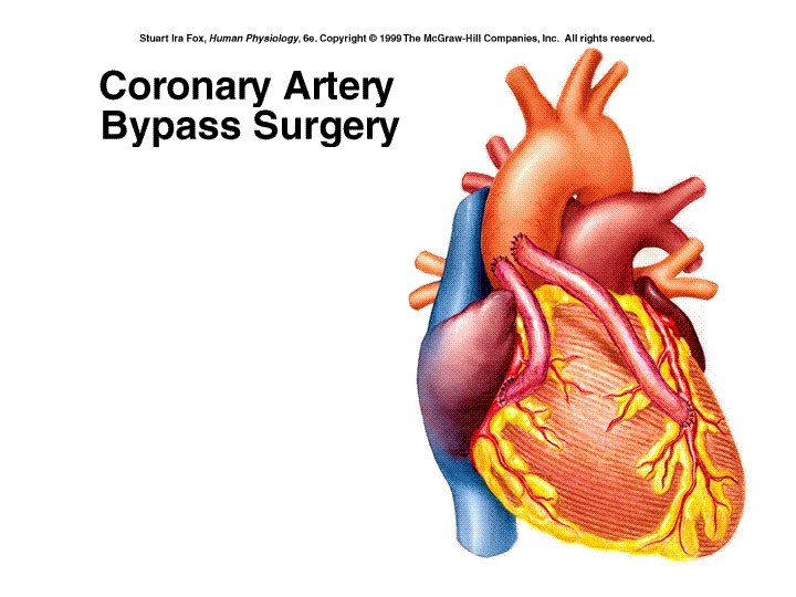
Coronary Arteries
Angioplasty Fig. 12. 66
Coronary Veins
Major Cardiac Valves
Heart Murmurs
Cardiac cycle Diastole: Period of Ventricular Filling
Systole: Isovolumetric Contraction
Systole: Ventricular Ejection
Diastole: Isovolumetric Relaxation
Conduction System of Heart
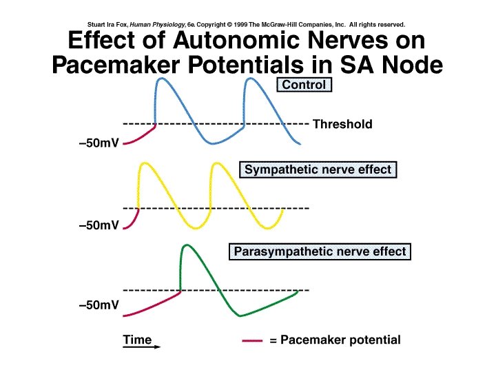
Pacemaker Potential
Einthoven’s Triangle (bipolar lead placement) Depolarizations are positive in front and negative behind wave. Repolarization is negative in front and positive behind wave Direction and type of wave within heart influences whether machine records an upward or downward deflection
ECG and electrical changes
Normal ECG Segments are the time between wave forms; Intervals include the space and the wave form.
ECG Normal Sinus Rhythm Junctional Rhythm (AV node rhythm)
Second Degree Heart Block Ventricular Fibrillation (V-fib)
Depolarization delayed between atria and ventricle; PR interval is prolonged. 2: 1, 3: 1 ratio between ventricle and atria rhythm PR interval increased with each beat until a QRS is skipped. No ration between ventricle and atria rhythm; P maybe buried in QRS complex
Heart Sounds • “Lub-dub” • Sound associated with valve closing producing turbulent blood flow
Cardiac Cycle
(ml/min)
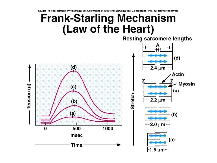
Factors Affecting SV • Stroke Volume (SV) = End Diastolic Volume – End Systolic Volume • SV = EDV – ESV (ml/beat) • EDV affected by: – Venous return which is dependent on venous tone, skeletal muscle pumps, etc. • ESV – As the heart fills it is stretched which allows for better overlap of the contractile proteins which will affect the force of contraction and the ESV (Starling’s Law of the Heart) – Increasing the force of contraction at any EDV will decrease the ESV and increase the SV (sympathetic stimulation and epinephrine)
Sympathetic Stimulation • Leads to increase HR • Increases in Ca++ release from SR, increase Ca++ through membrane and increase myosin crossbridge cycling • Increases force of contraction
Heart Rate Control • Sinus Rhythm = normal SA node control • Autonomic Activity – Sympathetic = accelerator (tachycardia) – Parasympathetic = brake (bradycardia) • Hormones – epinephrine • Drugs -caffeine, nicotine, atropine, etc.
Exercise Effects