Cardiovascular system Blood vascular system consists of a

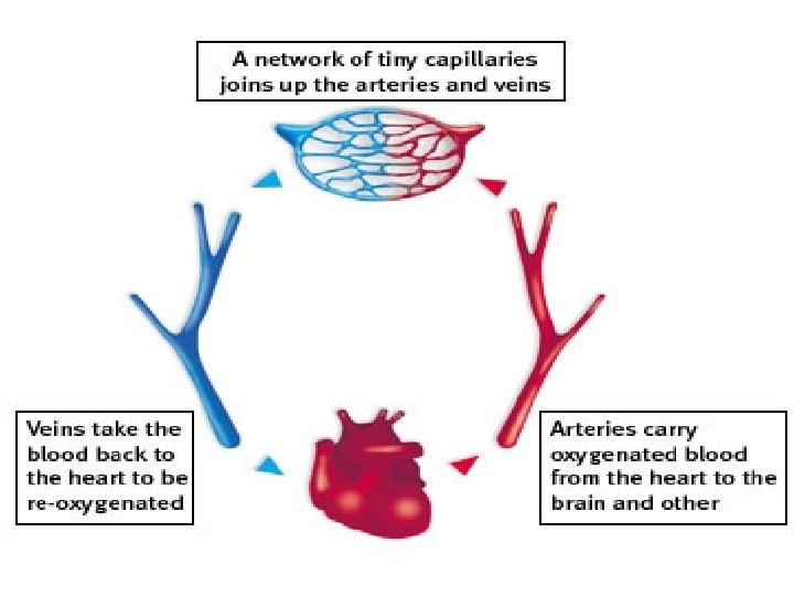
Cardiovascular system Blood vascular system consists of a system of a muscular pump (the heart) and two systems of blood vessels, which are pulmonary and systemic circulation. In both, the blood pumped from the heart passes successively through arteries of diminishing diameters, to networks of minute capillaries and then back to the heart through veins of increasing caliber

Structure of the heart: The heart is composed of two atria and two ventricles and is surrounded by a fibroserous sac called perichondrium. The cardiac wall in all chambers (atria and ventricles) consists of 3 layers 1. Endocardium 2. Myocardium 3. Epicardium
. It consists of : 1. Endothelium: lines the")
• Endocradium (the inner layer). It consists of : 1. Endothelium: lines the lumen of the heart and composed of simple squamous epithelium rests on basal lamina. 2. Subendothelial layer of fibero – elastic connective tissue. • Myocardium ( the middle layer ) : It forms the main bulk of the heart. It is composed of cardiac muscle fibers arranged in a spiral fashion. The myocardium contracts to propel blood into arteries for distribution to the body. • Epicardium : ( the outer layer ) : It consists of 1. Broad layer of adipose connective tissue. 2. Thin layer of fibero-elastic connective tissue, which supports the mesothelium. 3. The free surface of the heart is covered by mesothelium.

• Fibrous skeleton of the heart: It consists of thick bundles of collagen fibers oriented in various directions. • Vales of the heart: They are 4 valves: 1. Tricuspid valve between the right atrium and right ventricle. 2. Pulmonary valve between the right ventricle and pulmonary artery. 3. Mitral valve between the left atrium and left ventricle. 4. Aortic valve between the left ventricle and aorta. Structure of the valve: The valve is fold from the endocardium. It is formed of core of fibro-elastic fibers covered by endothelium


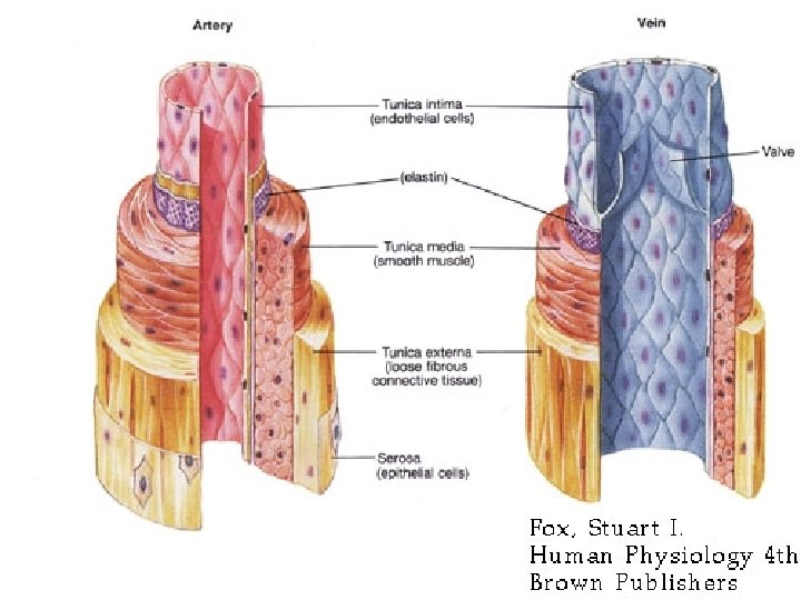
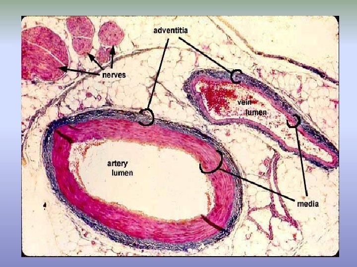
General structure of the blood vessels 1. Tunica intima: the innermost layer. It consists of: a) Endothelium, s. sq. epithelium rests on a basal lamina. b) Subendothelial layer of loose connective tissue. c) Internal elastic lamina, a layer of elastic f. (in arteries only). 2. Tunica media: (middle layer), the main bulk of the artery. It consists of : smooth muscle fibers, elastic, reticular and some collagen fibers. ± In large arteries thin external elastic lamina separating the media from the adventitia. 3. Tunica adventitia: it is the outermost layer. It consists of loose connective tissue rich in collagen fibers. Tiny blood vessels (vasa vasorum), lymphatic vessels and nerve fibers (vasa nervosa) are present in this layer. It merges with the surrounding connective tissue.

Types of blood vessels 1. Arteries. 2. Veins. 3. Connections between the arteries and veins. a) Capillaries b) Blood sinusoids c) Arterio-venous anastomosis (shunt)

Arteries are organs designed to propel and distribute blood from the heart to various tissues. Types of arteries: 1. Large or elastic arteries. 2. Medium sized or muscular arteries. 3. Small or arterioles.

Large arteries Called: elastic arteries & conducting arteries. They include aorta and its large branches: common carotid and subclavian. Structure: 1. Tunica intima: It is thicker than that of muscular arteries. It consists of: -Endothelium. 2. -Subendothelial layer of loose connective tissue. - The internal elastic lamina (difficult to recognize). 2. Tunica media: thick and consists chiefly of series layers of fenestrated elastic laminae between which smooth muscle cells are present. The large amount of elastic tissue present in the media allows the arterial wall to become distended by the blood ejected from the heart during the cardiac systole. 3. Tunica adventitia: It is a thin layer of loose connective tissue rich in collagen fibers. It contains vasa vasorum lymphatic vessels and nerve fibers.

Medium sized arteries Most of the arteries of the body are muscular arteries, make up the great majority of the arteries such as brachial, radial, ulnar, femoral and tibial arteries. They distribute blood throughout various organs. Structure: 1. Tunica intima: - Endothelium rests on basement membrane. - Subendothelial layer of loose connective tissue. - Internal elastic lamina, the layer of elastic fibers. 1. Tunica media: This layer consists chiefly of concentric layers of smooth muscle cells intermingled with few elastic fibers. External elastic lamina is present in large muscular arteries. It is a layer of elastic fibers separating the media from the adventitia. 1. Tunica adventitia: It consists of loose connective tissue rich in elastic and collagen fibers. It contains vasa vasorum, lymphatic vessels and nerve fibers.
. They regulate the")
Arterioles They are arteries of small caliber (less than 100 µ). They regulate the blood entering the organs. They have thick walls and narrow lumen. Structure: 1. Tunica intima: It consists of: - Endothelium. -Subendothelium, loose connective tissue. - Internal elastic lamina, it is a wavy prominent layer especially in larger arterioles. 2. Tunica media: The media is muscular and its thickness depends upon the size of the arteriole. It is composed of circularly arranged smooth muscle cells (about 1 -5 layers). 3. Tunica adventitia: It consists of thin layer of loose CT. • As arterioles branch they become smaller and the thickness of their coats diminish gradually until they disappear and extend with the capillaries. The terminal arteriole, has no internal elastic lamina. Its media consists of a continuous layer of smooth muscle cells. Metarterioles, the media consists of discontinuous layer of smooth

Specialized arteries Some arteries have some variations in their structure according to their location and function. • Cerebral arteries: e. g. basilar artery. They have thin walls because they are protected by the skull. Tunica intima: It consists of: -Endothelium. -Subendothelium. - Well-developed internal elastic lamina. Tunica media: It is thin and consists of smooth muscle cells. Tunica adventitia: Layer of loose connective tissue. • Coronary arteries: They have thick wall as they are subjected to high pressure. Tunica intima: It consists of: - Endothelium. - Subendothelium CT. - Layer of longitudinal smooth muscle cells. - Internal elastic fibers. Tunica media and tunica adventitia are similar to that of MSAs. External elastic lamina is present between the media and adventitia.

1. Umbilical artery: There is no internal elastic lamina. The media consists of inner longitudinal and outer circular smooth muscle cells. The adventitia is formed of mucoid connective tissue. Sensory organs of arteries: The carotid sinus and carotid body are specialized neural organs. They are of great importance in regulating respiration, heart rate and the vasomotor activities controlling blood pressure. 1. Carotid sinus (baroreceptor) 2. Carotid body (chemoreceptor):
: Carotid sinus is a slight dilatation close to the bifurcation")
1. Carotid sinus (baroreceptor): Carotid sinus is a slight dilatation close to the bifurcation of the common carotid artery or the beginning of the internal carotid artery. In this part the tunica media is thin and the tunica adventitia is thick and provided with numerous endings of afferent nerve fibers that enter the carotid branch of the glossopharyngeal nerve. An increase in the arterial blood pressure, impulses pass readily through the thin media to the sensory endings of the glossopharyngeal nerve in the tunica adventitia. This information is relayed to the control centers in the brain resulting in the slowing of the heart rate and vasodilatation of the peripheral blood vessels. 2. Carotid body (chemoreceptor): The carotid body is a small condensation of tissue that is situated in the terminal part of the common carotid artery. It contains groups of ovoid cells, called Glomus cells that are richly supplied with chemoreceptive nerve endings and fenestrated blood capillaries. These cells are sensitive to excess carbon dioxide, decrease oxygen tension in blood or changes in blood PH. When the chemoreceptor cells are stimulated, more afferent nerve impulses ascend the glossopharyngeal nerve and increase the heart and respiratory rates.

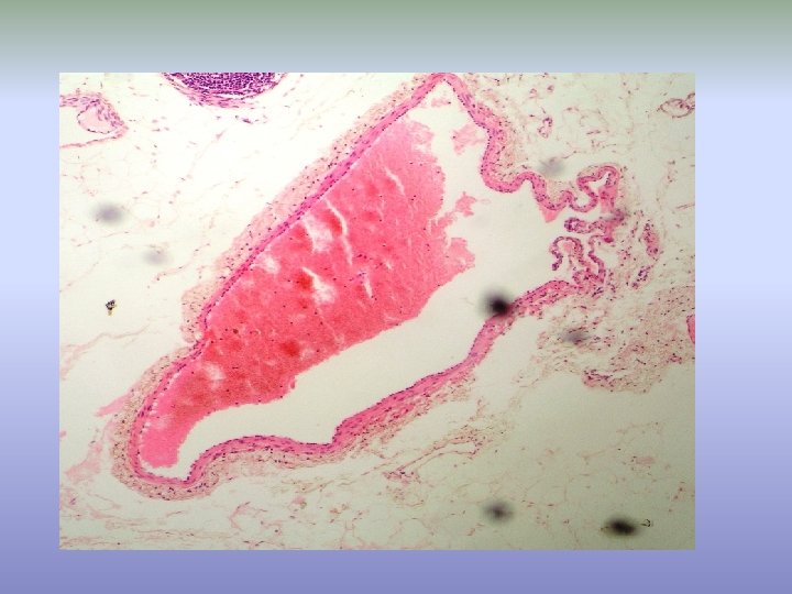
Veins Venous system returns blood from the capillary networks to the heart. They start at the capillary bed as 1. Post-capillary venules 2. venules 3. medium-sized veins 4. large veins. Compared with the arteries the veins have wide collapsed lumen, thin wall and full of blood. Structure: similar to those of the arteries, but the elastic and muscular components are less prominent. Venules: • The intima is composed of endothelium rest on a thin layer of connective tissue, which may be absent. • The media consists of few cells of smooth muscle • with thin layer of loose connective tissue.

Medium-sized veins: veins of the limbs and viscera. Tunica intima: It consists of: - Endothelium rests on basement membrane. - Subendothelial layer of loose connective tissue. Tunica media: It is thin and consists of few layers of smooth muscle cells intermixed with reticular fibers. Tunica adventitia: It is very thick and forms the main thickness. Such as: inferior and superior vena cava. Large veins: Tunica intima: It is thick and consists of endothelium and subendothelial connective tissue. Tunica media: It consists of few layers of circularly arranged smooth muscle fibers (similar to that of the medium-sized vein). Tunica adventitia: It is broad and well developed layer. It contains bundles of longitudinally arranged smooth muscle cells. The adventitia is rich in vasa vasorum.

Valves: - Valves are present mainly in veins of the extremities but they are generally absent from the veins of the abdomen and thorax. - They are found distal to the point of entrance of a tributary. Each valve consists of two leaflets preventing backflow of the blood from the heart. - Valves are projections from the intima. It consists of core of connective tissue covered by endothelium.

Connections between arteries and veins 1 - Capillaries the typical connections between the terminal ramifications of arterioles and venules. • They have round regular diameter (6 -9 µ). • They form a network and present elsewhere in the body. • They are elongated in nerves, muscles and tendons, while in organs they are irregular in shape.

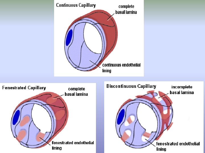
Structure of blood capillaries: • Capillaries are formed of endothelial cells rest on basal lamina supported by layer of reticular fibers. • Pericyte cells “mesenchymal cells” are present outside the wall of the capillary and supported by their own basement membrane. • These perivascular cells have great potential for transformation into other cells. The edges of endothelial cells are joined by fascia occludens. • The flattened endothelial cell nuclei bulge into the capillary lumen. • Pinocytotic vesicles are found in both surfaces of the endothelial cells indicating the interchange of materials. Types of capillaries: 1. Continuous capillaries 2. Fenestrated capillaries

1. Continuous capillaries: - They are the usual type found in most tissues. - Their endothelial cells are continuous and separated from each other by a thin space of 200 A°. - In the brain the blood capillaries have the following characters: • The endothelial cells are joined by zonula occludens. • Thick basement membrane. • The capillaries are surrounded by the processes of the pericytes. 2. Fenestrated capillaries: - The endothelial cells have rounded circular pores or fenestrae, which covered by thin diaphragm (capillaries in kidney glomeruli lacks these diaphragms, hence represent true openings). - The edges of the endothelial cells have wide gaps of 800 A°. - This type of capillaries is found in some tissues where there is much molecular exchange with the blood as in the small intestine, endocrine glands and kidney. Fenestrations permit the rapid passage of macromolecules smaller than plasma proteins.

2 - Blood sinusoids are other connections between the terminal branches of arterioles and venules. - They are very thin walled blood vessels having an irregular cross diameter. - The endothelial cells have many fenestrae. - The basement membrane is thin and incomplete or even absent. - Macrophages are present among or around the endothelial cells. - Blood sinusoids are found in the bone marrow, spleen and liver.
 This type of connections allows blood to pass directly")
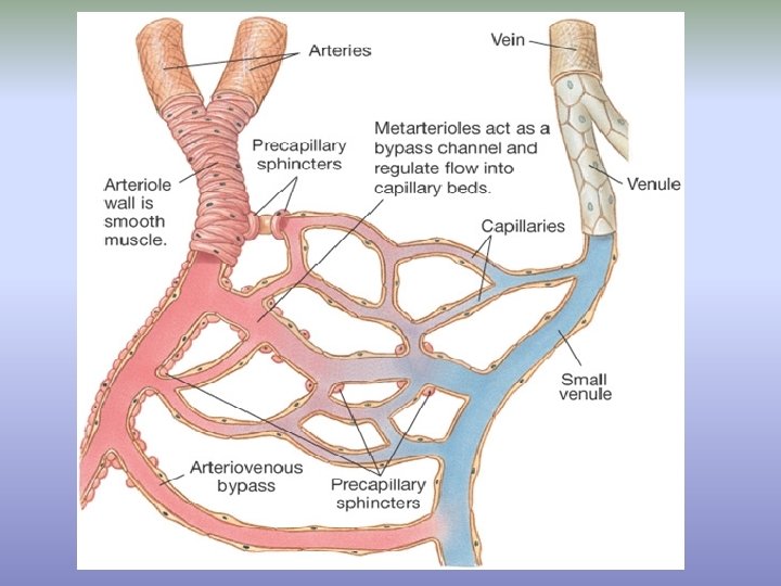
3 - Arterio-venous anastomosis (shunt) This type of connections allows blood to pass directly from arterioles to venules without having to circulate through a capillary network. Structure: - The wall of the connecting segment between the arterial and venule has 3 distinct segments: 1. The arterial end of the segment is similar in structure to the artery. 2. The venous end of the segment has the structure of the venule. 3. The intermediate segment lacks the internal elastic lamina. The media is well developed and contains more smooth muscle cells, which are highly innervated. Types: The A-V anastomosis may be: 1. Simple 2. Glomus. the A-V anastomosis are branched and tortuous. The glomus present in finger tips.

Sites: A-V anastomosis is most common in skin in certain regions such as finger tips, lips, nose, ears and toes. Function: • They play an important role in regulating blood flow to a region. Contraction of the thick smooth muscle layer, blood flows through the capillary bed. When it is relaxed, blood can flow directly from arterioles to venules by passing capillaries network. • Thermoregulation, closure of the anastomosis diverts blood into the dermal blood capillaries and permits heat loss, while opening of the vessel closes the capillary bed and conserves heat.

Nutrition of the blood vessels Walls of the small blood vessels can be easily nourished from their own blood by diffusion. In long arteries, only the intima and the inner part of the media are nourished by diffusion, while the outer part of the media and adventitia are nourished from vasa vasorum (vessels of the vessel). Vasa vasorum, can arise from the branches of artery they supply or from neighboring arteries. They branch profusely in the adventitia and the outer part of the media. Veins have abundant vasa vasorum. Lymphatic capillaries are also present in the adventitia of the blood vessels. Innervation of the blood vessels The blood vessels are supplied with network of unmyelinated vasomotor nerve fibers.

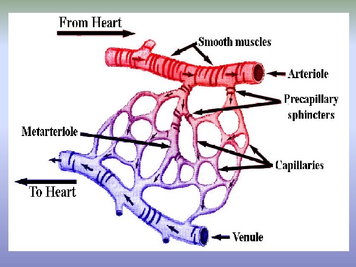
The microcirculation including arterioles, capillaries and venules. The capillaries lie between, or connect, the arterioles and venules. Capillaries form extensive branching networks that dramatically increase the surface areas available for the rapid exchange of molecules. • A metarteriole is a vessel that emerges from an arteriole and supplies a group of 10 to 100 capillaries. Both the arteriole and the proximal portion of the metarterioles are surrounded by smooth muscle fibers whose contractions and relaxations regulate blood flow through the capillary bed. Typically, blood flows intermittently through a capillary bed due to the periodic contractions of the smooth muscles (5 -10 times per minute, vasomotion), which is regulated both locally (metabolically) and by sympathetic control.
- Slides: 34