CARDIOVASCULAR SYSTEM Blood Blood Facts Blood is the

CARDIOVASCULAR SYSTEM Blood

Blood Facts Blood is the only fluid tissue The normal blood volume is 4 -5 L in females and 5 -6 L in males Normal blood p. H is 7. 35— 7. 45 Normal blood count is 4. 3— 5. 2 million cells/mm 3 in females and 5. 1— 5. 8 million cells/mm 3 in males

Blood Functions Oxygen delivery from lungs to cells Transport of metabolic wastes Transport of hormones Maintenance of body temperature Maintenance of p. H Maintenance of fluid volume Prevention of blood loss Prevention of infection

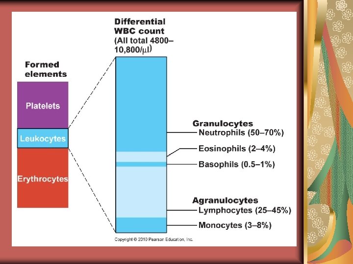
Blood Composition 55% of the blood consists of plasma 45% consists of formed elements 44% of the formed elements consist of erythrocytes <1% consists of leukocytes and platelets The leukocytes and platelets together make up the “buffy coat”
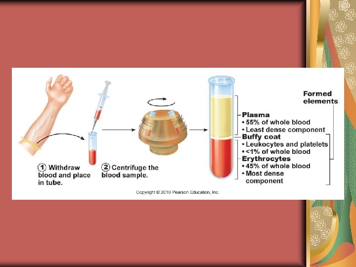

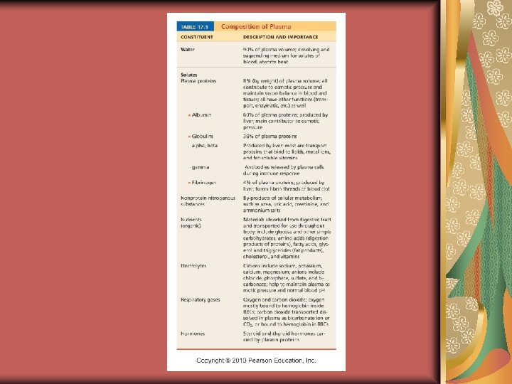
Plasma The liquid component of blood 90% of the plasma is made up of water 8% is made up of proteins 2% is made up of several ions

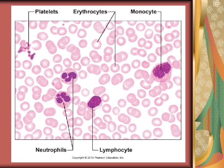
Blood Cells Erythrocytes—red blood cells; function to transport oxygen Leukocytes—white blood cells; function to protect the body Platelets—cell fragments that function to clot the blood

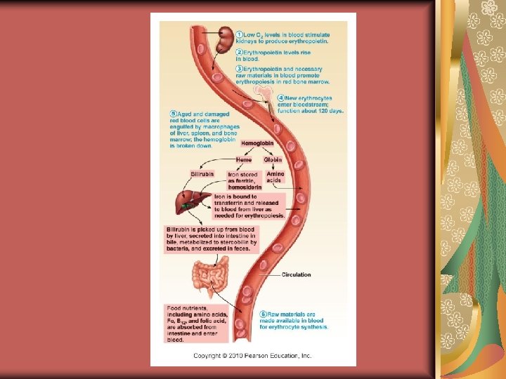
Erythrocytes No nucleus Few organelles Flexible, biconcave discs Hemoglobin Last 100 -120 days Destroyed by the liver; iron is recycled; the rest of the hemoglobin molecule is changed into bilirubin (a yellow pigment) and is excreted in the bile
 groups + iron Oxygen binds to the")
Hemoglobin Consists of 4 heme (red pigment) groups + iron Oxygen binds to the iron Has 4 globin (protein) groups— 2 alpha and 2 beta groups When oxygen combines with hemoglobin, it is referred to as oxyhemoglobin and appears bright red When there is no oxygen, it is referred to as deoxyhemoglobin (a. k. a. carbaminohemoglobin) and appears dark red
 Carbon dioxide binds to the globin chains Each hemoglobin carries 4")
Hemoglobin (cont. ) Carbon dioxide binds to the globin chains Each hemoglobin carries 4 oxygen molecules Each erythrocyte has about 250 million hemoglobin molecules, so 1 cell can carry about 1 billion oxygen molecules

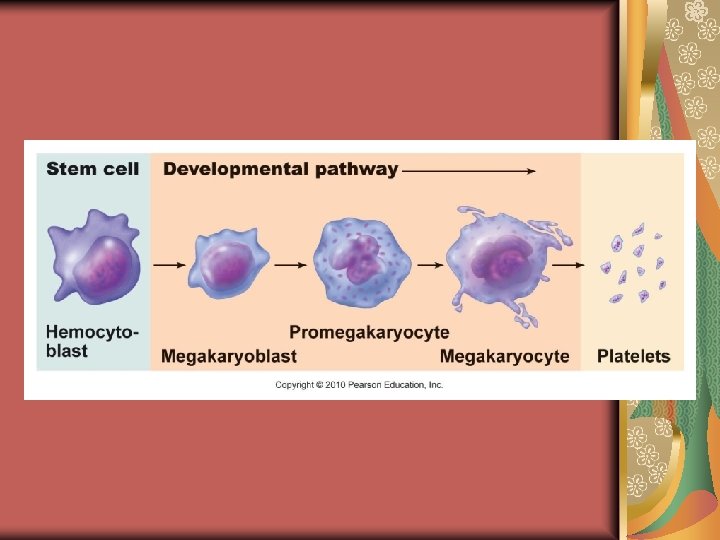
Hematopoiesis Blood cell formation Occurs in the red bone marrow Cells develop from blood stem cells called hemocytoblasts Red blood cell formation is referred to as erythropoiesis

Erythropoiesis Steps in erythropoiesis: 1. production of immature erythrocytes 2. synthesis of hemoglobin 3. ejection of nucleus and most organelles Stimulated by the hormone erythropoietin Release of erythropoietin depends on the concentration of oxygen, not on the number of RBCs present
 Decrease")
Erythropoietin Release Decrease in the number of RBCs (hemorrhage or excess RBC destruction) Decrease in the availability of oxygen (high altitude or pneumonia) Increased demand for oxygen by the tissues (aerobic exercise) Testosterone enhances erythropoietingenerating function of kidney; explains why men have more RBCs than women

Leukocytes Involved with allergic reactions Responsible for inflammation and antibody production There are 4, 000 -11, 000 cells/mm 3 2 types: Granulocytes Agranulocytes

Granulocytes Have lobed nuclei Have a granular cytoplasm 3 types: Neutrophils Basophils Eosinophils

Neutrophils Most common type of leukocyte Accounts for ½ of all the leukocytes Attracted to sites of inflammation Actively phagocytic

Basophils Make-up less than 1% of leukocytes Release histamine (a vasodilator that makes blood vessels “leaky”) and heparin (prevents blood clotting)

Eosinophils Released during allergic reactions Released during parasitic invasion

Agranulocytes Have a round or oval nucleus Have no granules These cells migrate into lymph tissues 2 types: Lymphocytes Monocytes

Lymphocytes 2 types: B cells—these cells will migrate from the bone marrow to lymph nodes T cells—these cells will migrate from the bone marrow to the thymus and then into the lymph nodes

Monocytes These cells will become macrophages Macrophages are phagocytic

Blood Cells B A C D E F H I G

Megakaryocytes & Platelets Megakaryocytes are large, multinucleated cells Megakaryocyte fragments form platelets Platelets last about 10 days
 A CBC is one of the most common types of")
Complete Blood Count (CBC) A CBC is one of the most common types of diagnostic tests performed It is a count of the formed elements It also includes a hematocrit (the percentage of RBCs to total blood volume) and testing for clotting factors
")
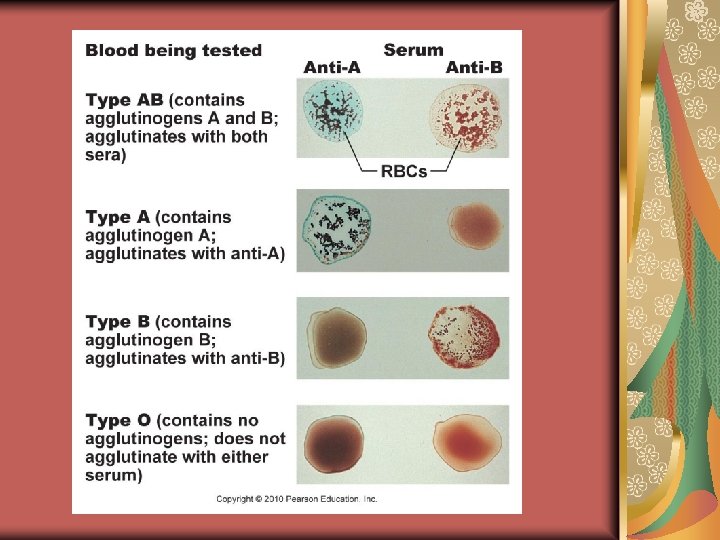
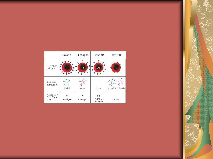
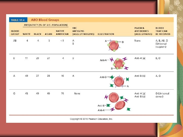
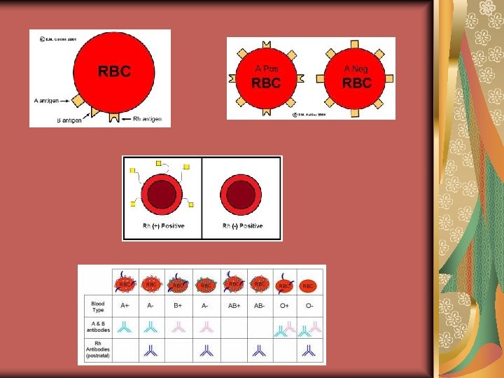
ABO Blood Groups Type A: has A antigens and anti-B antibodies (attacks B proteins) Type B: has B antigens and anti-A antibodies (attacks A proteins) Type AB: has A & B antigens and no antibodies (universal recipient) Type O: has no antigens and anti-A & anti-B antibodies (universal donor)

Rh Groups Rh+: have Rh antigen Rh-: doesn’t have the Rh antigen If an Rh- female is pregnant with an Rh+ baby, the baby will be healthy, but she will become sensitized and will produce antibodies to the Rh+ If she becomes pregnant again with another Rh+ baby, her antibodies will attack the babies RBCs (the baby becomes anemic and hypoxic, which can lead to brain damage and even death)
- Slides: 35