Cardiovascular System Block Cardiac Arrhythmias Physiology Ahmad Hersi
 Ahmad Hersi")
Cardiovascular System Block Cardiac Arrhythmias (Physiology) Ahmad Hersi

Lecture Objectives • Describe sinus arrhythmias • Describe the main pathophysiological causes of cardiac arrhythmias • Explain the mechanism of cardiac block • Explain the origin of an ectopic foci • Enumerate the common arrhythmias and describe the basic ECG changes

Depolarization and Repolarization
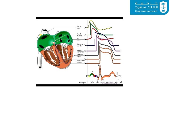

Electrical Conduction

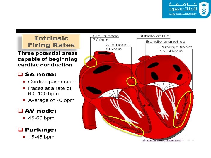
The Conduction System

Chest Leads

Limb leads

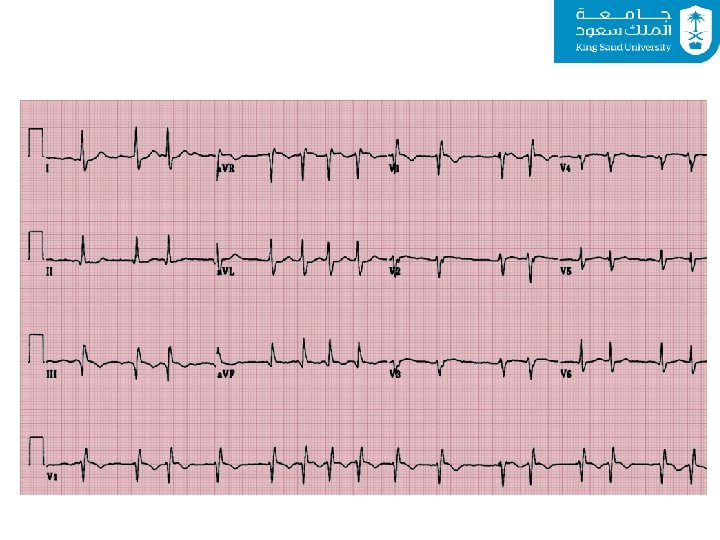
12 Leads EKG

Normal Sinus Rhythm • • Regular Single p-wave precedes every QRS complex P-R interval is constant and within normal range P-P interval is constant
. • Tachycardia: >100. • Bradycardia: <60.")
Rate • Normal sinus rate: (60 -100). • Tachycardia: >100. • Bradycardia: <60.

Causes of Cardiac Arrhythmias 1. Abnormal rhythmicity of the pacemaker 2. Shift of the pacemaker from the sinus node to another place in the heart 3. Blocks at different points in the spread of impulse through the heart 4. Spontaneous generation of impulses in any part of the heart

Causes of Cardiac Arrhythmias • Rate above or below normal • Regular or irregular rhythm • Narrow or broad QRS complex • Relation to P waves

Abnormal Sinus Rhythm • Tachycardia: an increase in the heart rate • Heart rate > 100 beats per minute • Causes: • Increased body temperature • Sympathetic stimulation • Drugs

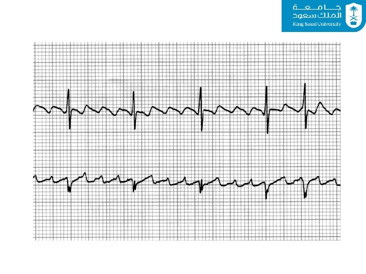
24 year-old pregnant woman with three days of frequent vomiting

Abnormal Sinus Rhythm • Bradycardia: – Slow heart rate < 60 beats per minute – Causes: • Parasympathetic stimulation

Sinus Arhythmia • Result from spillover of signals from the medullary respiratory center into the adjacent vasomotor center during inspiration and expiratory cycles of respiration • The spillover signals cause alternate increase and decrease in the number of impulses transmitted through the sympathetic and vagus nerves to the heart

Abnormal Cardiac Rhythms that Result from Impulse Conduction Block • Sinoatrial Block – The impulse from the S-A node is blocked before it enters the atrial muscle – Cessation of P waves

AVRT-Narrow Complex

Abnormal Cardiac Rhythms that Result from Impulse Conduction Block • A-V Block – When impulse from the S-A node is blocked – Causes: • • Ischemia of the A-V node Compression of the A-V node by scar formation Inflammation of the A-V node Strong vagal stimulation

Types of the A-V Block • First degree block • Second degree block • Third degree block

Heart Block st○ 1 • Constant PR prolongation without drop beat. nd○ 2 • Mobitz 1: Progressive PR prolongation + drop beat. • Mobits 2: Constant PR prolongation + drop beat. rd ○ 3 • Complete dissociation between P and QRS.

First Degree Heart Block
: • Mobitz (II):")
Second Degree Heart Block • Mobitz (I): • Mobitz (II):

Third Degree Heart Block

Types of the A-V block Second Degree Block • P-R interval > 0. 25 second • Only few impulses pass to the ventricles atria beat faster than ventricles “dropped beat” of the ventricles
 • Complete dissociation of P")
Types of the A-V block Third degree block (complete) • Complete dissociation of P wave and QRS waves The ventricle escape from the influence of S-A node • Stokes-Adams Syndrome: AV block comes and goes

Ventricular Fibrillation • The most serious of all arrhythmias • Cause: impulses stimulate one part of the ventricles, then another, then itself. Many part contracts at the same time while other parts relax (Circus movement) – – Tachycardia Irregular rhythm Broad QRS complex No P wave • Treatment : DC shock

Ventricular Fibrillation • Treatment : DC shock

Atrial Fibrillation • Cause: as ventricular fibrillation • It occurs more frequently in patients with enlarged heart • The atria do not pump if they are fibrillating • The efficiency of ventricular pumping is decreased 20 to 30% • A person can live for years with atrial fibrillation

Atrial Flutter • A single large wave travels around in the atria • The atria contracts at high rate (250 time per minute)

Ischemia and the ECG • One of the common uses of the ECG is in acute assessment of chest pain • Cause: restriction of blood flow to the myocardium, either: – Reversible: angina pectoris – Irreversible: myocardial infarction • Ischemia injury infarction

Reversible ischemia – Inverted T wave • ST segment depression

Myocardial Infarction • Complete loss of blood supply to the myocardium resulting in necrosis or death of tissue • ST segment elevation • Deep Q wave

Infero-Posterior MI

Antero-Lateral MI

57 year-old man with chest pressure and diaphoresis

Potassium and the ECG • Hypokalemia: – flat T wave • Hyperkalemia: – Tall peaked T wave

For Further Readings and Diagrams: Textbook of Medical Physiology by Guyton & Hall Chapter 10 (Cardiac Arrhythmias and their Electrocardiographic Interpretation)
- Slides: 44