Cardiovascular System Also known as the Circulatory System


- Slides: 28
Cardiovascular System
Also known as the Circulatory System • Consists of the heart, blood vessels, and blood. • Transports oxygen and nutrients to body cells. • Transports carbon dioxide and metabolic materials away from body cells.
• https: //www. youtube. com/watch? v=KSbb. D nb. SEy. M
Heart • • Muscular, hollow organ Pump Size of a closed fist Located in the mediastinal cavity, between the lungs, posterior to the sternum, and superior to the diaphragm
Three layers of tissue form the heart • Endocardium- Smooth, lines the inside of the heart. Allows for smooth blood flow. • Myocardium- Thickest layer. Muscular middle layer. • Pericardium- Double layered membrane or sac. Covers the outside of the heart.
Septum • Muscular wall • Separates the heart into a right and left side. • Prevents blood from moving between the right and left side of the heart.
Heart Chambers • Four chambers • Two upper chambers called atria • Two lower chambers called ventricles
Right Atrium • Receives blood as it is returns from body cells
Right Ventricle • Receives blood from the right atrium • Pushes blood into the pulmonary artery, which carries blood to the lungs for oxygen.
Left Atrium • Receives oxygenated blood from the lungs.
Left Ventricle • Receives blood from the left atrium. • Pushes the blood into the aorta so it can be carried to body cells.
Valves • One-way valves in the chambers of the heart keep the blood flowing in the right direction.
Tricuspid valve • Located between the right atrium and the right ventricle. • Closes when the right ventricle contracts and pushes blood into the lungs. • Prevents blood from flowing back into the right atrium.
Pulmonary Valve • Located between the right ventricle and pulmonary artery. • Closes when the right ventricle has finished contracting and pushing blood into the pulmonary artery. • Prevents blood from flowing back into the right ventricle.
Mitral or Bicuspid Valve • Located between the left atrium and the left ventricle. • Closes when the left ventricle is contracting and pushing blood into the aorta. • Prevents blood from flowing back into the left atrium.
Aortic Valve • Located between the left ventricle and the aorta (the largest artery in the body). • Closes when the left ventricle is finished contracting and pushing blood into the aorta. • Prevents blood from flowing back into the left ventricle.
http: //www. youtube. com/watch? v=Vs. LLP 0 ub. QH 8
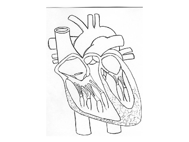
Activity Now you will label a heart diagram. In order to receive full credit describe the function of each part of the heart you are labeling and tell if has oxygenated or deoxygenated blood.
Right and Left sides of the heart work together in a cyclic manner even though they are separated by the septum
Electrical impulses originating in the heart causes the myocardium to contract in a cyclic manner. http: //www. youtube. com/watch? v=qi. IUr. Ce 2 Sxs http: //www. youtube. com/watch? v=f. ZT 9 vlb. L 2 u. A
Cycle consists of a brief period of rest called diastole followed by a period of ventricular contraction called systole
At the start of the cycle, the sinoatrial node creates an impulse that spreads across the atria causing them to simultaneously contract and push blood into the ventricles.
The impulse reaches the atrioventricular node where there is a brief pause causing the atria to relax and refill.
The impulse then travels through the bundle of His, down the left and right bundle branches and to the purkinje fibers causing the ventricles to contract. This active phase of ventricular contraction is called systole.
The right ventricle pushes blood into the pulmonary artery so it can go to the lungs for oxygen.
The left ventricle pushes blood into the aorta so it can be carried to all parts of the body.
Heartbeat Cycle youtube for heartbeat cycle merination. com