
Cardiovascular System A closed system of the heart
- Slides: 33
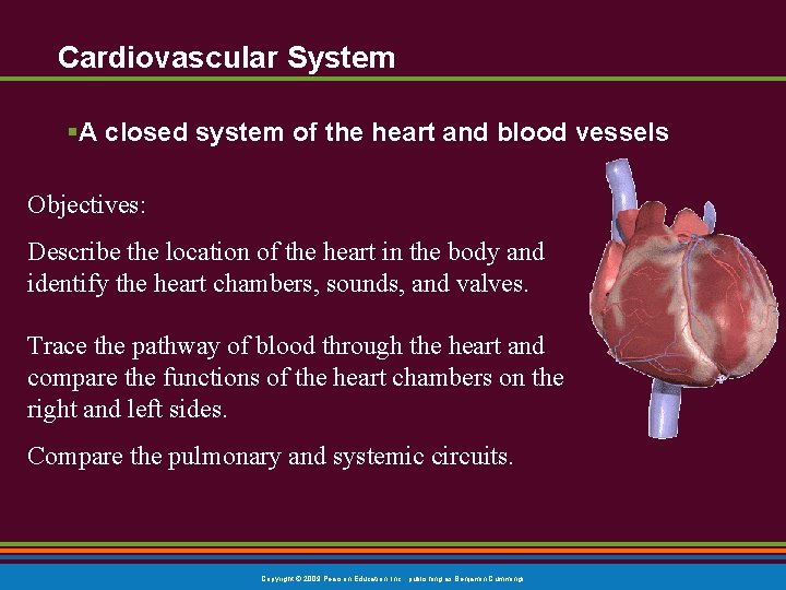
Cardiovascular System §A closed system of the heart and blood vessels Objectives: Describe the location of the heart in the body and identify the heart chambers, sounds, and valves. Trace the pathway of blood through the heart and compare the functions of the heart chambers on the right and left sides. Compare the pulmonary and systemic circuits. Copyright © 2009 Pearson Education, Inc. , publishing as Benjamin Cummings
The Cardiovascular System § The function of the cardiovascular system § TRANSPORTATION § deliver oxygen § deliver nutrients § movement of hormones § remove carbon dioxide § remove urea § remove other waste products Copyright © 2009 Pearson Education, Inc. , publishing as Benjamin Cummings
§ Blood: the mechanism for transport of these materials § Blood vessels: tubes through which materials are transported § Heart: the force that keeps materials circulating Copyright © 2009 Pearson Education, Inc. , publishing as Benjamin Cummings
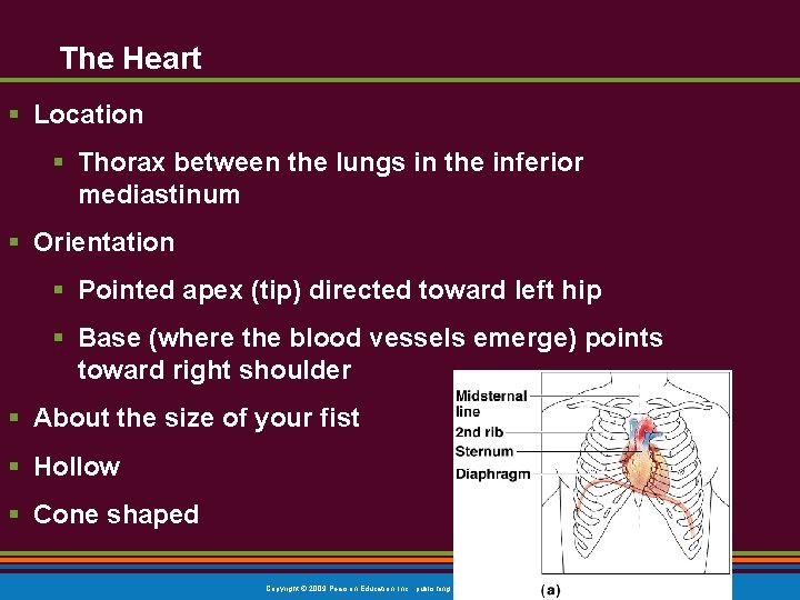
The Heart § Location § Thorax between the lungs in the inferior mediastinum § Orientation § Pointed apex (tip) directed toward left hip § Base (where the blood vessels emerge) points toward right shoulder § About the size of your fist § Hollow § Cone shaped Copyright © 2009 Pearson Education, Inc. , publishing as Benjamin Cummings
The Heart: Coverings § Pericardium—a double-walled sac 1. Fibrous pericardium is loose and superficial § for protection § to hold the heart in place Copyright © 2009 Pearson Education, Inc. , publishing as Benjamin Cummings
The Heart: Coverings 2. Serous pericardium (deep to the fibrous pericardium) 2 layers a. Visceral pericardium § Next to heart; also known as the epicardium b. Parietal pericardium § § Outside layer that lines the inner surface of the fibrous pericardium Serous fluid fills the space between the layers of pericardium Copyright © 2009 Pearson Education, Inc. , publishing as Benjamin Cummings
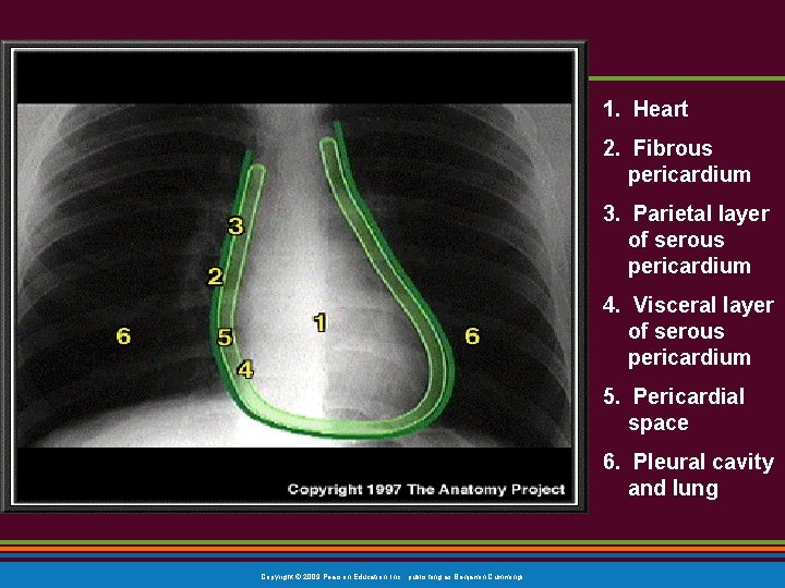
1. Heart 2. Fibrous pericardium 3. Parietal layer of serous pericardium 4. Visceral layer of serous pericardium 5. Pericardial space 6. Pleural cavity and lung Copyright © 2009 Pearson Education, Inc. , publishing as Benjamin Cummings
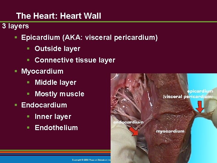
The Heart: Heart Wall 3 layers § Epicardium (AKA: visceral pericardium) § Outside layer § Connective tissue layer § Myocardium § Middle layer § Mostly muscle § Endocardium § Inner layer § Endothelium Copyright © 2009 Pearson Education, Inc. , publishing as Benjamin Cummings
The Heart: Chambers § Right and left side act as separate pumps § Four chambers § Atria (plural) § Receiving chambers § Right atrium § Left atrium § Ventricles § Discharging chambers § Right ventricle § Left ventricle Copyright © 2009 Pearson Education, Inc. , publishing as Benjamin Cummings
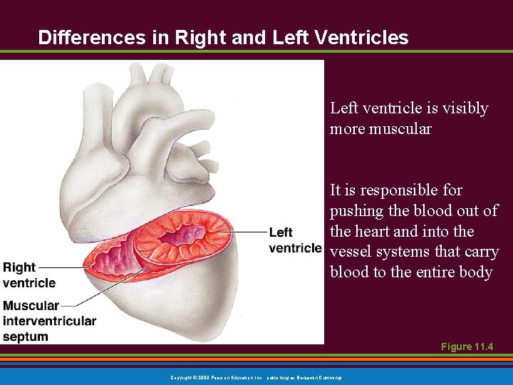
Differences in Right and Left Ventricles Left ventricle is visibly more muscular It is responsible for pushing the blood out of the heart and into the vessel systems that carry blood to the entire body Figure 11. 4 Copyright © 2009 Pearson Education, Inc. , publishing as Benjamin Cummings
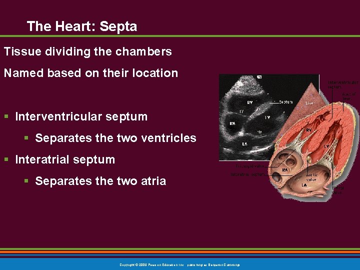
The Heart: Septa Tissue dividing the chambers Named based on their location § Interventricular septum § Separates the two ventricles § Interatrial septum § Separates the two atria Copyright © 2009 Pearson Education, Inc. , publishing as Benjamin Cummings
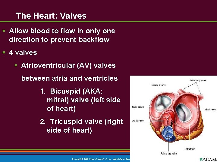
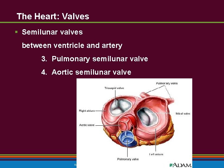
The Heart: Valves § Allow blood to flow in only one direction to prevent backflow § 4 valves § Atrioventricular (AV) valves between atria and ventricles 1. Bicuspid (AKA: mitral) valve (left side of heart) 2. Tricuspid valve (right side of heart) Copyright © 2009 Pearson Education, Inc. , publishing as Benjamin Cummings
The Heart: Valves § Semilunar valves between ventricle and artery 3. Pulmonary semilunar valve 4. Aortic semilunar valve Copyright © 2009 Pearson Education, Inc. , publishing as Benjamin Cummings
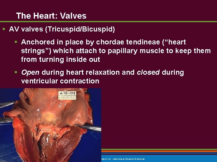
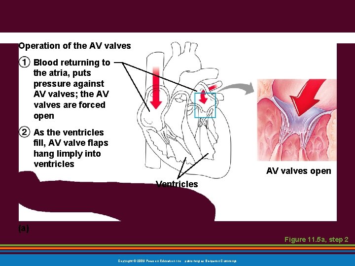
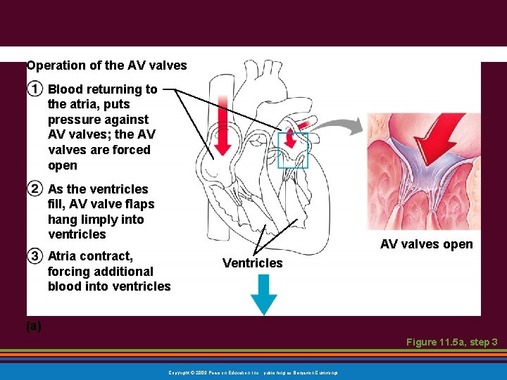
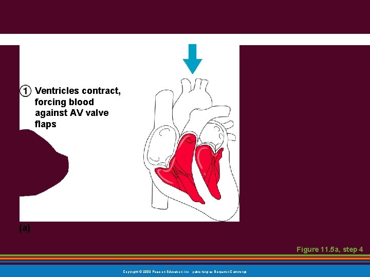
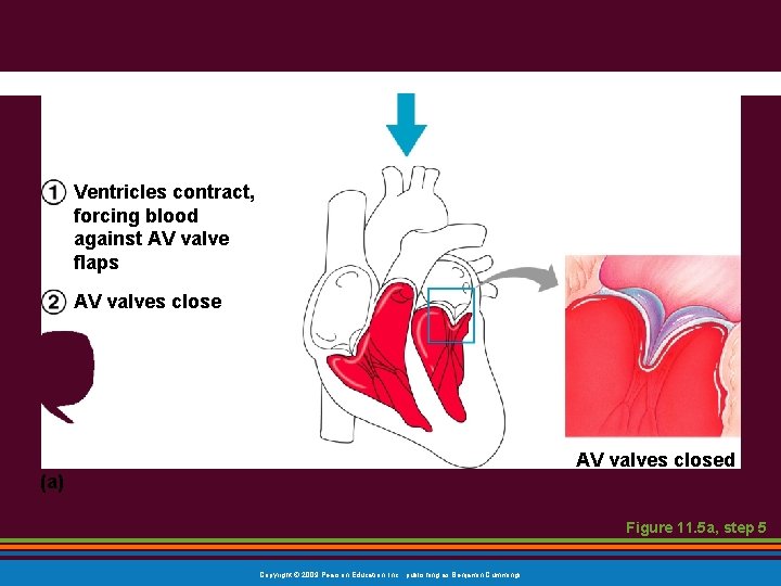
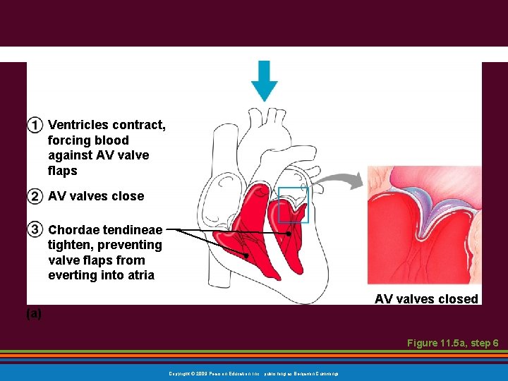
The Heart: Valves § AV valves (Tricuspid/Bicuspid) § Anchored in place by chordae tendineae (“heart strings”) which attach to papillary muscle to keep them from turning inside out § Open during heart relaxation and closed during ventricular contraction Copyright © 2009 Pearson Education, Inc. , publishing as Benjamin Cummings
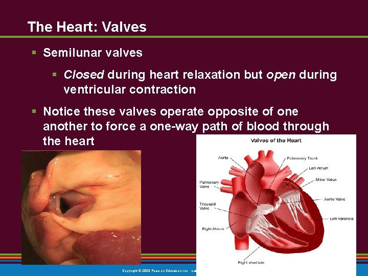
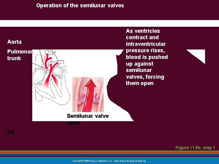
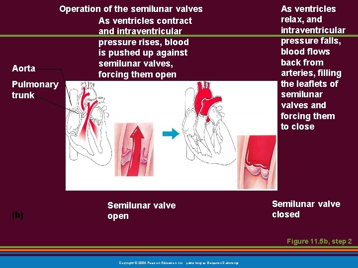
The Heart: Valves § Semilunar valves § Closed during heart relaxation but open during ventricular contraction § Notice these valves operate opposite of one another to force a one-way path of blood through the heart Copyright © 2009 Pearson Education, Inc. , publishing as Benjamin Cummings
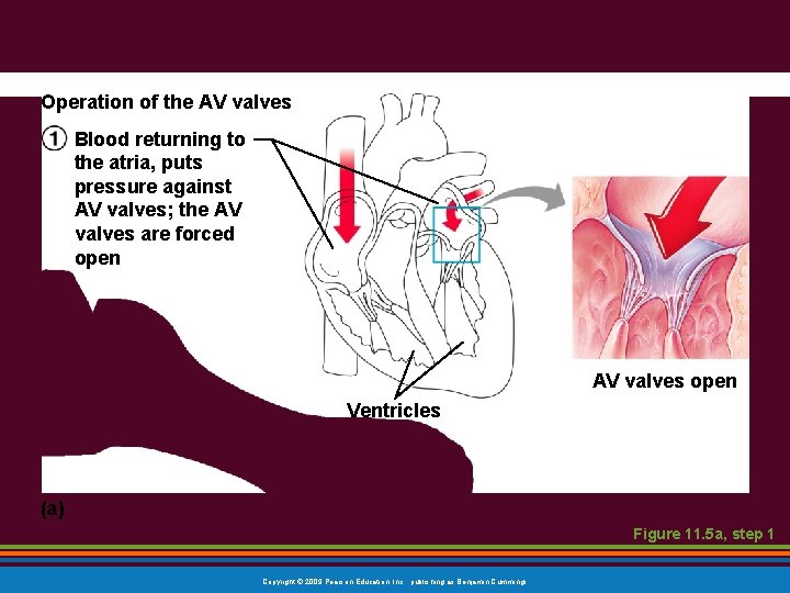
Operation of the AV valves Blood returning to the atria, puts pressure against AV valves; the AV valves are forced open AV valves open Ventricles (a) Figure 11. 5 a, step 1 Copyright © 2009 Pearson Education, Inc. , publishing as Benjamin Cummings
Operation of the AV valves Blood returning to the atria, puts pressure against AV valves; the AV valves are forced open As the ventricles fill, AV valve flaps hang limply into ventricles AV valves open Ventricles (a) Figure 11. 5 a, step 2 Copyright © 2009 Pearson Education, Inc. , publishing as Benjamin Cummings
Operation of the AV valves Blood returning to the atria, puts pressure against AV valves; the AV valves are forced open As the ventricles fill, AV valve flaps hang limply into ventricles Atria contract, forcing additional blood into ventricles AV valves open Ventricles (a) Figure 11. 5 a, step 3 Copyright © 2009 Pearson Education, Inc. , publishing as Benjamin Cummings
Ventricles contract, forcing blood against AV valve flaps (a) Figure 11. 5 a, step 4 Copyright © 2009 Pearson Education, Inc. , publishing as Benjamin Cummings
Ventricles contract, forcing blood against AV valve flaps AV valves closed (a) Figure 11. 5 a, step 5 Copyright © 2009 Pearson Education, Inc. , publishing as Benjamin Cummings
Ventricles contract, forcing blood against AV valve flaps AV valves close Chordae tendineae tighten, preventing valve flaps from everting into atria AV valves closed (a) Figure 11. 5 a, step 6 Copyright © 2009 Pearson Education, Inc. , publishing as Benjamin Cummings
Operation of the semilunar valves As ventricles contract and intraventricular pressure rises, blood is pushed up against semilunar valves, forcing them open Aorta Pulmonary trunk Semilunar valve open (b) Figure 11. 5 b, step 1 Copyright © 2009 Pearson Education, Inc. , publishing as Benjamin Cummings
Aorta Operation of the semilunar valves As ventricles contract and intraventricular pressure rises, blood is pushed up against semilunar valves, forcing them open Pulmonary trunk (b) Semilunar valve open As ventricles relax, and intraventricular pressure falls, blood flows back from arteries, filling the leaflets of semilunar valves and forcing them to close Semilunar valve closed Figure 11. 5 b, step 2 Copyright © 2009 Pearson Education, Inc. , publishing as Benjamin Cummings
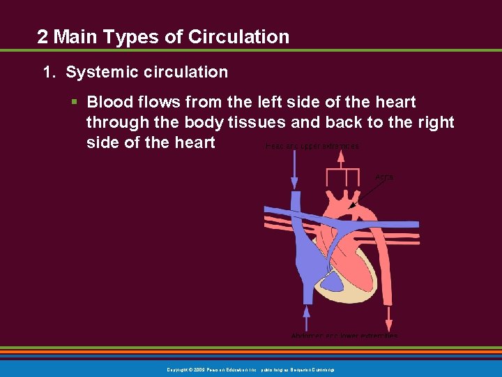
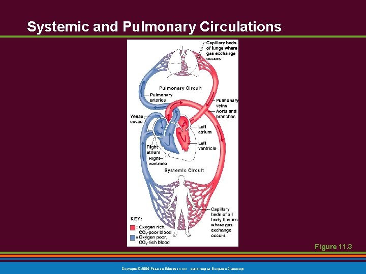
2 Main Types of Circulation 1. Systemic circulation § Blood flows from the left side of the heart through the body tissues and back to the right side of the heart Copyright © 2009 Pearson Education, Inc. , publishing as Benjamin Cummings
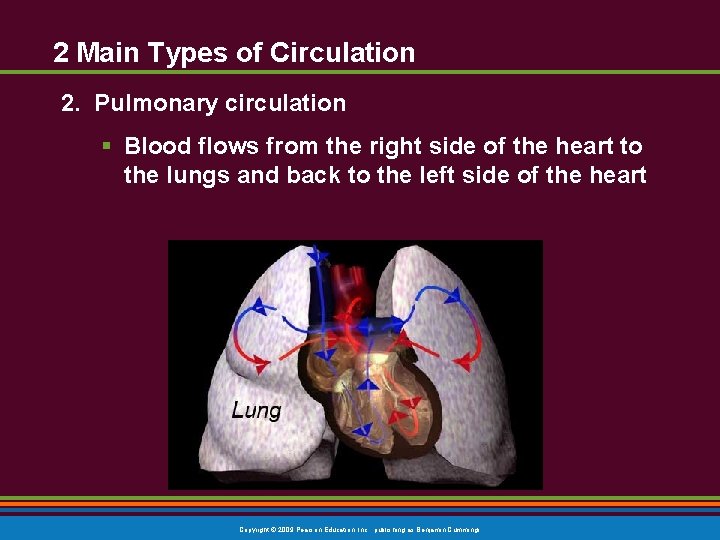
2 Main Types of Circulation 2. Pulmonary circulation § Blood flows from the right side of the heart to the lungs and back to the left side of the heart Copyright © 2009 Pearson Education, Inc. , publishing as Benjamin Cummings
Systemic and Pulmonary Circulations Figure 11. 3 Copyright © 2009 Pearson Education, Inc. , publishing as Benjamin Cummings
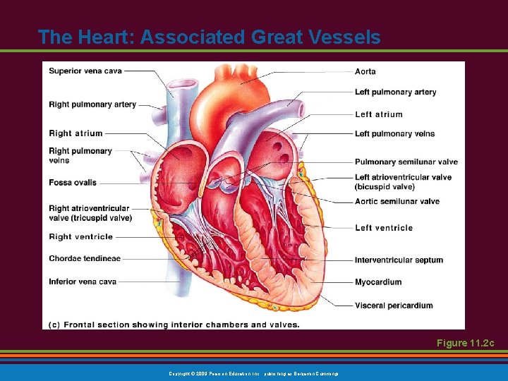
The Heart: Associated Great Vessels § Arteries § Aorta § Leaves left ventricle § Pulmonary arteries § Leave right ventricle Copyright © 2009 Pearson Education, Inc. , publishing as Benjamin Cummings
The Heart: Associated Great Vessels § Veins § Superior and inferior venae cavae § Enter right atrium § Pulmonary veins (four) § Enter left atrium Copyright © 2009 Pearson Education, Inc. , publishing as Benjamin Cummings
The Heart: Summary http: //www. mayoclinic. com/health/circulatory-system/MM 00636 Figure 11. 2 c Copyright © 2009 Pearson Education, Inc. , publishing as Benjamin Cummings
The Heart: Associated Great Vessels Figure 11. 2 c Copyright © 2009 Pearson Education, Inc. , publishing as Benjamin Cummings
Blood Flow Through the Heart § Superior and inferior venae cavae bring blood into the right atrium § From right atrium, through the tricuspid valve, blood travels to the right ventricle § From the right ventricle, blood leaves the heart as it passes through the pulmonary semilunar valve into the pulmonary trunk § Pulmonary trunk splits into right and left pulmonary arteries that carry blood to the lungs Copyright © 2009 Pearson Education, Inc. , publishing as Benjamin Cummings
Blood Flow Through the Heart § Oxygen is picked up and carbon dioxide is dropped off by blood in the lungs § Oxygen-rich blood returns to the heart through the four pulmonary veins § Blood enters the left atrium and travels through the bicuspid valve into the left ventricle § From the left ventricle, blood leaves the heart via the aortic semilunar valve and aorta Copyright © 2009 Pearson Education, Inc. , publishing as Benjamin Cummings
Practice!! § http: //highered. mcgrawhill. com/sites/0072495855/student_view 0/chapter 22/labeling _exercises. html § Practice Labeling Copyright © 2009 Pearson Education, Inc. , publishing as Benjamin Cummings