Cardiovascular disease is the leading cause of death
 Coronary disease")


")


,")


- Slides: 34
Cardiovascular disease is the leading cause of death among adults worldwide (1996) Coronary disease 7. 2 million Cancer 6. 3 Cerebrovascular disease 4. 6 Acute lower respiratory tract infections 3. 9 Tuberculosis 3. 0 COPD (chronic obstructive pulmonary disease) 2. 9 Diarrhea (including dysentery) 2. 5 Malaria 2. 1 AIDS 1. 5 Hepatitis B 1. 2
Coronary mortality: alarming worldwide forecasts
Atherosclerosis: a multifactorial disease
Arterial wall: structure and function
Different stages of atherosclerotic plaque development
Vascular endothelium modification in atherosclerosis
Plaque formation 1 — Fatty streak
Lipid core constitution LDL oxidation
Lipid core constitution Activated macrophages accumulate lipids
Plaque formation 2 — Fibrous cap
Plaque formation 3 — Lipid core
From plaque to thrombosis, key event: plaque rupture
Plaque vulnerability Key role of macrophages
Vulnerable plaque Key role of the macrophage in vascular wall inflammation
Vulnerable plaque Key role of the macrophage in the degradation of the fibrous cap
Parietal vascular inflammation The activated macrophage produces inflammatory cytokines
Parietal vascular inflammation NFk. B action in the inflammation process
Thrombus formation The macrophages release coagulation factors
Oxidized LDL and thrombogenesis
Plaque disruption (plaque cracking, fissuring, rupture – thrombosis start point)
Dyslipidemia and atherosclerosis
Diabetes and atherosclerosis
Tobacco and atherosclerosis
HTN, hemodynamic factor and atheroclerosis
Atherosclerosis
Inflammation links classic risk factors to altered cellular behavior within the arterial wall and secretion of inflammatory markers in the circulation.
Fibrinogen is an independent risk factor for atherosclerosis
Pathophysiology of atherosclerotic plaque § Plaque rupture § § inflammation markers (CRP (C-reactive protein), amyloid A) Intracoronary thrombus § § § increased coagulation factors and proteins (prothrombin fragments F 1+2; II – ATIII (thrombin – antithrombin III complex) increased soluble fibrin monomers increased P-selectin (a platelet membrane protein) reduced blood coronary flow § § § imaging changes fibrinolytic system activation (spontaneous / therapeutic) increased P-AP 2 (plamin – antiplasmin 2) complexes, fibrin degradation products (Ddimer) Myocardial ischemia § § § early ischemic indicators: glygogen phosphorylase BB ECG - ST depression § § Myocardial necrosis biochemical markers: CKMB, c. Tn. T, c. Tn. I ECG - ST elevation
Cardiac markers Enzymes: - CK, isoenzyme CK-MB, isoforms CK-MB 2 and CK-MM 3 - CK-MB mass - ASAT, ALAT - LDH, isoenzyme LDH 1 Non-enzymatic markers: - Troponins c. Tn. T, c. Tn. I - Myoglobin - H-FABP - NT-pro. ANP, NT-pro. BNP - Galectin - Inflammatory markers: - hs. CRP - IL 6 - VCAM 1 - Fibrinogen
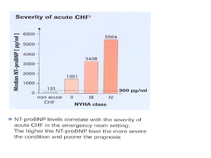
Fig. 1
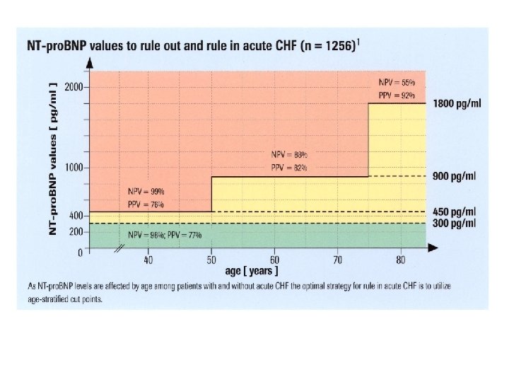
Fig. 2