Cardiovascular Assessment Heart and Circulation Location and Shape


 – Endocardium • Valves –")

 – Presystole (Atrial kick) – 25%")


- Slides: 23
Cardiovascular Assessment
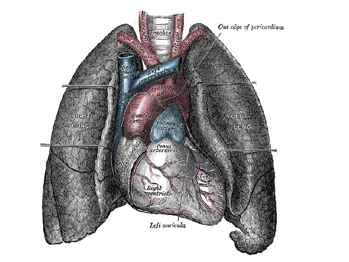
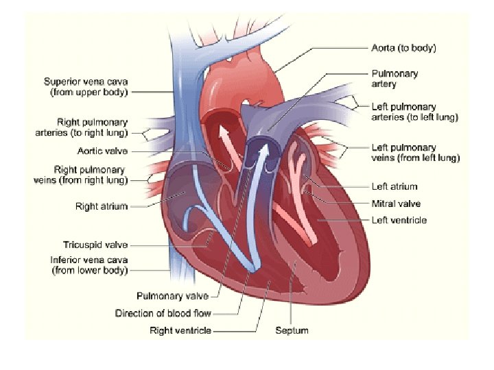
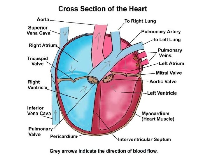
Heart and Circulation • Location and Shape – Precordium – Base – Apex • Great Vessels of the Heart – Superior and Inferior Vena Cava – Pulmonary Artery – Pulmonary Veins – Aorta
Heart Structure • Layers – Pericardium – Myocardium (subendocardium) – Endocardium • Valves – Semilunar • Aortic, Pulmonic – Atriventricular • Bicuspid (Mitral), Tricuspid
Conductance System • • • SA node AV node Bundle of HIS Purkinje fibers Myocardium
Cardiac Cycle • Diastole – Early filling (passive) – Presystole (Atrial kick) – 25% of stroke volume • Systole – AV valves shut (S 1) – Pressure builds, shoots through SL valves – SL valves shut (S 2)
Heart Sounds • Normal – S 1 – S 2 • Splitting • Extra – S 3 – S 4 • Murmur
Murmur • Causes – Increased blood velocity – Decreased blood viscosity – Structural defects, especially valve and septal • Grading: 1 – 6
Pumping action • Preload – volume of blood in heart before systole; also end-diastolic volume (EDV) • Afterload – diastolic blood pressure; pressure that the heart must overcome to pump blood • Stroke Volume (SV) – volume of blood pumped in one heartbeat • Ejection Fraction – SV/EDV
Developmental Concerns • Infants – foramen ovale, ductus arteriosus • Pregnant – Increase in blood volume • Aging adult – Systolic pressure increases – Myocardial thickening – Decreased exercise augmentation
History • Chest Pain – Acute – Angina pectoris • Stable • Unstable • Dsypnea – Paroxsymal • Orthopnea
History • Cough – Hemoptysis – Frothy pink sputum • • Fatigue Cyanosis, pallor Edema Nocturia
History • Past cardiac history – MI, HTN, cholesterol, murmur, congenital disease, rheumatic fever, valve problem, DM • Family history – HTN, cholesterol, CAD, DM, CVA • Personal habits – Nutrition, Smoking, ETOH, Exercise, Drugs
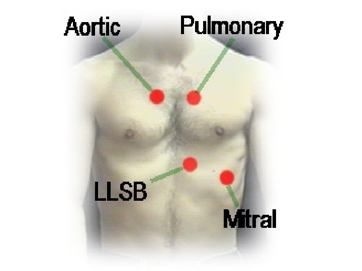
Physical Exam • • • Inspect chest Palpate Apical pulse Palpate precordium for thrills Percussion for heart size Auscultation – Rate – Rhythm – Murmurs, Rubs
Lub Dub • S 1 heard first • S 2 heard second – Exception - Tachycardia – Splitting • Abnormal – S 3 – fluid overload: HF – S 4 – ventricular resistance (hardened ventricle)
Heart Failure Findings • • Daily weight BP HR Orthopnea, PND S 3 Breath Sounds DOE Stages 1 – 4
Peripheral Vascular System
Peripheral Vascular System • Inspect: JVD • Auscultate – – – Temporal Carotid Aorta Renal Iliac Femoral • Palpate – – – – Carotid Brachial Radial Ulnar Femoral Popliteal Posterior Tibial Dorsalis Pedis
Other • Veins – Know difference between deep and superficial veins in arms and legs. Know what perforators do. – Homan’s sign • Skin – Color, warmth – Edema
Vocabulary • Doppler ultrasound • Peripheral lymph nodes to palpate – Epitrochlear, axillary, femoral • • Raynaud Lymphedema Ischemic ulcer Venous stasis ulcer Varicose veins DVT PAD, PVD