CARDIAC XRAY PRESENTED BY DR SANDEEP R 1

CARDIAC XRAY PRESENTED BY DR SANDEEP. R

� 1. BASICS OF CXR � 2. STRUCTURES IN ANTERIOR VIEW � 3. STRUCTURES IN LATERAL VIEW � 4. CHAMBER ENLARGEMENT � 5. PULMONARY CIRCULATION � 6. CONGENITAL HEART DISEASE � 7. PERICARDIAL DISEASE � 8. PACEMAKER & ICD � 9. CARDIAC CALCIFICATION � 10. PROSTHETIC HEART VALVE � 11. DISEASES OF AORTA � 12. MISCELLENOUS

�R – ROTATION � I -- INSPIRATION � P -- PROJECTION � E -- EXPOSURE

� The medial ends of both clavicles should be equidistant from the spinous process of the vertebral body projected between the clavicles

The increase in blackness of one hemithorax is always on the side to which the patient is rotated, irrespective of whether the CXR has been taken PA or AP

� It is ascertained by counting either the number of visible anterior or posterior ribs � Adequate inspiratory effort - six complete anterior or ten posterior ribs are visible � Poor inspiratory effort - fewer than six anterior ribs � Hyperinflated lung-more than six anterior ribs
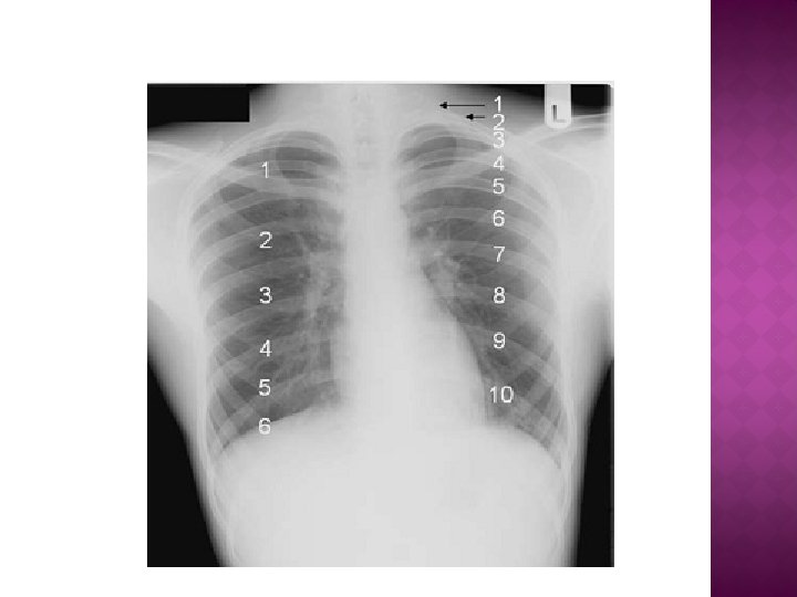

POOR INSPIRATORY FILM NORMAL INSPIRATORY FILM 1 3 2 1. MEDIASTINAL WIDENING 2. CMGLY 3. LOWER LOBE PATCHY OPACIFICN

� Projection is defined as the direction of x-ray with relation to the patient � If the direction of x-ray projection is from front – AP projection � If the direction of x-ray projection is from behind –PA projection
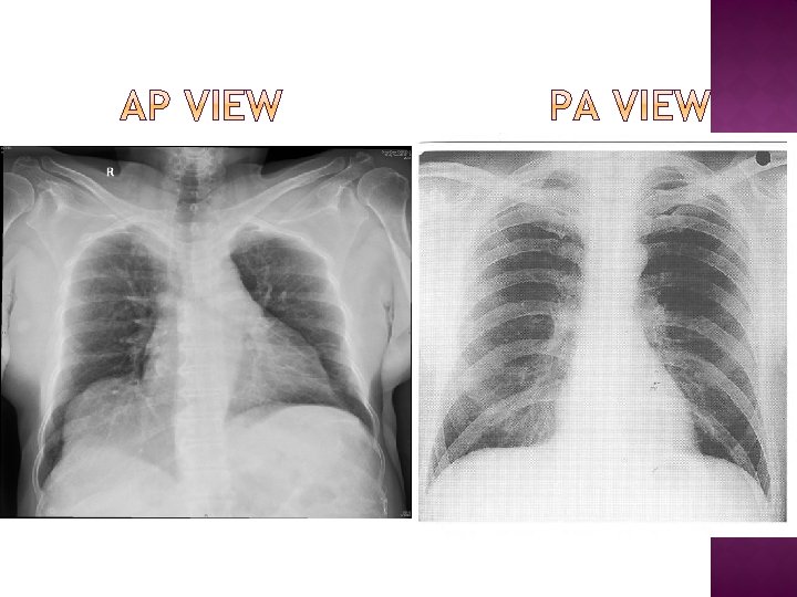

� In erect patients � Vertebral spines more prominent � Scapulae clear of lungs � Clavicles are horizontal � In supine patients � Vertebral bodies clear � Apparent cardiomegaly � Scapulae overlap � Clavicles are oblique

� Gas bubble in fundus with a clear air fluid level � Gas bubble in antrum � Apparent cardiomegaly

Normal exposure - the vertebral bodies should just be visible at the lower part of cardiac shadow Underexposed -If the vertebral bodies are not visible , insufficient number of x-ray photons have passed through the patient to reach the xray film Similarly, if the film appears too ‘black’, then too many photons have resulted in overexposure of the x-ray film.

OVERPENETRATION UNDERPENETRATION NORMAL

� 1. BASICS OF CXR � 2. STRUCTURES IN ANTERIOR VIEW � 3. STRUCTURES IN LATERAL VIEW � 4. CHAMBER ENLARGEMENT � 5. PULMONARY CIRCULATION � 6. CONGENITAL HEART DISEASE � 7. PERICARDIAL DISEASE � 8. PACEMAKER & ICD � 9. CARDIAC CALCIFICATION � 10. PROSTHETIC HEART VALVE � 11. DISEASES OF AORTA � 12. MISCELLENOUS

� Technical factors � Skeletal abnormalities and hardware � Situs: gastric air bubble, cardiac apex, and knob � Heart: position, size, and shape � Great vessels: position, size, and shape � Lung fields and vascularity by zone � Search for calcifications aortic
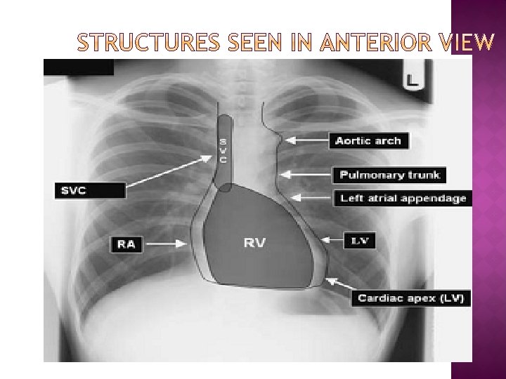

AA PT LA RV LV

ium br ula pa ls ste rna um Re tro tern of s Bod y ce Ma nu Aorta LPA RPA LA RV IVC Right upper lobe bronchus Left upper lobe bronchus Pulmonary outflow tract LV Scap Brachiocephalic vessels Trachea Confluence of pulmonary veins Right hemidiaphragm Gastric air bubble Left hemidiaphragm
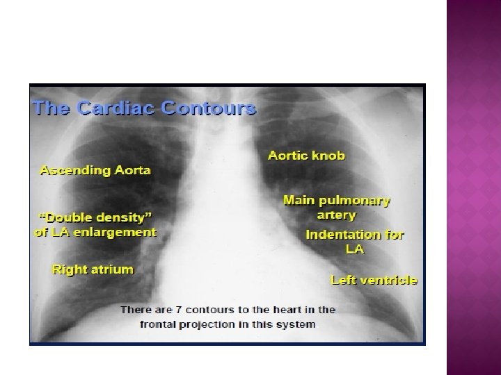
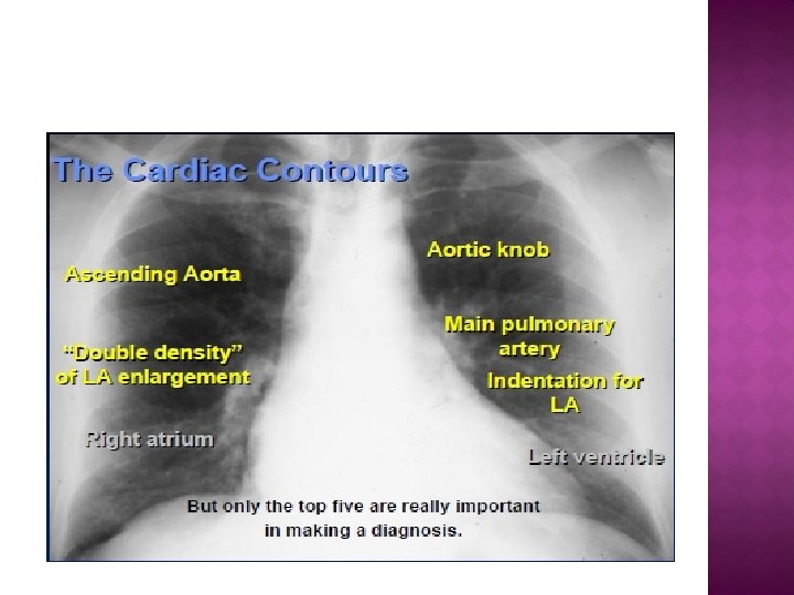
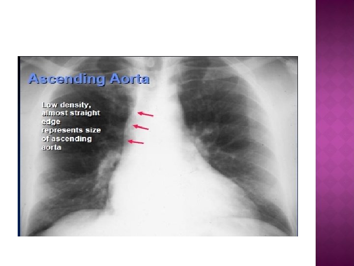
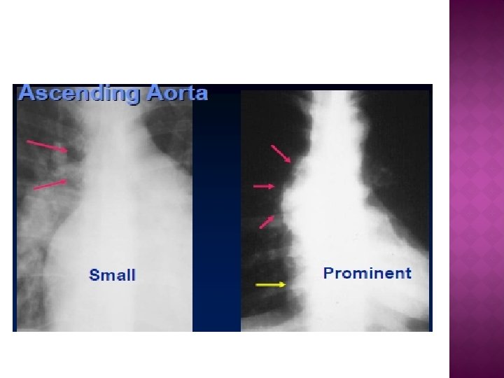

Aortic knob is the junction formed by the arch and descending aorta

LPA

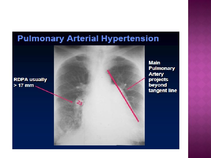
If we draw a tangent line from the apex of the left ventricle to the aortic knob(red line) and measure along a perpendicular to that tangent line (yellow line) The distance between the tangent and the main pulmonary artery (between two small green arrows) falls in a range between 0 mm (touching the tangent line) to as much as 15 mm away from the tangent line
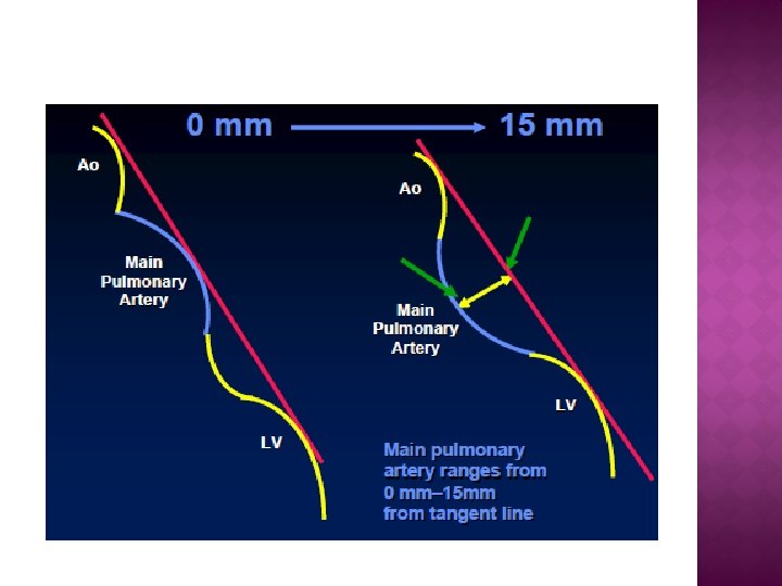

� Main pulmonary artery projects more than the tangent � Causes: 1. Increased pressure 2. Increased flow

� MPA > 15 mm from the tangent � Concave PA segment � Causes: 1. TOF 2. TRUNCUS ARTERIOSUS

L LA ENLARGEMENT HYPERTROPHIED LA APPENDAGE

� The cardiothoracic ratio should be less than 0. 5. i. e. A+B/C<0. 5 � A cardiothoracic ratio > 0. 5 suggests cardiomegaly in adults �A A B C cardiothoracic ratio > 0. 6 suggests cardiomegaly in newborn

CTR is more than 50% but heart is normal Extracardiac causes of cardiac enlargement � Portable AP films � Obesity � Pregnant � Ascites � Straight back syndrome � Pectus excavatum

� CTR is less than 50% but heart is abnormal Obstruction to outflow of the ventricles � Ventricular hypertrophy � Must look at cardiac contours < 50% ASCENDING AORTA DILATED LV CONTOUR

� Cardiothoracic ratio >0. 5 in adults � Cardiothoracic ratio >0. 6 in newborn � Any increase in transcardiac diameter > 2 cm compared to old x-ray � In old age and emphysema a transcardiac diameter more than 15. 5 cm in males &>12. 5 cm in females

� RA enlargement � LA enlargement � RV enlargement � LV enlargement

� Rt. Cardiac border becomes more convex > 50% of right border � Rt. Atrial border extends >3 intercostal spaces � Measurement from mid vertical line to max. convexity in rt. Border>5 cm in adult & >4 cm in children � Lateral view – fullness in space between sternum and front of upper part of cardiac silhouette

Ab > 5 cm a b
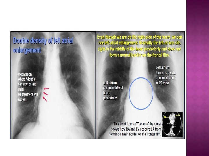
 � Elevation of left bronchus �")
� Widening of carina( normal 45 -75 degree) � Elevation of left bronchus � Straightening of left border � Double atrial shadow( shadow within shadow) � Grade 1 –double cardiac contour � Grade 2 - LA touches RA border � Grade 3 – LA overshoots the Rt. Cardiac border � Displaces the descending aorta to the left and esophagus to right seen in barium swallow

HYPERTROPHIED LA APPENDAGE

WIDENING OF CARINA ELEVATION OF LEFT BRONCHUS LAA enlargement DOUBLE ATRIAL SHADOW
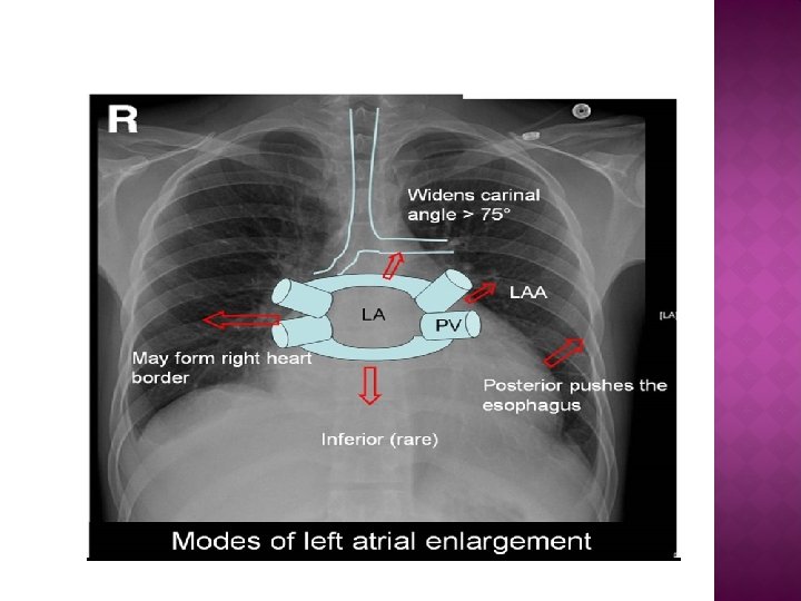

Widening of carina Aneurysmal LA Elevation of lt. bronchus Aneurysmal LA – When La enlarges to left and right and approaches within an inch of lateral chest wall

LA pushing the Esophagus posterior
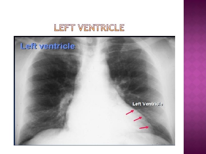
Left cardiac border gets enlarged and becomes more convex resulting")
� PA view � (a)Left cardiac border gets enlarged and becomes more convex resulting in cardiomegaly � (b)Lt. cardiac border dips into lt. dome of diaphragm � (c) rounded apical segment � (d) cardiophrenic angle is obtuse
 Left ventricle enlarges inferiorly and posteriorly � (b)Rigler’s measurement A")
Lateral view � (a) Left ventricle enlarges inferiorly and posteriorly � (b)Rigler’s measurement A is >17 mm � (c)Rigler, s measurement B is< 7. 5 mm � (d) Eyeler’s ratio becomes > 0. 42

� Rigler’s A & B used to differentiate left ventricular and right ventricular enlargement � Possible only when IVC shadow is present � Jn. Of IVC with Lt. Atrium – J point � Rigler’s A- from J point along line of IVC draw a line of 2 cm above and mark the point X.

� Draw a horizontal line from pt. A to posterior Cardiac border and mark that pt. y � Distance between points x & y is Rigler’s measurement A NORMAL<17 mm � � Rigler’s B-from the pt. J drop a perpendicular line to the dome and this distance is Rigler’s measurement B � NORMAL>7. 5 mm

� � � When LV enlarges, Posterior cardiac border gets displaced posteriorly & IVC shadow gets included in cardiac shadow, without getting displaced posteriorly Rigler’s measurement A >17 mm in lt. ventricular enlargement

� To differentiate lt. & rt. Ventricular enlargement � Valid when IVC shadow is absent or cannot be visualised � Mark the pt. of jn. where postero inferior cardiac border meets the dome as B � From this pt. B draw a horizontal line to the posterior border of sternum-AB

� From pt. B - draw another horizontal line posteriorly to the inner border of the rib-BC � Ratio of AB/BC is Eyeler’s ratio < 0. 42
 (a)Mild lt. Ventricular enlargement-obliteration")
LA Oblique view � There is a retrocardiac space( prevertebral) (a)Mild lt. Ventricular enlargement-obliteration of retrocardiac space (b) mod. Lt. ventricular enlargement-cardiac shadow overlaps vertebral column (c)Marked Lt. ventricular enlargement-cardiac shadow overshoots vertebral column

Chest X ray shows left ventricular enlargement. Left heart border is displaced leftward, inferior and posteriorly. Rounding of the cardiac apex.

PA VIEW Cardiophrenic angle is acute Clockwise rotation of heart causes RV to form the middle portion of the left heart border. RIGHT LATERAL VIEW Obliteration of retrosternal space LEFT LATERAL VIEW Rigler’s measurement will be 17 mm or less Rigler’s measurement will be 7. 5 mm or more Eyeler’s ratio is 0. 42 or less

� Narrow vascular pedicle � Cardiomegaly directly proportional to severity of pericardial effusion � This shadow has a rounded, globular appearance with no particular chamber enlargement � Cardiophrenic angle become more and more acute � Oligaemic pulmonary vascular markings � Marked change in cardiac silhouette in decubitus posture � ‘Epicardial fat pad sign’- anterior pericardial strip bordered by epicardial fat post. and mediastinal fat ant. >2 mm

Pulmonary oligaemia ‘Water bottle’ appearence Acute cardiophrenic angle Narrow vascular pedicle

� Chambers can be identified � Cardiophrenic angle is obtuse � Increased pulmonary venous hypertension � No change in cardiac silhouette in decubitus � Vascular pedicle is dilated or normal � Fluoro shows cardiac pulsation

1. Straightening of the right border 2. Pericardial thickening > 4 mm 3. Pericardial calcification (50% cases) 4. Dilatation of SVC and azygous vein � Pericardial calcification

� Focal bulge in area of main pulmonary artery � Sharply marginated � Absent right cardiac border � Increased distance between sternum and heart due to absence of sterno pericardial ligament

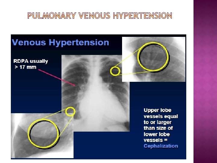
NORMAL PULMONARY CIRCULATION – 3 FEATURES 1. RDPA<17 MM


� LARRY ELLIOT’S CLASSIFICATION OF PVH RADIOGRAPHIC GRADE OF PVH ACUTE DISEASE PCWP CHRONIC DISEASE PCWP 1 13 -17 MMHG 2 18 -25 MMHG 18 -30 MMHG 3 >25 4 HEMOSIDEROSIS AND OSSIFICATION MMHG >30 MM HG LONG STANDING PVH

GRADE 0 -PCWP< 12 MM HG � Upper lobe pulmonary veins are less prominent than lower lobe veins GRADE 1 - PCWP 13 -17 MMHG Redistribution of blood flow with cephalization-’ANTLER SIGN’ � � � 1) increased resistance to flow due to interstitial odema 2) alveolar hypoxia in lower lobes causes reflex vasoconstriction 3) vasoconstriction of the arterioles due to LA or pulmonary vein reflex

� � � � GRADE 2 - PCWP 18 -25 mm hg Interstitial edema Peribronchial cuffing Kerley A, B, C lines Interlobular effusion Pleural effusion Hilar haze Peribronchial cuffing

� � � Distended lymphatic channels within edematous septa coursing from peripheral lymphatics to central hilar nodes Towards the hilum Less specific for Pulmonary venous hypertension KERLEY A LINES

� KERLEY B � � Horizontal lines 1 -3 mm thick Perpendicular to pleural surface � Towards the costophrenic angle � Accumulation of fluid in interlobular septa and lymphatics � Highly specific for PVH

KERLEY C LINES Crisscross lines seen between A &B � � GRADE 3 – pcwp > 25 mm hg Alveolar odema B/l diffuse patchy cotton wool opacities

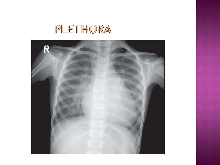
� � Pulmonary plethora – features Enlargement of central pulmonary artery , lobar and segmental artery � Prominent nodular vascular shadows in frontal CXR- shunt vessels that course ventral to dorsal � Upper & lower lobe vessels prominent � RPDA > 17 mm � Right descending pulmonary artery> tracheal diameter Ratio of RPDA to diameter of trachea > 1 � Plethora seen if shunt size >2: 1

� Pulmonary oligaemia � Decreased flow proximal to orgin of main pulmonary artery � Small pulmonary artery � Empty pulmonary bay � Pulmonary vessels small � Lung hypertranslucent � Lateral view shows diminution of hilar vessels

Empty pulmonary bay

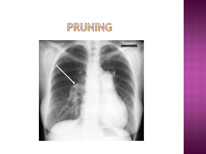
� Pruning � High pressure left to right shunts are associated with obliterative changes in the smaller pulmonary arteries & arterioles � Large main & large central pulmonary arteries taper down rapidly to very small vessels � Seen in Eisenmenger’s syndrome Precapillary PAH �

� � � Hamptons hump Wedge opacity Westermark sign Hampton’s hump Fleischner’s sign- prominent central pulmonary artery Palla’s sign-dilated rt. Descending pulmonary artery Chang’s sign – dilatation and abrupt change in calibre of the rt. Descending PA

MITRAL STENOSIS Small aortic knob from decreased cardiac output � Features of left atrial enlargement � Right atrial enlargement from tricuspid insufficiency � Pulmonary venous hypertension � Enlarged MPA � Calcified mitral valve �

� Mitral regurgitation � LA enlargement � LV enlargement � Pulmonary venous hypertension

AORTIC STENOSIS � Ascending aorta dilated � Left ventricular hypertrophy � Aortic valve calcification

AORTIC REGURGITATION � Dilated ascending aorta � LV enlargement

VALVULAR PS � No obvious cardiomegaly � Enlarged � Dilated PA left pulmonary artery � Normal to decreased pulmonary vasculature

INFUNDIBULAR PS � Concave segment � RVH PA

� ASD PULMONARY PLETHORA MPA DILATED RV APEX

DISEASE ASD VSD PDA LEFT ATRIUM AORTA

� VSD PLETHORA MPA DILATED RPA DILATED LV APEX

� PDA � Linear or railroad AORTIC KNOB track PROMINENT calcification at MPA site of ductus may be seen in PLETHORA adults with PDA LV APEX

� COARCTATION OF AORTA � “FIGURE OF 3” in CXR � “REVERSE 3” or SUBCLAVIAN “E sign” in ARTERY DILATION Barium meal COARCTATION POSTSTENOTIC AORTIC DILATION RIB NOTCHING
Aortic obstruction- Takayasu arteritis Coarctation of aorta � 2) Subclavian artery obstruction –Classic")
� 1)Aortic obstruction- Takayasu arteritis Coarctation of aorta � 2) Subclavian artery obstruction –Classic BT shunt Takayasu arteritis � 3)Chronic Svc obstruction � 4)Intercostal Av fistula � 5)Neurofibromatosis

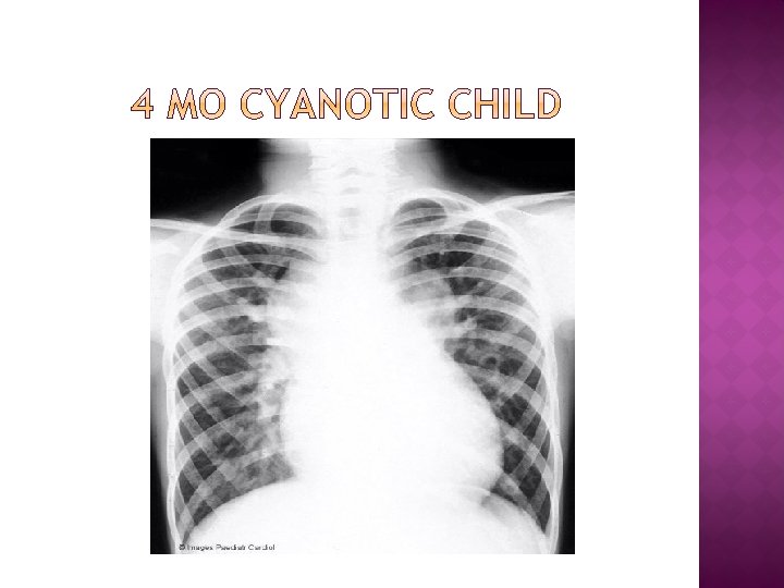
� Cyanosis With Decreased Vascularity Tetralogy of Fallot Truncus-type IV Tricuspid atresia Transposition of great arteries Ebstein’s anomaly � Cyanosis With Increased Vascularity Truncus types I, III TAPVC Tricuspid atresia Transposition Single ventricle
Rv apex 2)Underfilled LV 3)Concave pulmonary artery 4)Pulmonary")
TOF ‘Cour en sabot’ Due to 1)Rv apex 2)Underfilled LV 3)Concave pulmonary artery 4)Pulmonary oligaemia
Narrow pedicle � 2)Rt. Border RA �")
� TGA � ‘Egg on string’ � 1)Narrow pedicle � 2)Rt. Border RA � 3)Lt. border LV � 4)Increased vascularity � 5)Hypoplastic thymus
 � ‘figure of 8’ “snowman” � Rt border-SVC � Upper border-left innominate")
TAPVC (supracardiac) � ‘figure of 8’ “snowman” � Rt border-SVC � Upper border-left innominate � Left border-left vertical vein � Body of snowman-RA
 � The scimitar sign is produced by an anomalous pulmonary vein")
� PAPVC(Scimitar sign) � The scimitar sign is produced by an anomalous pulmonary vein that drains any or all of the lobes of the right lung. � Scimitar vein empties into the inferior vena cava

� EBSTEIN � ‘Box shaped’heart � Enlarged RA � Hypoplastic pulmonary trunk � Decreased vascularity

� � TRUNCUS ARTERIOSUS LV apex � Rt pulmonary artery has a superior orgin (20%) � ‘waterfall sign’ � ‘Hilar comma sign’ � Associated right aortic arch (33%) � Concave PA segment ELEVATED RIGHT HILUM

� Eisenmenger’s syndrome
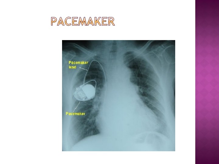
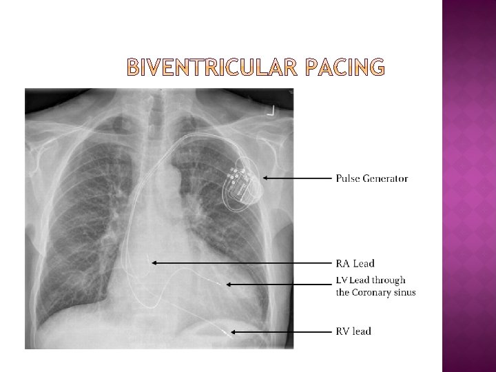

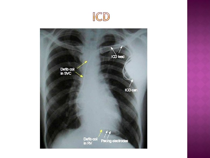
RV LEAD RA LEAD

LEAD FRACTURE DISPLACED RV LEAD DISPLACED RA LEAD
 Valvular – Mitral , aortic valve � � B)Pericardial – constrictive pericarditis")
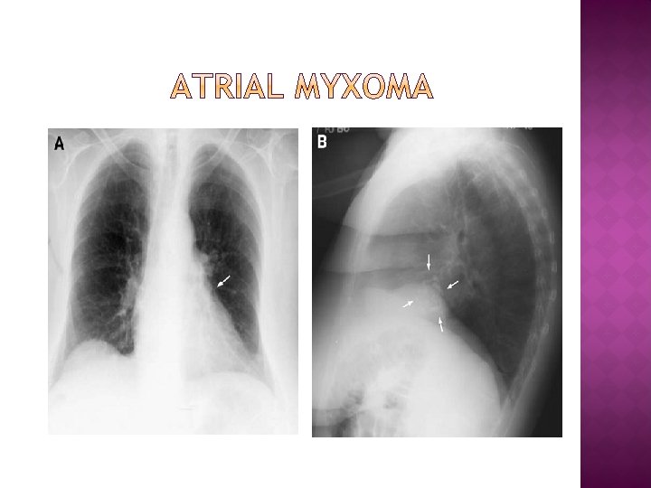
� A) Valvular – Mitral , aortic valve � � B)Pericardial – constrictive pericarditis � C) Myocardial – left atrial wall, LV aneurysm � D)Endocardial – Endomyocardial fibrosis � E) Intraluminal – La thrombus , La myxoma , Lv thrombus � F) Vascular – aortic calcification , coronary artery

MITRAL VALVE CALCIFICATION

CALCIFIED LV APICAL ANEURYSM

CALCIFIED PERICARDIUM

PERICARDIAL � � � SEEN IN BOTH SIDES OF HEART MOST COMMONLY IN AV GROOVE DIFFUSE CALCIFICATION AROUND THE HEART CALCIFICATION IS CHUNKY & UGLY MYOCARDIAL � SEEN IN ONLY LEFT SIDE MOST COMMON SITE IS ANT. WALL � LOCALIZED TO THE LEFT � CALCIFICATION IS FINE & CURVILINEAR �

GIANT LA CALCIFICATION

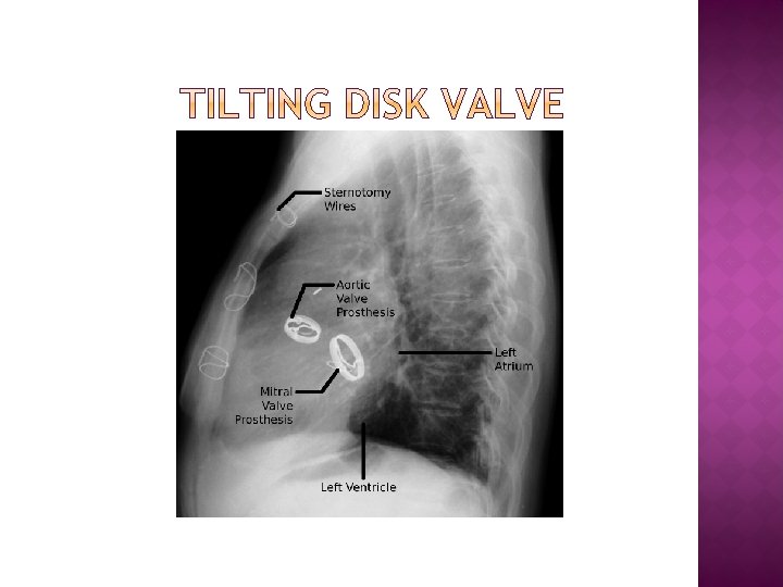
TILTING DISK VALVE

MITRAL STAR EDWARD

MITRAL PROSTHESIS

ST. JUDE MITRAL PROSTHETIC

Aneurysm of ascending & Descending aorta

Egg shell sign

LEFT SVC � � � LEFT SVC Occurs in less than 0. 5% of people Failure of regression of L common and Ant. Cardinal veins Drains left jugular and left subclavian vein Most patients also have right sided SVC Drains into dilated coronary sinus

� Leftward displacement of barium filled esophagus � Rt. � � Indentation of trachea Aortic knob is absent from left side Aorta descends on right � Associated RT. AORTIC ARCH with TOF Truncus arteriosus

Left superior intercostal vein � Seen in 5% of cases � To be differentiated from a mass � Also called pseudo dissection � It drains into hemiazygous vein Hartman T. Pearls & Pitfalls in Thoracic imaging, Variants and other difficult diagnosis

CERVICAL AORTIC ARCH � Left sided cervical aortic arch � Aortic knob at apex of lung � Descend on the left

SITUS INVERSUS WITH DEXTROCARDIA SITUS INVERSUS WITH LEVOCARDIA

DEXTROCARDIA HYDROPNEUMOPERICARDIUM

� PDA CLIP
Jefferson K, Rees S. Clinical cardiac radiology, 2 nd edition Butterworths; 1980. (2) Lipton")
(1)Jefferson K, Rees S. Clinical cardiac radiology, 2 nd edition Butterworths; 1980. (2) Lipton MJ. Plain film diagnosis of heart disease: cardiac enlargement. Contemporary Diagnostic Radiology 1988; 11: 1 -6. (3) Boxt LM, Reagon K, Katz J. Normal plain film examination of the heart and great arteries in the adult. J Thorac Imaging 1994; 9: 208 -18. (4)Murray G. Baron, Wendy M. Book. Congenital heart disease in the adult. North American Clinics of Radiology 2004; 3 (5) Ramesh M. Gowda, Lawrence M. Boxt. Calcifications of the heart. North American Clinics of Radiology 2004; 4 (6) Martin J. Lipton, Lawrence M. Boxt. How to approach cardiac diagnosis from the chest radiograph. North American Clinics Of Radiology 2004; 5 (7) Murray G. Baron. PLAIN FILM DIAGNOSIS OF COMMON CARDIAC ANOMALIES IN THE ADULT. North American Clinics Of Radiology 2004; 6 (8) Radiology imaging – sutton 6 th edition (9) Pediatric cardiology- Perloff’s clinical recognition of congenital heart disease (10)Radiology of congenital heart disease-Amplatz (11)Grainger & Allisons- diagnostic radiology vol 1 , 4 th edition (12)Cardiac Xrays- v. Chockalingam (13)Braunwald heart diseases 9 th edition (14) Emma C. Ferguson, Rajesh Krishnamurthy, Sandra A. A. Oldham. Classic Imaging Signs of Congenital Cardiovascular Abnormalities; Radio. Graphics 2007; 27: 1323– 1334 (15)www. learningradiology. com

EBSTEIN’S ANOMALY

ATRIAL SEPTAL DEFECT

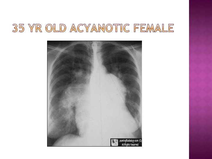
MITRAL STENOSIS

PERICARDIAL EFFUSION

ASD WITH PAH

SITUS INVERSUS WITH DEXTROCARDIA

RT AORTIC ARCH WITH RT DESCENDING AORTA

TAPVC SUPRACARDIAC
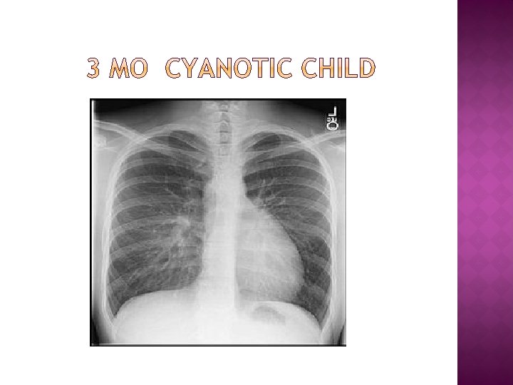

D-TGA
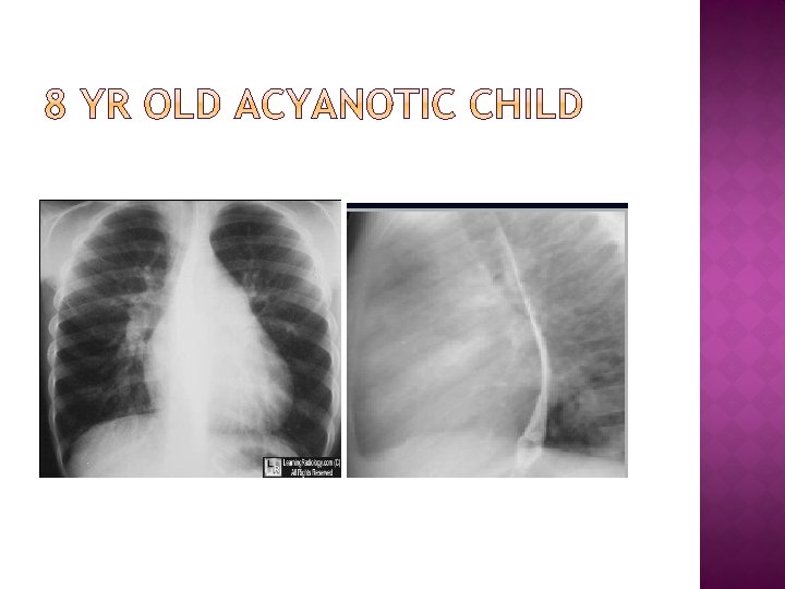

VSD
- Slides: 144