Cardiac Physiology The heart chambers the valves Cardiac

Cardiac Physiology • The heart: chambers, the valves • Cardiac muscle cells – Some cardiac muscle cells are autorhythmic – Arrangement of cardiac muscle cells – Excitation-contraction coupling
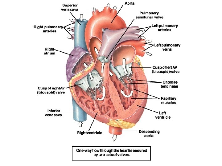
 Valves • guard the passageway between the atria and the ventricles –")
Atrioventricular (AV) Valves • guard the passageway between the atria and the ventricles – Tricuspid valve between right atrium and right ventricle – Bicuspid (mitral) valve between left atrium and left ventricle

Semilunar valves • Between ventricles and arteries – Pulmonary valve between right ventricle and pulmonary artery – Aortic valve between left ventricle and aorta
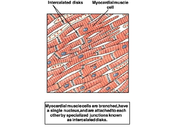

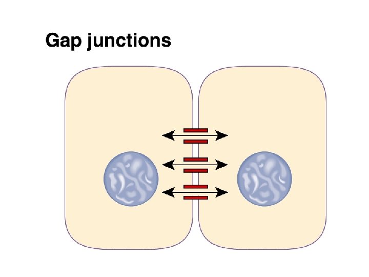
All myocardial cells • Gap junctions at intercalated discs, waves of depolarization spread from one cell to another
 are small myocardial cells with few contractile fibers •")
Autorhythmic myocardial cells • (pacemakers) are small myocardial cells with few contractile fibers • Spontaneously generate action potentials • Enables the heart to contract without any outside signal • The heart is myogenic: signal for contraction originates from heart muscle itself
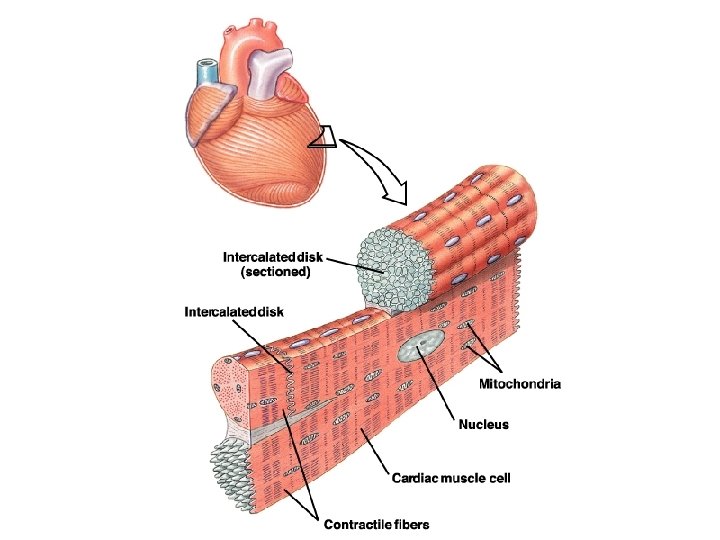

Most myocardial cells • • Remaining myocardial cells are striated Have sarcomeres Much smaller than skeletal muscle fibers Connected by gap junctions at intercalated discs • Lots of mitochondria • Lots of blood flow to myocardial cells

More facts about myocardial cells • Large branching t-tubules • Sparse sarcoplasmic reticulum • Source of Ca++ is largely extracellular

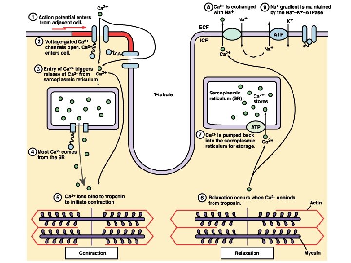
Excitation-contraction coupling • Depolarization cell membrane voltage gated Ca++ channels open • Ca++ enters cell • calcium-induced calcium release: Ca++ released from SR • Ca++ binds to troponin contraction

Myocardial cell relaxation • Ca++ dissociates from troponin • Ca++ returns to SR by Ca++ ATPase • Ca++ also transported from cell by Na+Ca++ indirect active transport protein: Ca++ is exchanged for Na+, which moves in along its electrochemical gradient Na+ removed by active transport

Regulation of cardiac muscle contraction • Graded contractions • Effect of cardiac muscle stretching • Channel activity during action potentials – In myocardial contractile cells – In autorhythmic pacemakers

Graded contraction • The amount of force varies with the number of cross-bridges formed • Low Ca++ few cross-bridges • High Ca++ more cross-bridges

The effect of epinephrine and norepinephrine of contraction • NE and E bind to beta 1 receptors on contractile myocardial cells • The beta 1 receptor is coupled to a G protein • Cyclic AMP is formed

The effect of epinephrine and norepinephrine of contraction • cyclic AMP is formed • 1. Voltage gated Ca++ channels are phosphorylated stay open longer more intracellular Ca++ stronger contractions • 2. A regulatory protein, phospholamban, is phosphorylated increased activity on SR Ca++ ATPase contractions shorten duration

Effect of phospholamban on Ca++ release • NE and E activity increase phospholamban activity increase Ca++ ATPase activity on SR more Ca++ is sequestered into the SR more Ca++ is available for Ca++ release during stimulation stronger force of contraction

Effect of NE and E on contraction • Stronger, more frequent contractions

When myocardial cells elongate • The amount of Ca++ entering the myocardial cells may increase the force of contraction increases

Myocardial contractile cell action potentials • Resting potential is stable -90 m. V • Wave of depolarization through gap junctions • Voltage gated Na+ channels open • Voltage gated K+ channels open • Slow voltage gated Ca++ channels open and K+ channels close • Ca++ channels close and K+ channels open
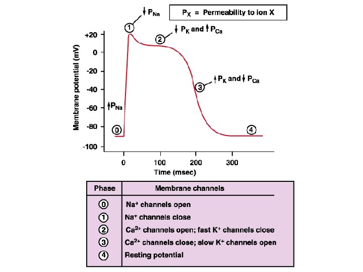
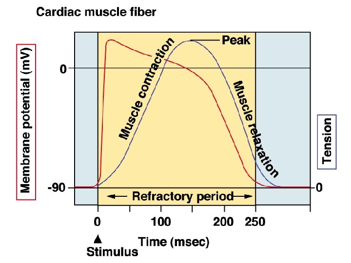
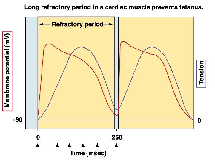

Long action potential • Myocardial cell refractory period and contraction end simultaneously

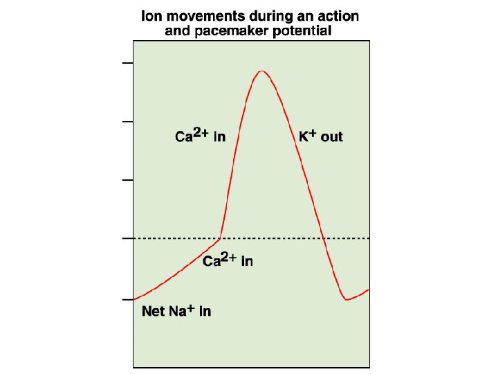
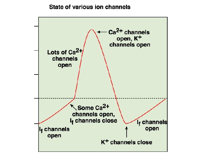
Action potentials in myocardial autorhythmic cells • The channels: – If channels allow passage of Na+ and K+ – Ca++ channels
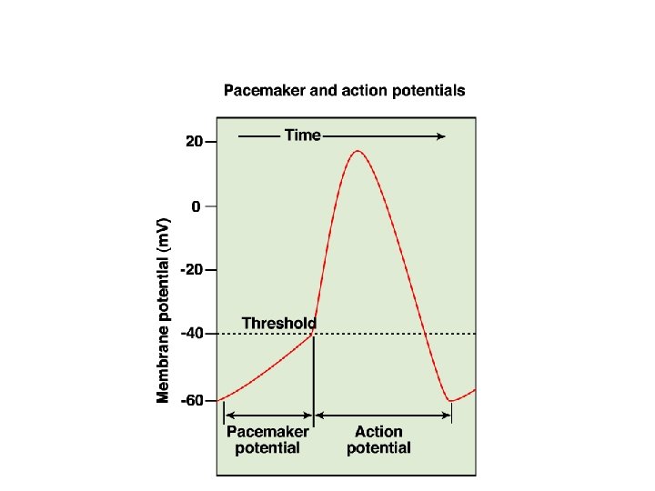

Action potentials in myocardial autorhythmic cells • Unstable resting membrane potential • Pacemaker potential • At a membrane potential of -60 m. V Na+ enters through the If channels • mb depolarizes • Ca++ channels open • Ca++ channels close • K+ leaves
 and E (adrenal hormone) • Autorhythmic cells")
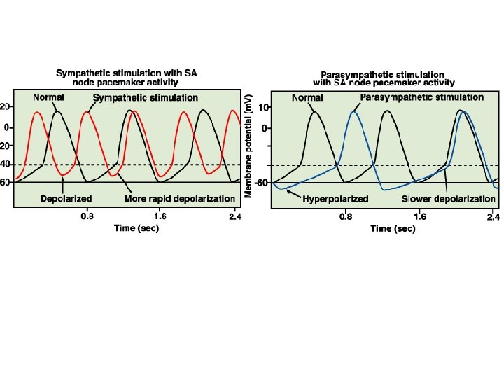
Modulation of autorhythmic cells • NE (sympathetic) and E (adrenal hormone) • Autorhythmic cells have beta 1 receptors • Cyclic AMP levels increase • Properties of If and Ca++ channels altered • More rapid Na+ and Ca++ entry • Rapid action potential • Rapid contractions

Modulation of autorhythmic cells • Parasympathetic, acetyl choline • Muscarinic receptors • K+ channels open mb hyperpolarizes cell less excitable • Ca++ channel less likely to open slower depolarization cell is less excitable
- Slides: 37