CARDIAC EMBRYOLOGY EARLY DEVELOPMENT OF HEART Dr Santhosh

CARDIAC EMBRYOLOGY EARLY DEVELOPMENT OF HEART Dr. Santhosh Narayanan

• • FORMATION OF TRILAMINAR EMBRYO CARDIOGENESIS MOLECULAR DEVELOPMENT OF HEART DEVELOPMENTAL ABNORMALITIES

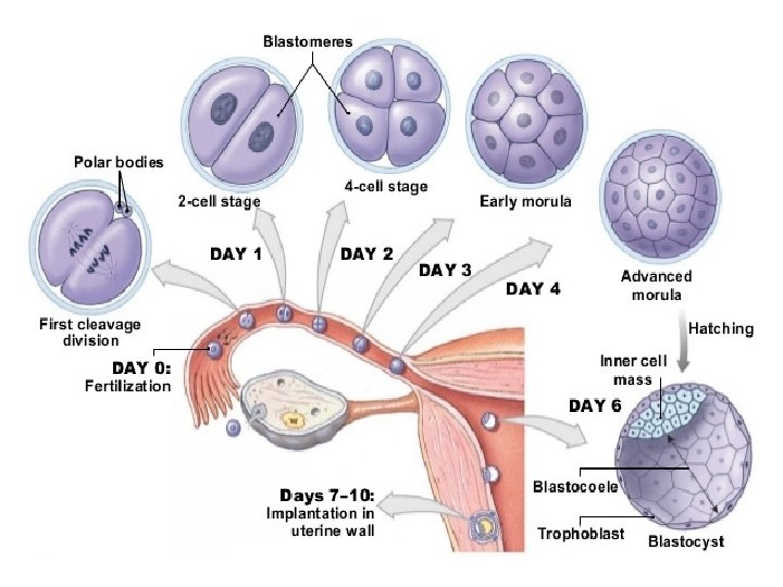
The Beginning DAY 0 - FERTILISATION DAY 1 -4 - CELL DIVISION- MORULA DAY 5 -6 - BLASTOCYST DAY 7 -10 - IMPLANTATION
 EPIBLAST HYPOBLAST")
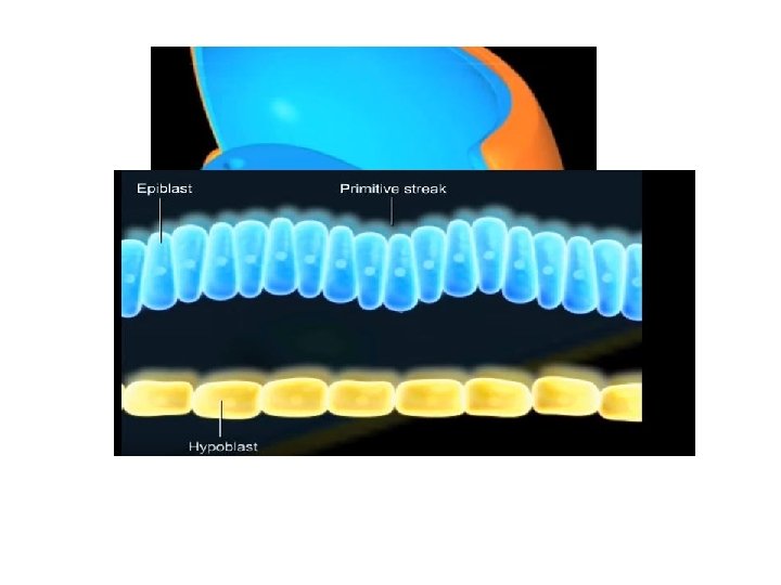
Bilaminar embryo (Days 11 -14) EPIBLAST HYPOBLAST

The Amnion and Yolk sac

Primitive streak week 3

Gastrulation - Week 3

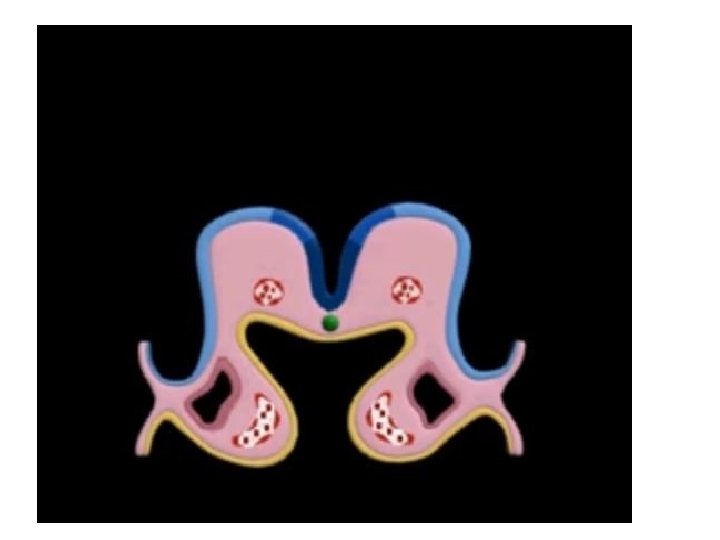
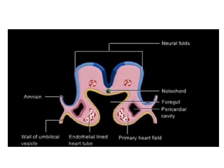
Neurulation

Neurulation

Derivatives of Germ layers

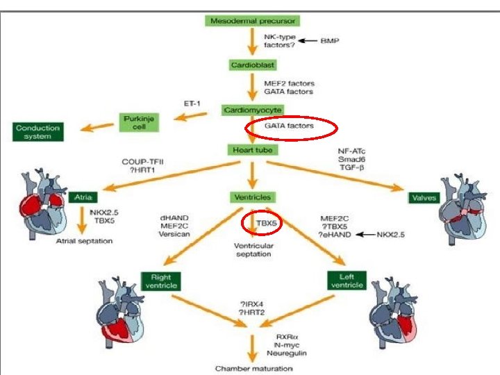
The Cardiogenesis

CARDIOGENESIS • Formation of bilateral heart fields • Formation of the heart tube • Folding of the heart tube • Looping of the heart tube • Chamber specification and compaction

Cardiac precursor cells • • First heart field Second heart field Neural crest cells Pro epicardium
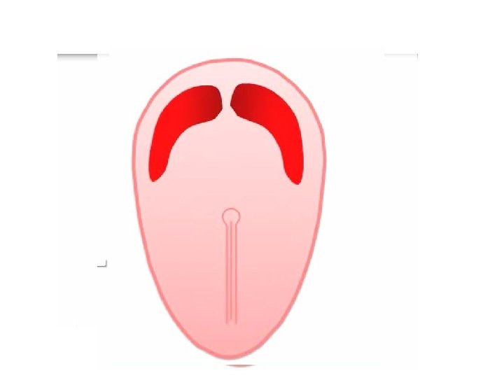

The "Cardiac Crescent"

The second heart field

3 rd Heart field

The Cardiac Neural crest

Sequence of Events • Day I 8 - Cardiac precursor cells seen in the form of blood islands • Day 20 - First intraembryonic blood vessels • Day 21 - Folding, heart tube formation, looping • Day 22 – heart starts to beat • Day 28 – embryonic circulation established

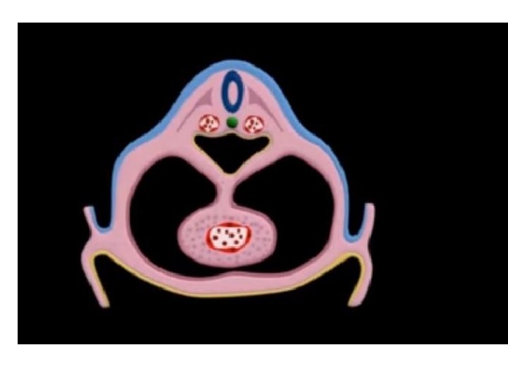
Folding of Embryo CRANIOCAUDAL AXIS • More rapid growth of the neural tube forming the brain at its cephalic end. • Growth in this direction will cause the embryo to become convex shaped. LATERAL FOLDING Two lateral edges of the germ disk fold forming a tube-like structure

Folding of embryo
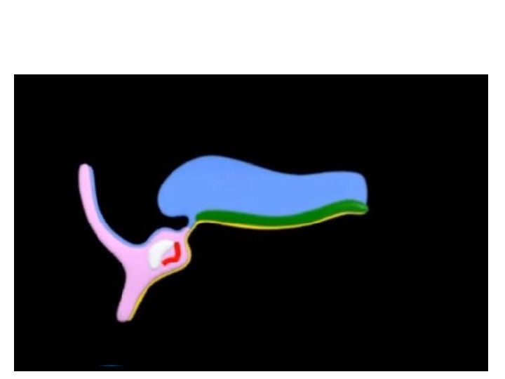
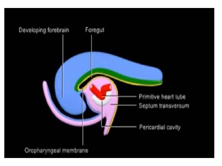
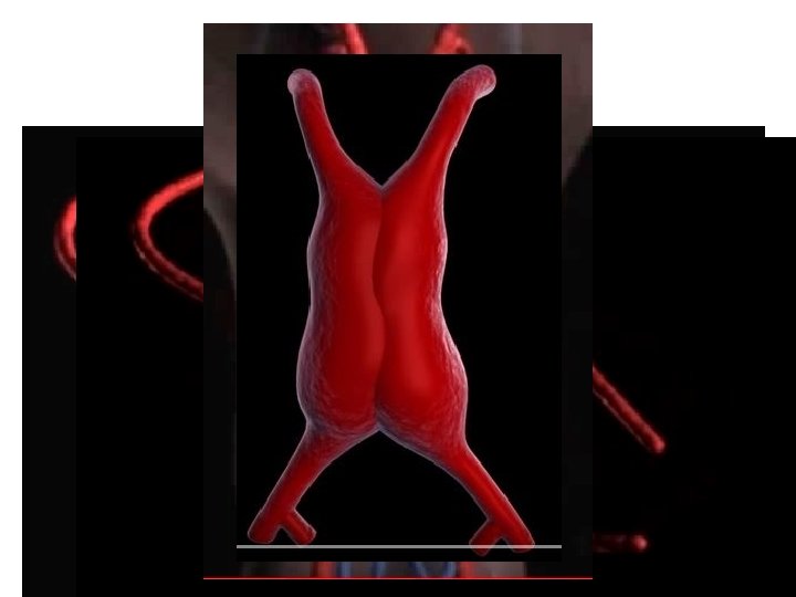

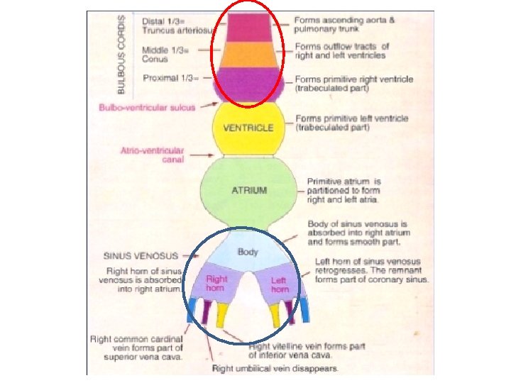
Arterial end of the heart tube • BULBUS CORDIS – proximal part called the conus – a distal part called truncus arteriosus. – The truncus continues distally with the aortic sac.

Venous end • The sinus venosus • vitelline vein from the yolksac • umbilical vein from the placenta • common cardinal vein from the bodywall

Looping

Looping
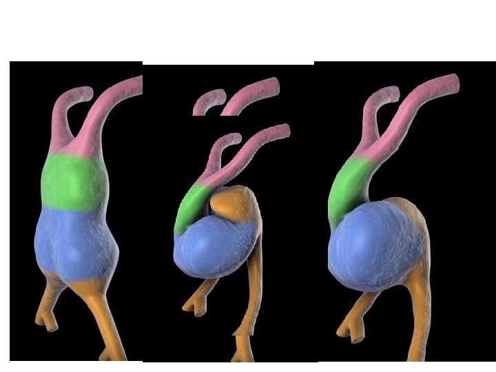

Mechanism of looping • Differential growth of the heart tube in comparison with foregut • Differential growth within the heart tube itself • Posterior, leftward, slower growth and anterior, rightward, faster growth resulting in RIGHTWARD LOOPING

• Bending of the heart tube at the inflow as well as within the ventricular segment • eventually positions the inflow and future left ventricular segments posteriorly and to the left, with the future right ventricle and outflow segments anteriorly and to the right

b

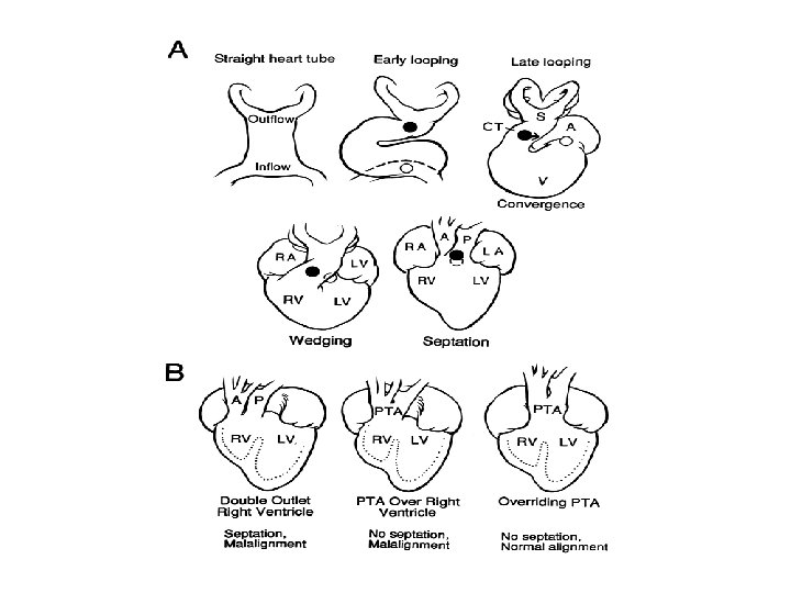
Convergence • Movement of the outflow tract and the atrioventricular canal into a more midline position • Alignment
 of the primitive ventricles and outflow tract into systemic (aorta)")
Wedging • Seperation (septation) of the primitive ventricles and outflow tract into systemic (aorta) and pulmonary (pulmonary artery) trunks is created by a process called wedging • The counterclockwise rotation of the outflow tract with movement of the future aortic valve position behind the pulmonary trunk

Convergence and Wedging

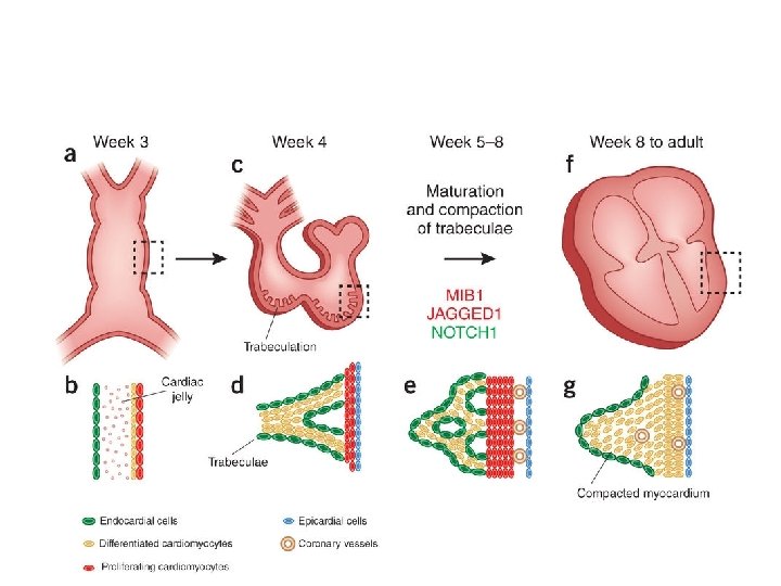
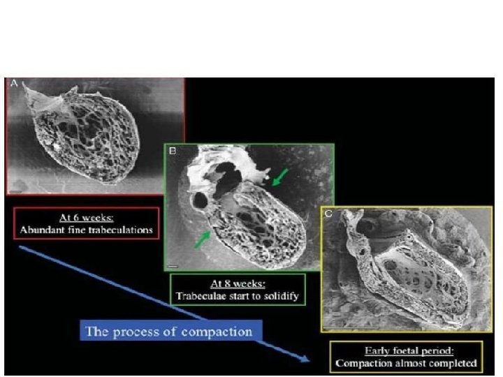
Myocardial Compaction • Emergence of trabeculations • Trabecular remodelling

Emergence of trabeculations • Emerge after looping of the primitive heart tube • At the end of the fourth week of gestation • Trabeculation patterns are ventricle-specific • trabeculations in the LV are generally thicker and the corresponding intertrabecular spaces larger

Trabecular remodeling • After completion of ventricular septation at 8 weeks of gestation in • Increase in ventricular volumes results in compression of the trabeculations with an increase in the thickness of the compacted myocardium • Compaction process coincides with the invasion of epicardial coronary arteries

Evolution of human heart • Progressive displacement of the inflow structures from a caudal position to a dorsal (fishes) and cephalad (reptiles) position. • Septation of the atrium in a right and a left cavity (amphibians). • Development of the right ventricle (RV) from the proximal part of the conus arteriosus.
.")
• Septation starting at the interventricular groove separating the left from RV (crocodiles). • Development of a high-pressure left ventricle (LV) and a low-pressure RV. • Disappearance of the sinus venosus and the conus arteriosus (birds and mammals).

Molecular development of heart
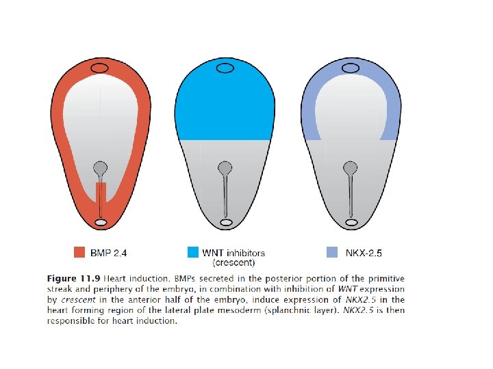
 endoderm induces a heart-forming region by")
Genes in induction of crescent • Anterior (cranial) endoderm induces a heart-forming region by inducing the transcription factor NKX 2. 5. • The signals require secretion of BMPs 2 and 4 secreted by the endoderm and lateral plate mesoderm. • Concomitantly, the activity of WNT proteins (3 a and 8) secreted by the neural tube, must be blocked because they normally inhibit heart development. • The combination of BMP activity and WNT inhibition by crescent and cerberus causes expression of NKX 2. 5.

Genes in differentiation

Right left asymmetry

Signalling networks NOTCH BMP WNT FGF SEMAPHORIN

Developmental abnormalities

• From Fertilization to Primitive Heart Tube -Abnormal development at this stage of embryogenesis results in embryonic death because of the critical nature of the early circulation to the further growth

Abnormal left right signalling • HETEROTAXY SYNDROMES -Right Isomerism -Left Isomerism -Situs inversus with dextrocardia

Heterotaxy spectrum
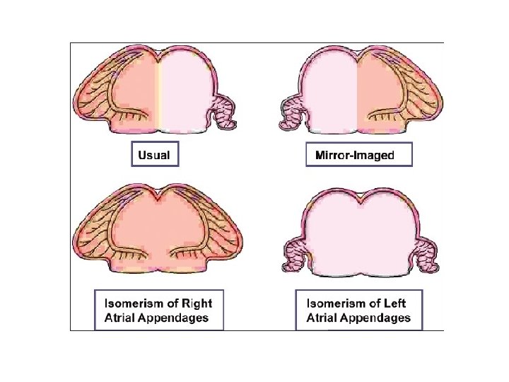
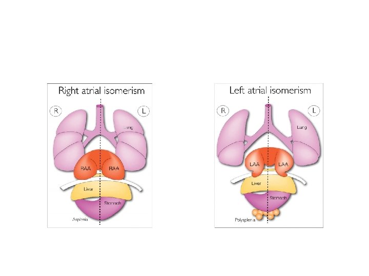
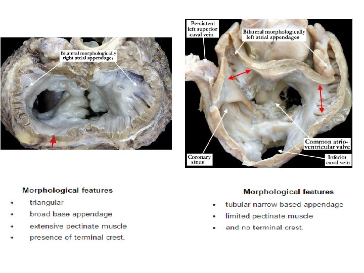
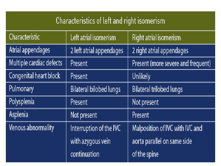

Right Isomerism • Cardiac : – The sinus nodes are paired because bilateral superior vena cavae are attached to bilateral morphologic right atria. – The P-wave axis is normal because the right sinus node is usually the dominant atrial pacemaker – The atrioventricular conduction system is equipped with 2 nodes often connected by a sling of tissue – Supraventricular tachycardia is attributed to reentry between paired atrioventricular nodes.

Right Isomerism- Associations – – – – – common atrium, common atrioventricular valve, Morphologic single ventricle, Functional single ventricle (hypoplasticright or left), pulmonary stenosis or atresia, absent coronary sinus total anomalous pulmonary venous connection bilateral ductus arteriosus. Ventricular and great arterial connections are usually discordant.

Left Isomerism • More prevalent in women • The pulmonary veins can be connected in a symmetric fashion, 2 to the right-sided atrium and 2 to the left-sided atrium • The most distinctive and the most diagnostically useful clinical feature is inferior vena caval interruption with azygous continuation, in which the suprarenal segment of the inferior cava is absent, and the infrarenal segment continues as the azygos or hemiazygous vein • Fetal complete heart block is presumptive evidence of in utero left isomerism

Left Isomerism • Sinus node is absent or hypoplastic. • The atrial pacemaker is therefore ectopic, and the P-wave axis is abnormal. • The ectopic atrial pacemaker can shift from 1 site to another or may fire slowly (ectopic atrial bradycardia

Malpositions due to looping defects

Situs solitus with Dextrocardia • Isolated dextrocardia with AV concordance and NRGA • The base-to-apex axis points to the right, right hemidiaphragm is lower • The embryonic straight heart tube initially bends rightward (dloop) but fails to move into the left chest. • Congenital heart defects: – ventricular septal defect – left SVC to the coronary sinus – coarctation of the aorta – secundum atrial septal defect – anomalous pulmonary venous connections – complete AV septal defect

• ECG – atrial activation is normal, and the P-wave frontal plane axis is 70 to 80 – the frontal plane QRS axis exhibits a rightward shift or right-axis deviation – a gradual and progressive reduction in the QRS Rwave voltage is observed

Corrected Transposition of the Great Arteries • Isolated Dextrocardia with AV and VA Discordance and Left Anterior Aorta • most common form of dextrocardia

Situs Inversus

Situs inversus with Dextrocardia • Incidence 1 in 8, 000 • The thoracic and abdominal viscera are mirror images of normal • morphologic right bronchus is concordant with the morphologic right atrium and a trilobed lung – usually occurs with a structurally normal heart

ECG • Situs inversus with Dextrocardia: reversed ventricular activation and reversed repolarization. – lead 1: QRS negative & the T wave inverted, – lead a. VR resembles a. VL and vice versa, – right precordial leads resemble leads from corresponding left precordial sites. – Septal Q waves appear in right precordial leads because septal depolarization is from right to left.
 • Situs")
Situs inversus with Dextrocardia Inverted Normally Related Great Arteries (Left Posterior Aorta) • Situs inversus totalis with persistence of normal AV and VA connections • CHD : – VSD – TOF – pulmonary atresia – complete AV septal defect – OS ASD.

Situs Inversus with Dextrocardia, Atrioventricular Concordance, and Ventriculoatrial Discordance with Left Anterior Aorta • Inverted form of complete TGA • The hemodynamics and physiology identical to TGA • ECG : – inverted P-wave axis because of the atrial inversion – more evidence of associated right and left ventricular hypertrophy because of transposition physiology

Situs Inversus with Dextrocardia Atrioventricular and Ventriculoatrial Discordance, with Right Anterior Aorta • Inverted form of corrected TGA • Rare

Situs Inversus with Dextrocardia, AV Discordance, and VA Concordance with Inverted NRGA • Represents the inverted form of isolated ventricular inversion • extremely rare • cardiac abnormalities – common atrium – common AV valve – severe right ventricular hypoplasia.

MCQ'S

1. Coronary vessels develop from • • A. FHF B. SHF C. Neural Crest D. Proepicardium • Answer D

2. All of the following are false except • A. Right isomerism is associated with ectopic pacemaker • B. Lungs are trilobed in left isomerism • C. Complete heart block is presumptive evidence of right isomerism • D. Interrupted IVC is seen in left isomerism • Answer D

3. Intraembryonic blood vessels are seen on • • A. Day 18 B. Day 19 C. Day 20 D. Day 22 • Answer C

4. All of the following are correctly matched except • • A- NKX 2. 5 - Cardiac crescent induction B. e. Hand- RV myocyte C. TBX 5 - Septation D. GATA factors -Heart tube formation • Answer B

5. Which of the following is false • A. Mesocardia is always associated with congenital heart defects • B. CCTGA-AV concordance with VA discordance • C. Situs inversus with dextrocardia usually occurs with a structurally normal heart • D. Right hemidiaphragm is lower in situs solitus with dextrocardia • Answer B

THANK YOU House model of HEART
- Slides: 90