Cardiac Disease Risk Factors Prevention Treatment Cardiac Anatomy


 is a graphic produced by an electrocardiograph machine, which records")


 occurs when")


- Slides: 18
Cardiac Disease Risk Factors Prevention Treatment
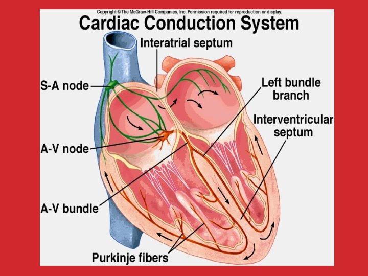
Cardiac Anatomy Name interior anatomy Trace blood flow From the Vena Cava > Right Atrium > Through Tricuspid Valve > Right Ventricle > Pulmonary Valve > Pulmonary Artery > LUNGS (pick up oxygen) > Pulmonary Veins > Left Atrium > Mitral Valve > Left Ventricle > Aortic Valve > Aorta > BODY (to deliver oxygen to all cells) > then start from the beginning again!
The arteries are divided into the right and left main. LCA: LAD (left anterior descending) supplies blood to front and left side of the heart. CX (circumflex) supplies to the lateral and back side of the heart. RCA (right coronary artery) Supplies blood to the right atrium, ventricle, Sino atrial node (cluster of cells regulates heart’s rhythmic rate), atrioventricular node. Smaller branches include; acute marginal, posterior descending (PDA), obtuse marginal (OM), septal perforator, and diagonals.
Electrocardiogram An electrocardiogram (ECG) is a graphic produced by an electrocardiograph machine, which records the electrical activity of the heart over time. Key waveforms of the ECG used for diagnostic purposes include the: P wave- beginning of P-R interval –atrial contraction QRS complex – ventricle contraction S-T segment – ventricle depolarization T wave – ventricle recovery The exercise ECG test A graded exercise treadmill test paired with electrocardiographic recording The test is considered “positive” if there is a specific standard level of change in the S-T segment component of the ECG. Can be employed for diagnostic or functional assessment
Controllable Risk Factors Smoking – Decreases oxygen in blood raises heart rate and blood pressure Hypertension – tears arterial walls • • • Diabetes (type II) – Obesity – Increases BP, cholesterol, increased work load on the heart, diabetes type II. High Cholesterol – Increases fatty build up of arteries. Physical Inactivity – Leads to obesity, high BP, weakens the heart muscle Response to stress – some studies show stress causes people to overeat, smoke, or drink too much alcohol. Drinking too much alcohol – can raise BP and triglycerides. The top number is the systolic blood pressure reading. It represents the maximum pressure exerted when the heart contracts. The bottom number is the diastolic blood pressure reading. It represents the pressure in the arteries when the heart is at rest. Long term high blood pressure can result in: – heart attack – stroke – kidney failure
Non Controllable Risk Factors • Age – 83% of people who die of MI are over 65 yrs. Sex – Men at more risk until women go through menopause then equal risk. Women have less risk due to less overall stress, less incidence of smoking/alcohol use, and increased estrogen levels increasing HDL. Family History – Mom, dad, grandmother, grandfather, siblings under 50 -55 highest. Plaque and Blockage
The Process of CAD • CAD – The end result of atherosclerosis within the walls of the coronary arteries that supply the myocardium – A chronic inflammatory response due in part to the deposition of lipoproteins • Atherogenesis – The process of the development of arterial plaques – Injury to the endothelium causes inflammation that results in molecules that facilitate atherosclerosis. • An acute coronary event is more often caused by rupture of an atherosclerotic plaque than by a gradual closure of the coronary blood vessel. • The formation, progression, and rupture of the plaque are collectively viewed as a process related directly to inflammation
Angina Pectoris • Discomfort in the chest, arms, shoulders, and jaw that results from inadequate blood flow to the heart • Stable angina – Occurs regularly with activity, upon awakening, or at other predictable times • Unstable angina – Changes in intensity, character, or frequency – May precede myocardial infarction and requires urgent medical attention – Individuals who have unstable angina are not appropriate clients and should always be referred back to their physicians.
So what is a Heart Attack? • Acute myocardial infarction (MI) occurs when the blood supply to the heart muscle is interrupted. • MI symptoms may include various combinations of pain in the chest, upper extremity, or jaw, or mid-back discomfort with exertion or at rest. – The discomfort usually lasts at least 20 minutes. • Women often experience different symptoms from men. – Shortness of breath – Weakness – Fatigue • Approximately one-third of all myocardial infarctions are “silent. ”
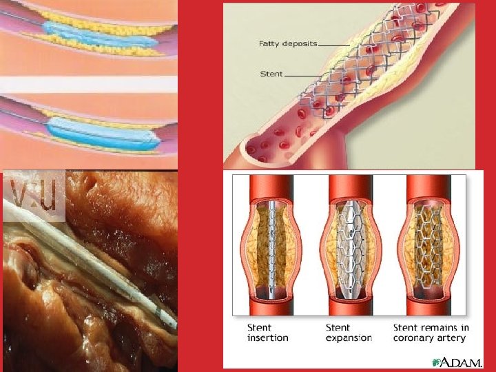
Coronary Angiography • • Involves inserting a catheter into the groin area and routing it into the coronary arteries of the heart A radio contrast agent is passed into the catheter and is visualized on a fluoroscope. The cardiologist can perform a percutaneous transluminal coronary angioplasty (PTCA). This technique has several goals: – To confirm the presence of a suspected blockage in a coronary artery – To quantify the severity of the disease and its effect on the heart – To seek the cause of a symptom such as angina, shortness of breath, or other signs of cardiac insufficiency – To make a patient assessment prior to heart surgery
Cardiac Catherization
Cardiac Output – Liters/minute the heart pumps blood (5 for rest – 30 for exercise). Stroke Volume – Volume ejected with each beat. Low Density Lipoproteins (LDL) – the bad cholesterol the liver produces in response to a poor diet, help to add deposits to walls of arteries. High Density Lipoproteins (HDL) – the good cholesterol the liver produces in response to exercise and estrogen levels. Help to remove fat build up in arteries.
http: //www. youtube. com/watch? v=WM_tcf 5 Ogy 0 http: //www. youtube. com/watch? v=dee 8 CMa mo. Q 0