Cardiac cycle and heart sounds Arwa Rawashdeh Blood

Cardiac cycle and heart sounds Arwa Rawashdeh

Blood flow and Laplace's law
 q.")
Blood flow Laminar flow : normal blood flow in the blood vessels (physiological) q. As you go toward the edges the velocity the blood is going to be slower and the velocity in the middle is highest q. So imagine you are looking to blood vessels as a circle, and you are looking at the flow from the back you are going to notice that is flow is very concentric and this type of flow is silent Turbulent flow : pathological and physiological one q. Inside our heart you have a valves mitral valve and aortic valve whenever blood is being pumped upward right it can hit mitral valve as it hits mitral valve it can develop turbulent flow q Imagine a blood vessels and plaques inside ; as the normal flow gets to the occlusion it start developing a turbulence and that gives a lot of heat and changes the action of perfusion pressure and produce what called brutes and can be heard at carotid artery so if you take a stethoscope and put it over carotid artery you can hear it as actual sounds that caused by turbulent flow. It also can produce murmurs

If you look at the graph here ; as you increase the pressure the flow is increasing in laminar or turbulent flow, but you get to the point where the flow veers off and the flow start decreasing as the perfusion pressure start increasing If there is a turbulent flow it decreases the actual flow the volume of blood that circulating through an area of blood vessel per a minute and increase the perfusion pressure and the resistance is going to be very high

Implication of Laplace's law

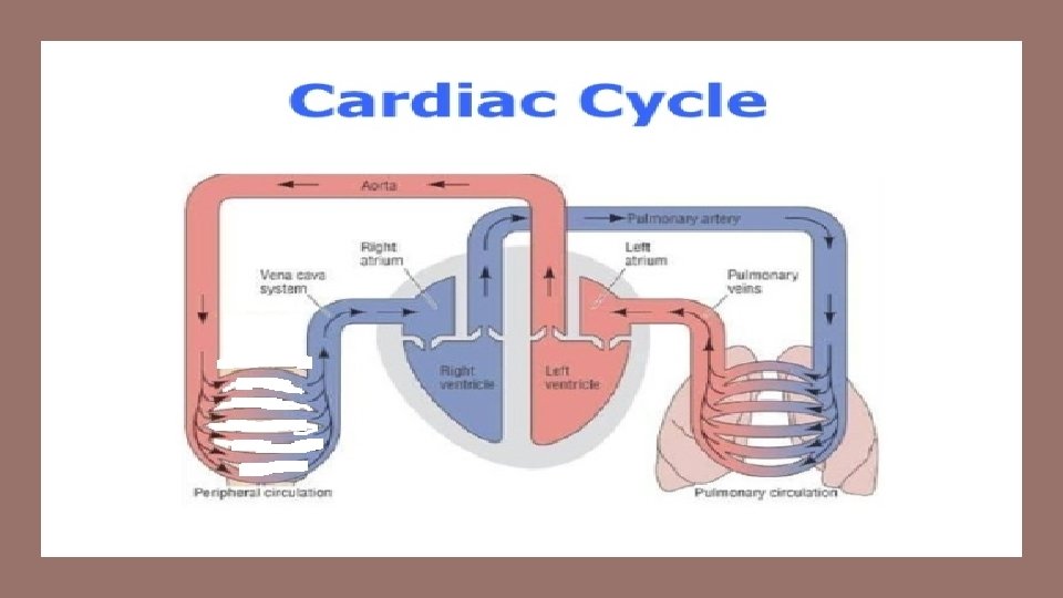
Cardiac cycle

Mid to Late ventricular diastole • Atrial pressure > ventricular pressure • Arterial pressure > ventricular pressure • AV valves Open • SLV valves closed

Isovolumetric contraction • Atrial pressure < ventricular pressure • Arterial pressure > ventricular pressure • AV valves closed Lub sound “S 1” • SLV valves closed

Mid to late ventricular systole or ventricular ejection • Atrial pressure < ventricular pressure • Arterial pressure < ventricular pressure • AV valves closed • SLV valves open

Isovolumetric relaxation • Atrial pressure < ventricular pressure • Arterial pressure < ventricular pressure • AV valves closed • SLV valves closed “Dub”sound

Positioning of stethoscope
 • First component due to turbulent rushing of blood")
FIRST HEART SOUND (S 1) • First component due to turbulent rushing of blood towards A-V valves • 2 nd component occurs due to the closure of the A -V valves • The mitral component heard at the apex beat area [left 5 th intercostal space at midclavicular line] • The tricuspid component is best heard in the 4 th intercostal space at the left sternal border
 • This sound is produced by the vibration associated")
SECOND HEART SOUND (S 2) • This sound is produced by the vibration associated with the closure of the semilunar valves (aortic and pulmonary) at the end of ventricular systole. • This sound is sharp and loud and described as “DUB. ” • Two subcomponents • Pulmonary component heard at the level of 2 nd left intercostal space. • Aortic component is heard at the level of the 2 nd right interscostal space near the right border of the sternum.
- Slides: 14