Cardiac Arrest Alireza Sedaghat MD FCCM Cardiac Arrest


 increases survival from VF/pulseless VT. Because these")


, go to")


- Slides: 86
Cardiac Arrest Alireza Sedaghat MD - FCCM
Cardiac Arrest Ventricular Fibrilation Pulseless Ventricular tachycardia Pulseless Electrical activity Asystole
Cardiac Arrest: VF – Pulseless VT Shockable Rhythm Ventricular Fibrilation
Cardiac Arrest: VF – Pulseless VT Shockable Rhythm Pulseless Ventricular tachycardia
Defibrillation Resume CPR While Manual Defibrillator Is Charging. Shortening the interval between the last compression and the shock by even a few seconds can improve shock success. Thus, it is reasonable for healthcare providers to practice efficient coordination between CPR and defibrillation to minimize the hands-off interval between stopping compressions and administering the shock.
Defibrillation For example, after verifying a shockable rhythm and initiating the charging sequence on the defibrillator, another provider should resume chest compressions and continue until the defibrillator is fully charged. The defibrillator operator should deliver the shock as soon as the compressor removes his or her hands from the patient’s chest and all providers are “clear" of contact with the patient.
Defibrillation It is important that healthcare providers be knowledgeable of how their defibrillator operates and if possible: limit pauses in chest compressions to rhythm analysis and shock delivery
Defibrillation Step 3: directs you to deliver 1 shock. The appropriate energy dose is determined by the identity of the defibrillator: monophasic or biphasic. If you are using a monophasic defibrillator, give a single 360 -J shock. Use the same energy dose for subsequent shocks.
Defibrillation Biphasic defibrillators use a variety of waveforms, each of which is effective for terminating VF over a specific dose range. When using biphasic defibrillators, providers should use the manufacturer’s recommended energy dose (eg, initial dose of 120 to 200 J). If you do not know the effective dose range, deliver the maximal energy dose for the first and all subsequent shocks.
Defibrillation If the initial shock terminates VF but the arrhythmia recurs later in the resuscitation attempt, deliver subsequent shocks at the previously successful energy level. Immediately after the shock, resome CPR, beginning with chest compressions. Give 2 minutes of CPR.
Defibrillation does not restart the heart. Defibrillation stuns the heart and briefly terminates all electrical activity, including VF and p. VT. If the heart is still viable, its normal pacemakers may eventually resume electrical activity (return of spontaneous rhythm) that ultimate results in a perfusing rhythm (ROSC).
Defibrillation In the first minutes after successful defibrillation, however, any spontaneous rhythm is typically slow and may not create pulses or adequate perfusion. The patient needs CPR (beginning with chest compressions) for several minutes until adequate heart function resumes. This is why it is important to resume high-quality CPR, beginning with chest compressions immediately after a shock
Defibrillation The interval from collapse to defibrillation is one of the most important of survival from cardiac arrest. Early defibrillation is critical for patients with sudden cardiac arrest for the following reasons When VF is present, CPR can provide a small amount of blood flow to the heart and brain but cannot directly restore an organized rhythm.
Defibrillation A common initial rhythm in out-of-hospital witnessed sudden cardiac arrest is VF. Pulseless VT rapidly deteriorates to VF. Electrical defibrillation is the most effective way to treat VF (delivery of a shock to stop the VF). The probability of successful defibrillation decreases quickly over time. VF deteriorates to asystole if not treated.
Defibrillation The likelihood of restoring a perfusing rhythm is optimized with immediate CPR and defibrillation within a few minutes of the initial arrest The earlier defibrillation occurs, the higher the survival rate.
Defibrillation For every minute that passes between collapse and defibrillation, the chance of survival from a witnessed VF sudden cardiac arrest declines by 7% to 10% per minute if no bystander CPR is provided. When bystanders perform CPR, the decline is more gradual and averages 3% to 4% per minute.
Clearing for Defibrillation To ensure safety during defibrillation, always announce the shock warning. State the warning firmly and in a forceful voice before delivering each shock Check to make sure you are clear of contact with the patient, the stretcher, or other equipment.
Resume CPR Immediately resume CPR, beginning with chest compressions Establish IV/IO access. Do not perform a rhythm or pulse check at this point unless the patient is showing signs of life or advanced monitoring indicates ROSC.
Resume CPR ACLS providers functioning with in a high-performance team can choose the optimal approach for minimizing interruptions in chest compressions A default compression-to-ventilation ratio of 30: 2 Use of different protocols, such as 3 cycles of 200 continuous compressions with passive ventilation, compression-only CPR in the first few minutes after arrest, and continuous chest compressions with asynchronous ventilation once every 6 seconds with the use of a bag-mask device,
Resume CPR Use of different protocols, such as: 3 cycles of 200 continuous compressions with passive ventilation, compression-only CPR in the first few minutes after arrest, and continuous chest compressions with asynchronous ventilation once every 6 seconds with the use of a bagmask device,
Resume CPR Progression from lay rescuers to highly trained healthcare providers for CPR delivery.
Rhythm Check Conduct a rhythm check after 2 minutes of CPR. Be careful to minimize interruptions in chest compressions. The pause in chest compressions to check the rhythm should not exceed 10 seconds. If a non-shockable rhythm is present and the rhythm is organized, a team member should try to palpate a pulse. If there is any doubt about the presence of a pulse, Immediately resume CPR
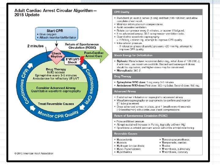
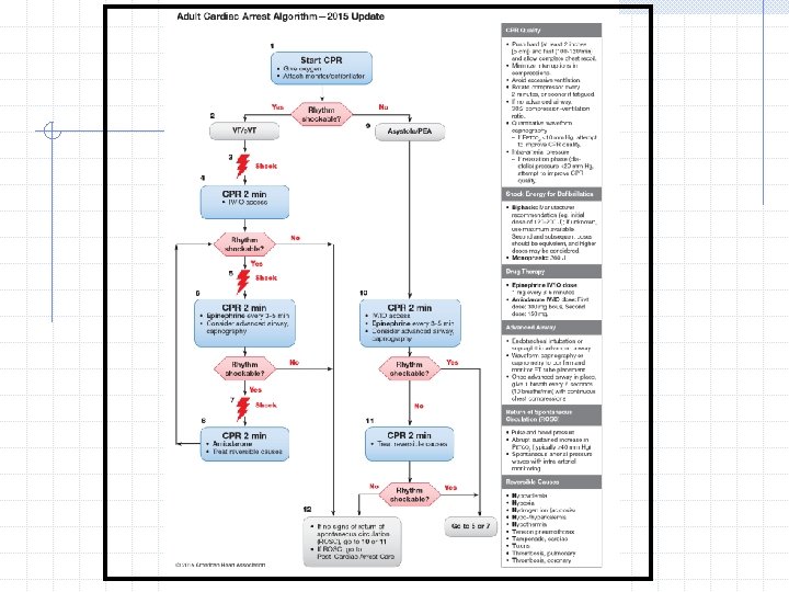
Rhythm Check If the rhythm is organized and there is a palpable pulse, proceed to post-cardiac arrest care. If the rhythm check reveals a non-shockable rhythm and there is no pulse, proceed along the asystole/PEA pathway on the right side of the Cardiac Arrest Algorithm (Steps 9 through 11). If the rhythm check reveals a shockable rhythm, give 1 shock and resume CPR immediately for 2 minutes after the shock (Step 6).
self-adhesive pads The AHA recommends routine use of self-adhesive pads. Using conductive materials (gel pads or self-adhesive pads) during the defibrillation attempt reduces transthoracic impedance, or the resistance that chest structures have on electrical current.
Shock & Vasopressors For persistent VF/pulseless VT, give 1 shock and resume CPR immediately for 2 minutes after the shock. Immediately after the shock, resume CPR, beginning with chest compressions. Give 2 minutes of CPR. When IV/IO access is available, give epinephrine during CPR after the second shock as follows: Epinephrine 1 mg IV/IO : repeat every 3 to 5 minutes
Shock & Vasopressors Epinephrine is used during resuscitation primarily for its beta-adrenergic effects: vasoconstriction. Vasoconstriction increases cerebral and coronary blood flow during CPR by increasing mean arterial pressure and aortic diastolic pressure. In previous studies, escalating and high-dose epinephrine administration did not improve survival to discharge or neurologic outcome after resuscitation from cardiac arrest.
Shock & Vasopressors No known vasopressor (epinephrine) increases survival from VF/pulseless VT. Because these medications can improve aortic diastolic blood pressure, coronary artery perfusion pressure, and the rate of ROSC, the AHA continues to recommend their use.
Shock & Vasopressors Vasopressin has been removed from the 2015 AHA Guidelines Update for CPP and ECC. The 2015 AHA Guidelines Update for CPR and ECC states that: vasopressin offers no advantage as a substitute for epinephrine in cardiac arrest.
Rhythm Check Conduct a rhythm check after 2 minutes of CPR. Be careful to minimize interruptions in chest compressions. The pause in chest compressions to check the rhythm should not exceed 10 seconds. If a nonshockable rhythm is present and the rhythm is organized, a team member should try to palpate a pulse. If there is any doubt about the presence of a pulse, immediately resume CPR
Rhythm Check If the rhythm is organized and there is a palpable pulse, proceed to post-cardiac arrest care. If the rhythm check reveals a non-shockable rhythm and there is no pulse, proceed along the asystole/PEA pathway on the right side of the Cardiac Arrest Algorithm (Steps 9 through 11). If the rhythm check reveals a shockable rhythm, resume chest compressions if indicated while the defibrillator is charging (Step 8).
Shock & Antiarrhythmics Healthcare providers may consider giving antiarrhythmic drugs, either before or after the shock. Research is still lacking on the effect of antiarrhythmic drugs given during cardiac arrest on survival to hospital discharge. If administered, amiodarone is the first-line agent given in cardiac arrest because it improves the rate of ROSC and hospital admission in adults with refractory VF/pulseless VT.
Shock & Antiarrhythmics Amiodarone 300 mg IV/IO bolus, then consider an additional 150 mg IV/IO once (in 3 -5 minutes) Amiodarone is considered a class III antiarrhythmic drug, but it possesses electrophysiologic characteristics of the other classes: Amiodarone blocks sodium channels at rapid pacing frequencies (class I effect) and exerts a noncompetitive antisympathetic action (class II effect). lengthening of the cardiac action potential (class III effect).
Shock & Antiarrhythmics If amiodarone is not available, providers may administer lidocaine Lidocaine 1 to 1. 5 mg/kg IV/IO first dose, then 0. 5 to 0. 75 mg/kg IV/IO at 5 – 10 minute intervals, to a maximum dose of 3 mg/kg If no IV/IO access is available, the dose for ET administration is 2 to 4 mg/kg.
Shock & Antiarrhythmics Lidocaine suppresses automaticity of conduction tissue in the heart, by increasing the electrical stimulation threshold of the ventricle, His-Purkinje system, and spontaneous depolarization of the ventricles during diastole by a direct action on the tissues. Lidocaine blocks permeability of the neuronal membrane to sodium ions, which results in inhibition of depolarization and the blockade of conduction.
Shock & Antiarrhythmics Providers should consider magnesium sulfate for torsades de pointes associated with a long QT interval. Magnesium sulfate for torsades de pointes, loading dose 1 to 2 g IV/IO diluted in 10 m. L given as IV/IO bolus, typically over 5 to 20 minutes. Routine administration of magnesium sulfate in cardiac arrest is not recommended unles torsades de pointes
Search for and treat any treatable underlying cause of cardiac arrest. See the column on the right of the algorithm. (H’s and T’s).
Physiologic Monitoring during CPR The AHA recommends using quantitative waveform capnography in intubated patients to monitor CPR quality , optimize chest compressions, and detect ROSC during chest compressions. Although placement of invasive monitors during CPR is not generally warranted, physiologic parameters such as intra-arterial relaxation pressures and central venous oxygen saturation (SCVO, ), when available, may also be helpful for optimizing CPR and detecting ROSC.
Physiologic Monitoring during CPR End-Tidal CO 2 The main determinant of PETCO 2 during CPR is blood delivery to the lungs. Persistently of PETco 2, values less than 10 mm Hg during CPR in intubated patients suggest that ROSC is unlikely. If PETco 2 abruptly increases to a normal value of 35 to 40 mm Hg, It is reasonable to consider this an indicator of ROSC.
Physiologic Monitoring during CPR End-Tidal CO 2 If the PETCo is less than 10 mm Hg during CPR, it is reasonable to try to improve chest compressions and vasopressor therapy.
Physiologic Monitoring during CPR End-Tidal CO 2 ROSC is recognized by the abrupt increase in PETCO, to greater than 50 mm Hg, which is consistent with a substantial improvement in blood flow
Physiologic Monitoring during CPR Coronary Perfusion Pressure or Arterial Relaxation Pressure Increased CPP correlates with both myocardial blood flow and ROSC. A reasonable surrogate for CPP during CPR is arterial relaxation (“diastolic") pressure, which can be measure by using an intra-arterial catheter. If the arterial relaxation pressure is less than 20 mm Hg it is reasonable try to improve chest compressions and vasopressor therapy.
Physiologic Monitoring during CPR Coronary Perfusion Pressure or Arterial Relaxation Pressure
Physiologic Monitoring during CPR Central Venous Oxygen Saturation If oxygen consumption, arterial oxygen saturation, and hemoglobin are constant, changes in Scvo, reflect changes in oxygen delivery due to changes in cardiac output. Scvo can be measured continuously by using oximetric tipped central venous catheters placed in left superior vena cava or pulmonary artery. Normal range is 60% to 80%. If it is less than 30%, it is reasonable to try to improve chest compressions and vasopressor therapy
Routes of Access for Drugs Priorities during cardiac arrest are high-quality CPR and early defibrillation. Insertion of an advanced airway and drug administration are of secondary importance. No drug given during cardiac arrest has been studied adequately to show improved survival to hospital discharge or improved neurologic function after cardiac arrest.
Routes of Access for Drugs Historically in ACLS, providers have administered drugs either via the IV or ET route. ET absorption of drugs is poor and optimal drug dosing is not known. For this reason, the l. V or IO route is preferred. A peripheral IV is preferred for drug and fluid administration unless central line access is already available.
Routes of Access for Drugs Central line access is not necessary during most resuscitation attempts. Central line access may cause interruptions in CPR and complications during insertion, including vascular laceration, hematomas, and bleeding. Insertion of a central line in a noncompressible vessel is a relative (not absolute) contraindication to fibrinolytic therapy in patients with ACS.
Routes of Access for Drugs peripheral line Establishing a peripheral line does not require interruption of CPR. Drugs, however, typically require 1 to 2 minutes to reach the central circulation when given by the peripheral IV route. §Give the drug by bolus injection unless otherwise specified. §Follow with a 20 -m. L bolus of IV fluid. §Elevate the extremity for about 10 to 20 seconds to facilitate delivery of the drug to the central circulation.
Routes of Access for Drugs Interosseous access IO access can be established in all age groups. IO access often can be achieved in 30 to 60 seconds. The IO route of administration is preferred over the ET route and may be easier to establish in cardiac arrest. Any ACLS drug or fluid that is administered IV can be given IO.
Routes of Access for Drugs Interosseous access IO cannulation provides access to a noncollapsible marrow venous plexus, which serves as a rapid, safe, and reliable route for administration of drugs, crystalloids, colloids, and blood during resuscitation. The technique uses a rigid needle, preferably a specially designed IO or bone marrow needle from an IO access kit.
Routes of Access for Drugs Endotracheal Route IV and IO administration routes are preferred over the ET administration route. The optimal dose of most drugs given by the ET route is unknown. The typical dose of drugs administered via the ET route is 2 to 2. 5 times the IV route. CPR will need to be stopped transiently so drug does not regurgitate up the ET tube.
Routes of Access for Drugs Endotracheal Route Studies demonstrate that epinephrine, vasopressin and lidocaine are absorbed into the circulatory system after administration via the ET route. When giving drugs via the ET route, dilute the dose in 5 to 10 m. L of sterile water or normal saline. Inject the drug directly into the ET tube.
Fluid Administration The optimal post cardiac arrest blood pressure remains unknown; however, a mean arterial pressure 65 mm Hg or greater is a reasonable goal. In hypovolemic patients, the ECF volume is typically restored with normal saline or lactated Ringer. Avoid DW because it will reduce serum sodium too rapidly. Serum electrolytes should be appropriately monitored.
Steroids in Cardiac Arrest The use of steroids in cardiac arrest has been assessed in both the out-of-hospital and in-hospital settings. In IHCA, steroids were combined with a vasopressor bundle or cocktail of epinephrine and vasopressin In light of the data presented, no recommendation can be made on the routine use of steroids alone in IHCA. For OHCA patients, use of steroids during CPR is of uncertain benefit
Extracorporeal CPR The use of ECPR may allow providers additional time to treat reversible underlying causes of cardiac arrest or serve as a bridge for LV assist device implantation or cardiac transplantation. 2015 AHA Guidelines Update suggests a benefit to survival and favorable neurologic outcome with the use of ECPR when compared with conventional CPR in patients with refractory cardiac arrest.
For patients with known or suspected opioid overdose who are in respiratory arrest, healthcare providers should give naloxone as soon as it is available. It may be gived at a dose of 2 mg IN or 0. 4 mg IM/IV, which may be repeated every 4 minutes if necessary.
Cardiac Arrest: Pulseless Electrical Activity
Cardiac Arrest: Pulseless Electrical Activity PEA encompasses a heterogeneous group of rhythms that are organized or semi-organized but lack a palpable pulse: • ldioventricular rhythms • Ventricular escape rhythms • Postdefibrillation idioventricular rhythms • Sinus rhythm • Other rhythms
Cardiac Arrest: Pulseless Electrical Activity
Cardiac Arrest: Pulseless Electrical Activity Any organized rhythm without a pulse is defined as PEA. (Even sinus rhythm without a detectable pulse). Pulseless rhythms that are excluded by definition include VF, pulseless VT, and asystole Previously, high-performance teams used the term electromechanical dissociation (EMD) to describe patients who displayed electrical activity on the cardiac monitor but lacked apparent contractile function because of an undetectable pulse.
Cardiac Arrest: Pulseless Electrical Activity This is the most common initial condition present after successful defibrillation. PEA also includes other conditions where the heart is empty because of inadequate preload. This may occur as a result of severe hypovolemia, or as a result of decreased venous return from PE or pneumothorax.
Cardiac Arrest: Pulseless Electrical Activity High-performance team initiate and perform high-quality CPR throughout the BLS Assessment and the Primary and Secondary Assessments. The team interrupts CPR for 10 seconds or less for rhythm and pulse checks. This patient has an organized rhythm on the monitor but no pulse. The condition is PEA (Step 9). Chest compressions resume immediately. The team leader now directs the team in the steps outlined in the PEA pathway of the Cardiac Arrest Algorithm (Figure 39), beginning with Step 10.
Cardiac Arrest: Pulseless Electrical Activity Conduct a rhythm check and give 2 minutes of CPR after administration of the drugs. Be careful to minimize interruptions in chest compressions The pause in CPR to conduct a rhythm check should not exceed 10 seconds. Give epinephrine as soon as IV/IO access becomes available: 1 mg IV/IO , repeat every 3 to 5 minutes Consider advanced airway and capnography
Cardiac Arrest: Pulseless Electrical Activity If no electrical activity is present (asystole), go to Step 10. If organized electrical activity is present, try to palpate a pulse. Take at least 5 seconds but do not take more than 10 seconds to check for a pulse. If no pulse is present, or if there is any doubt about the presence of a pulse, immediately resume CPR for 2 minutes, starting with chest compressions. Go back to Step 10. If a palpable pulse is present and the rhythm is organized, begin post cardiac arrest care.
Cardiac Arrest: Pulseless Electrical Activity If the rhythm check reveals a shockable rhythm, resume CPR with chest compressions while the defibrillator is charging if possible. Switch to the left side of the algorithm and perform steps according to the VF/p. VT sequence starting with Step 5 or 7.
Cardiac Arrest: Pulseless Electrical Activity Treatment of PEA is not limited to the interventions outlined in the algorithm. Healthcare providers should attempt to identify and correct an underlying cause if present.
Cardiac Arrest: Pulseless Electrical Activity Common Underlying Causes of PEA Hypovolemia and hypoxia are the 2 most common underlying and potentially reversible causes of PEA. Be sure to look for evidence of these problems as you assess the patient.
Cardiac Arrest Asystole
Cardiac Arrest: Asystole The team interrupts CPR for 10 seconds or less for a rhythm check. This patient has no pulse and the rhythm on the monitor is asystole. Chest compressions resume immediately. The team leader now directs the team in the steps outlined in the asystole pathway of the Cardiac Arrest Algorithm
Cardiac Arrest: Asystole You will need to recognize the following rhythms: • Asystole • Slow PEA terminating in bradyasystolic rhythm This case involves these drugs: • Epinephrine • Other medications, depending on the causes
Cardiac Arrest: Asystole is a cardiac arrest rhythm associated with no discernible electrical activity on the ECG (also referred to as flat line). You should confirm that the flat line on the monitor is indeed “true asystole" by validating that the flat line • Not another rhythm masquerading as a flat line • Not the result of an operator error
Cardiac Arrest: Asystole The term flat line is nonspecific and can result from several possible conditions, including absence of cardiac electrical activity, lead or other equipment failure, and operator error. Some defibrillators and monitors signal the operator when a lead or other equipment failure occurs. For a patient with cardiac arrest and asystole, rule out any other causes of an isoelectric ECG
Cardiac Arrest: Asystole The management of asystole consists of the following components: • Implementing the steps in the Cardiac Arrest Algorithm • Identifying and correcting underlying causes • Terminating efforts as appropriate
Cardiac Arrest: Asystole Conduct a rhythm check and give 2 minutes of CPR after administration of the drugs. Be careful to minimize interruptions in chest compressions The pause in CPR to conduct a rhythm check should not exceed 10 seconds. Give epinephrine as soon as IV/IO access becomes available: 1 mg IV/IO , repeat every 3 to 5 minutes Consider advanced airway and capnography
Non-shockable Rhythm If no electrical activity is present , go back to Step 10 or 11. If electrical activity is present and organized, try to palpate a pulse. If no pulse is present or if there is any doubt about the presence of a pulse, continue CPR, starting with chest compressions for 2 minutes. Go back to Step 10 and repeat the sequence. If a good pulse is present and the rhythm is organized, begin post cardiac arrest care
Shockable Rhythm If the rhythm check reveals a shockable rhythm, prepare to deliver a shock Resuming chest compressions during charging if appropriate. Refer to the left side of the algorithm and perform steps according to the VF/p. VT sequence, starting with Step 5 or 7.
Defibrilation There is no evidence that attempting to “defibrillate” asystole is beneficial. If it is unclear whether the rhythm is fine VF or asystole, an initial attempt at defibrillation may be warranted. Fine VF may be the result of a prolonged arrest. At this time, the benefit of delaying defibrillation to perform CPR before defibrillation is unclear.
Terminating Resuscitative Efforts ØTime from collapse to CPR ØTime from collapse to first defibrillation attempt ØComorbid disease ØPre arrest state ØInitial arrest rhythm ØResponse to resuscitative measures ØETCO less than 10 after 20 minutes of CPR However, the duration of resuscitative efforts is an important factor associated with poor outcome.
Continue out-of-hospital resuscitative efforts until one of the following occurs: ØRestoration of effective, spontaneous circulation and ventilation ØTransfer of care to a senior emergency medical professional ØThe presence of reliable criteria indicating irreversible death ØThe healthcare provider is unable to continue ØA valid DNAR order is presented ØOnline authorization from the medical control physician or by prior medical protocol for termination of resuscitation