
Carcinoma Stomach Liver tumours Portal hypertension Chronic liver

- Slides: 65
Carcinoma Stomach Liver tumours Portal hypertension Chronic liver disease Dr Amit Gupta Associate Professor Dept Of Surgery
Carcinoma Stomach
Introduction • Gastric cancer is endemic in Japan • Late stage at diagnosis because of – Low incidence – Non specific symptoms – Risk factors not definable • Biologically more aggressive • Increasing incidence of adenocarcinomas of proximal stomach and distal esophagus
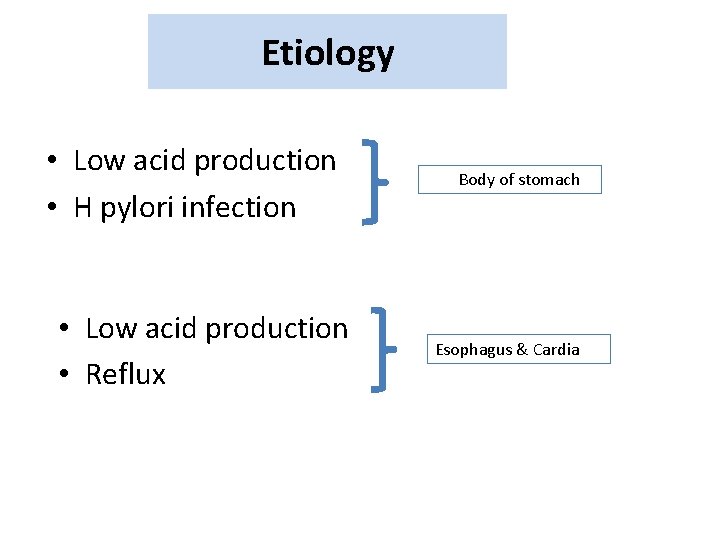
Etiology • Low acid production • H pylori infection • Low acid production • Reflux Body of stomach Esophagus & Cardia
• Obesity • High BMI • High glycaemic load diet • GERD • Smoking • Alcohol • Tobacco
Although no normal lymphoid tissue is found in the gastric mucosa, the stomach is the most common site for lymphomas of the gastrointestinal tract
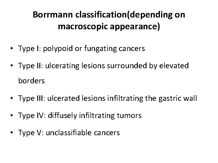
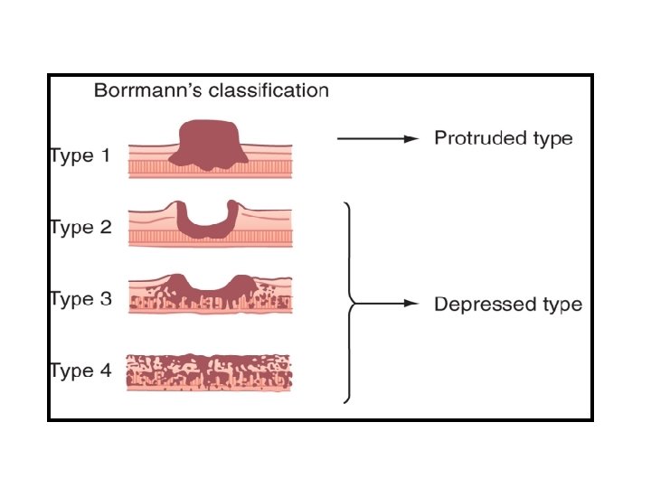
Borrmann classification(depending on macroscopic appearance) • Type I: polypoid or fungating cancers • Type II: ulcerating lesions surrounded by elevated borders • Type III: ulcerated lesions infiltrating the gastric wall • Type IV: diffusely infiltrating tumors • Type V: unclassifiable cancers
Patterns of Spread • Local extension • Lymphatic metastases – left supraclavicular fossa (Virchow’s node) – left axilla (Irish’s node) – S/c periumbilical tumour deposits (Sister Mary Joseph’s nodes) • Peritoneal metastases • Distant metastases
Clinical Presentation • • • Weight Loss Anorexia Early satiety (Diffusely infiltrative type) Recurrent Vomiting (pyloric involvement ) Dysphagia Bleeding Anemia Fatigue Epigastric pain
• Ascites, jaundice, or palpable mass indicates incurable disease • Transverse colon is a potential site of malignant fistulization and obstruction from gastric primary tumor • Diffuse peritoneal spread of disease frequently produces other sites of intestinal obstruction • Large ovarian mass ( krukenberg’s tumor) • Large peritoneal implant in the pelvis ( blumer’s shelf)
• Macroscopic description of the tumor • Extent of peritoneal metastases (P 0 -1) • Extent of hepatic metastases (H 0 -1) • Peritoneal cytology findings
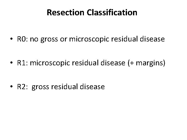
Resection Classification • R 0: no gross or microscopic residual disease • R 1: microscopic residual disease (+ margins) • R 2: gross residual disease
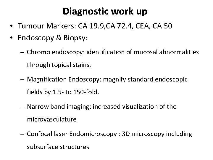
Diagnostic work up • Tumour Markers: CA 19. 9, CA 72. 4, CEA, CA 50 • Endoscopy & Biopsy: – Chromo endoscopy: identification of mucosal abnormalities through topical stains. – Magnification Endoscopy: magnify standard endoscopic fields by 1. 5 - to 150 -fold. – Narrow band imaging: increased visualization of the microvasculature – Confocal laser Endomicroscopy : 3 D microscopy including subsurface structures
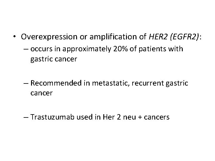
• Overexpression or amplification of HER 2 (EGFR 2): – occurs in approximately 20% of patients with gastric cancer – Recommended in metastatic, recurrent gastric cancer – Trastuzumab used in Her 2 neu + cancers
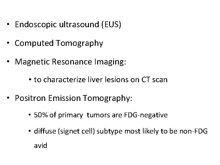
• Endoscopic ultrasound (EUS) • Computed Tomography • Magnetic Resonance Imaging: • to characterize liver lesions on CT scan • Positron Emission Tomography: • 50% of primary tumors are FDG-negative • diffuse (signet cell) subtype most likely to be non-FDG avid
• Staging Laparoscopy and Peritoneal Cytology: • directly inspects the peritoneal and visceral surfaces • done to spare nontherapeutic operations
Treatment • Surgery (Gastrectomy with lymph node dissection) ` – Radical Gastrectomy (Proximal gastric cancer) – Subtotal Gastrectomy (mid & distal gastric cancer) When the oncologic goal of an R 0 resection can be achieved by a gastricpreserving approach, partial gastrectomy is preferred over total gastrectomy
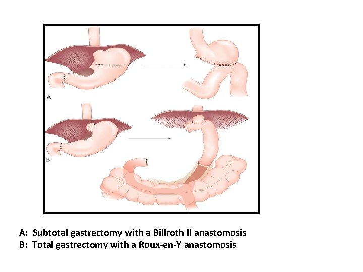
A: Subtotal gastrectomy with a Billroth II anastomosis B: Total gastrectomy with a Roux-en-Y anastomosis
• Lymphadenectomy – 15 nodes for adequate pathologic staging • Partial Pancreatectomy and Splenectomy—Resect or Preserve? Splenectomy: – Intraoperative evidence of direct tumor extension into the spleen – Primary tumor is located in the proximal stomach along the greater curvature Partial Pancreatectomy: • direct tumor extension into the pancreas
Stage specific Survival rates for gastric adenocarcinoma are higher in Japan than in Western countries because: • Better-prognosis intestinal-type tumors are more common • Poorer-prognosis proximal gastric cancers are less frequent • Widespread use of extensive D 2 or D 3 lymphadenectomy
Chemotherapy • Adjuvant • Neoadjuvant: • Higher rate of R 0 resections • Early treatment of micro metastatic disease • Periperative
Adjuvant Intraperitoneal Chemotherapy • Hyperthermic intraoperative peritoneal chemotherapy (HIPEC) • Normothermic intraoperative chemotherapy (NIIC) • Early postoperative intraperitoneal chemotherapy (EPIC) • Delayed postoperative intraperitoneal chemotherapy (DPIC)
Chemotherapy Drugs • Cisplatin • 5 -Fluorouracil • Taxanes • Epirubicin • Irinotecan • Trastuzumab (targeted therapy in Her 2 neu + )
Radiotherapy • Preoperative • Intraoperative • Postoperative
Liver Tumours
Benign tumours • Liver Cell Adenoma • Focal Nodular hyperplasia • Hemangioma • Mesenchymal hamartomas
Liver Cell Adenoma – – – – – benign proliferation of hepatocytes young women (aged 20 -40 years) associated with use of oral contraceptive pills (OCPs) usually singular Upper abdominal pain tumor markers are normal MRI scans of LCA have specific imaging characteristics two major risks are rupture & malignant transformation Hepatic artery embolization Resection of the mass
Focal nodular hyperplasia • Second most common benign tumor • young women • small (<5 cm) nodular mass • involves the right and left liver equally • Related to developmental vascular malformation • Nonspecific symptoms • Contrast-enhanced CT and MRI scan • Symptomatic pts: resection
Hemangioma • Most common benign tumor of the liver • Women more common than men (3: 1 ratio) • Mean age of about 45 years • Occur equally in the right and left liver • Usually single • Lesions > 5 cm are arbitrarily called giant hemangiomas • Tumor markers are usually normal • CT and MRI scan • Resection: enucleation with inflow control
Hepatocellular carcinoma
Introduction • Fifth most common malignancy worldwide • Male-to-female ratio 2. 4: 1 • Poor prognosis
Etiology • Infections with hepatitis B and C viruses • Ethanol abuse • Obesity • Type 2 diabetes • Non-alcoholic fatty liver disease • Aflatoxin B 1
Pathology • Malignant epithelial neoplasms: 85% to 95% • Benign: 6 %to 12% • Malignant mesenchymal tumors: 1% to 3% • Metastatic tumors
• Tumors metastatic to the liver are: – Peripheral – Multiple – Cause umbilication of surface of the liver • Primary liver tumors are: – Central – Solitary – Exophytic
• HCC spreads most commonly to – Lymph nodes around the liver – Peritoneal cavity – Lung Characteristic feature of HCC is invasion of the portal vein
Clinical Features Symptoms Signs • Abdominal pain • Hepatomegaly • Weight loss • Hepatic bruit • Weakness • Fullness • Anorexia • Ascites • Splenomegaly • Jaundice • Wasting • Abdominal swelling • Fever • Jaundice • Virchow-Troisier nodes • Vomiting • Cutaneous metastases (red-blue nodules)
Paraneoplastic Syndromes Hypoglycemia Erythrocytosis Hypercalcemia Hypercholesterolemia Dysfibrinogenemia Carcinoid syndrome Increased thyroxin-binding globulin Sexual changes (gynecomastia, testicular atrophy, and precocious puberty) • Porphyria cutanea tarda • •
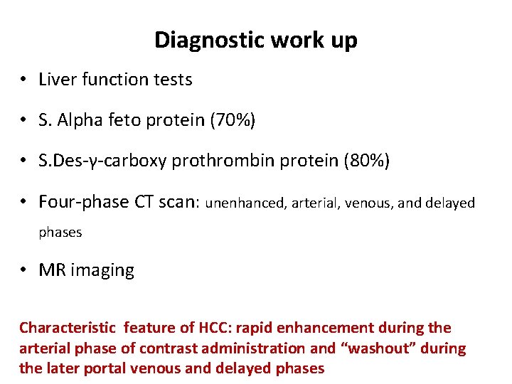
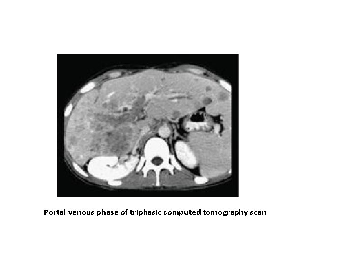
Diagnostic work up • Liver function tests • S. Alpha feto protein (70%) • S. Des-γ-carboxy prothrombin protein (80%) • Four-phase CT scan: unenhanced, arterial, venous, and delayed phases • MR imaging Characteristic feature of HCC: rapid enhancement during the arterial phase of contrast administration and “washout” during the later portal venous and delayed phases
Portal venous phase of triphasic computed tomography scan
• PET CT Scan: not recommended • Core biopsy: hallmark feature of HCC is stromal invasion
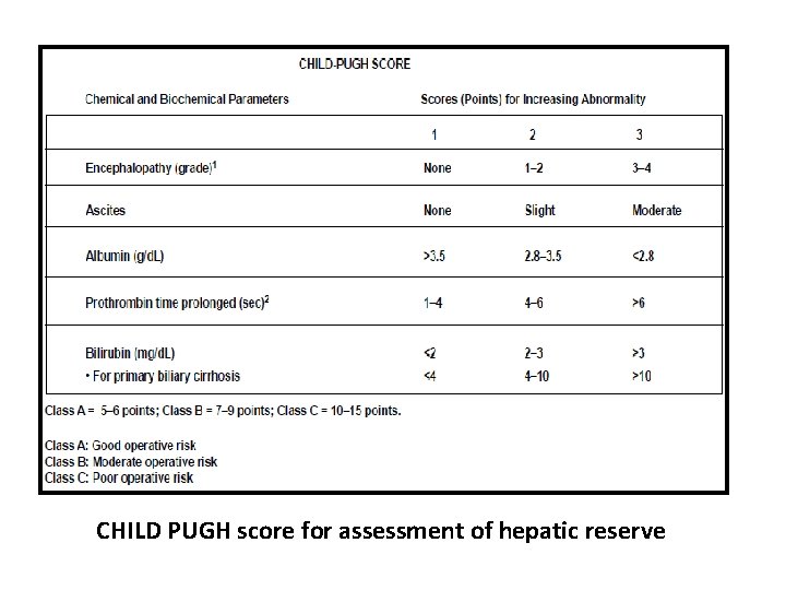
CHILD PUGH score for assessment of hepatic reserve
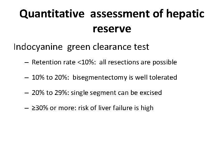
Quantitative assessment of hepatic reserve Indocyanine green clearance test – Retention rate <10%: all resections are possible – 10% to 20%: bisegmentectomy is well tolerated – 20% to 29%: single segment can be excised – ≥ 30% or more: risk of liver failure is high
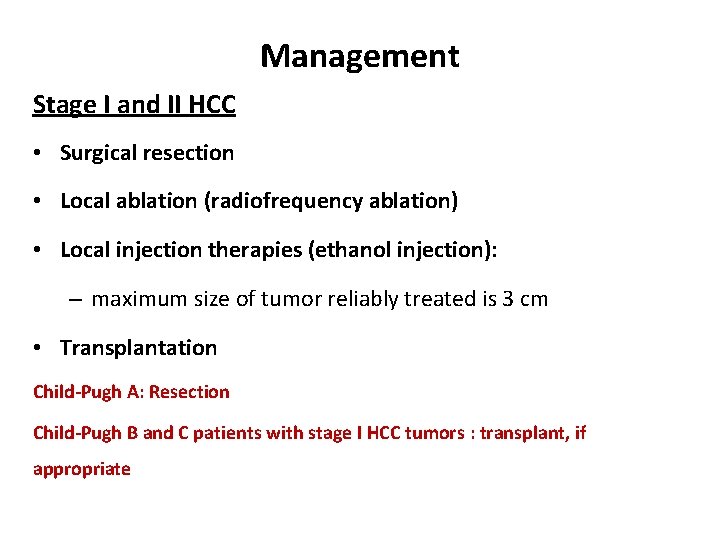
Management Stage I and II HCC • Surgical resection • Local ablation (radiofrequency ablation) • Local injection therapies (ethanol injection): – maximum size of tumor reliably treated is 3 cm • Transplantation Child-Pugh A: Resection Child-Pugh B and C patients with stage I HCC tumors : transplant, if appropriate
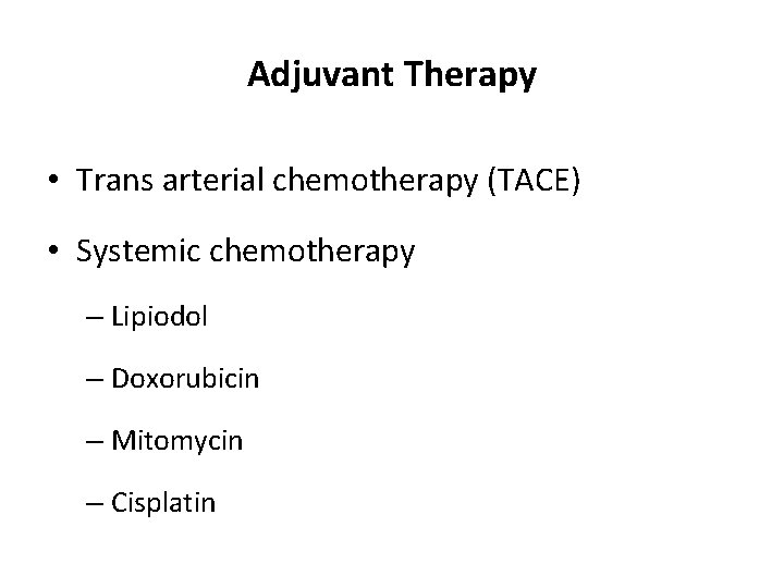
Adjuvant Therapy • Trans arterial chemotherapy (TACE) • Systemic chemotherapy – Lipiodol – Doxorubicin – Mitomycin – Cisplatin
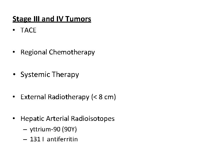
Stage III and IV Tumors • TACE • Regional Chemotherapy • Systemic Therapy • External Radiotherapy (< 8 cm) • Hepatic Arterial Radioisotopes – yttrium-90 (90 Y) – 131 I antiferritin
Portal Hypertension
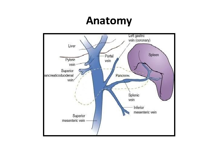
Anatomy
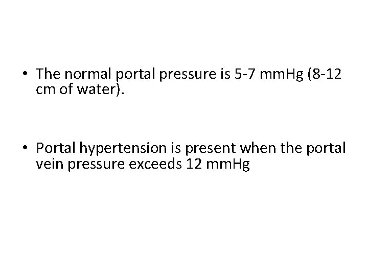
• The normal portal pressure is 5 -7 mm. Hg (8 -12 cm of water). • Portal hypertension is present when the portal vein pressure exceeds 12 mm. Hg
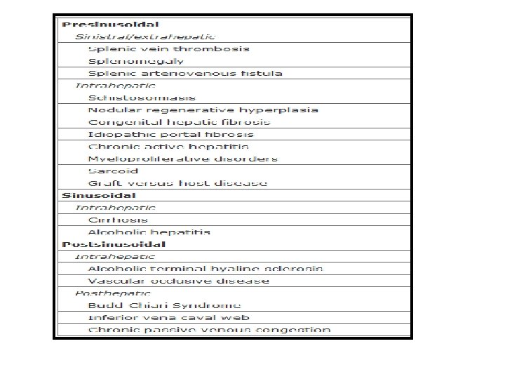
Causes • Presinusoidal • Sinusoidal • Postsinusoidal
Sequelae & Clinical picture • Porto-systemic collaterals • Splenomegaly • Congestion of the whole GIT • Bleeding varices • Ascites
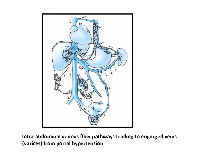
Intra-abdominal venous flow pathways leading to engorged veins (varices) from portal hypertension
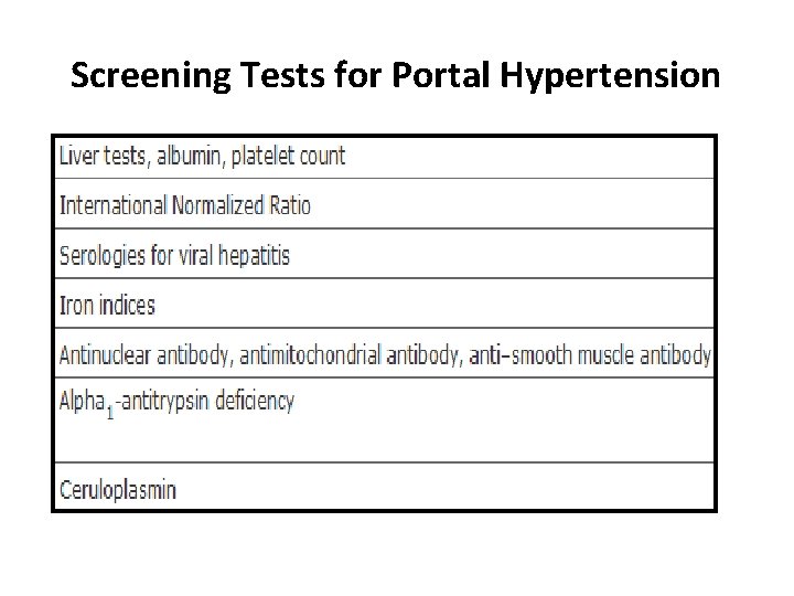
Screening Tests for Portal Hypertension
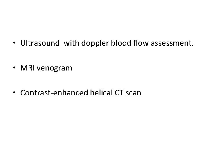
• Ultrasound with doppler blood flow assessment. • MRI venogram • Contrast-enhanced helical CT scan
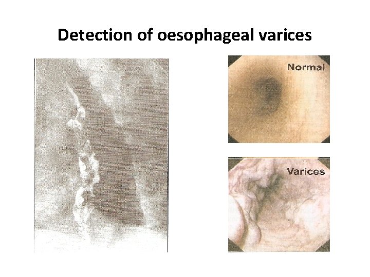
Detection of oesophageal varices
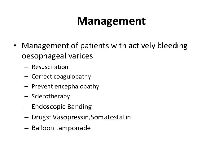
Management • Management of patients with actively bleeding oesophageal varices – – Resuscitation Correct coagulopathy Prevent encephalopathy Sclerotherapy – Endoscopic Banding – Drugs: Vasopressin, Somatostatin – Balloon tamponade
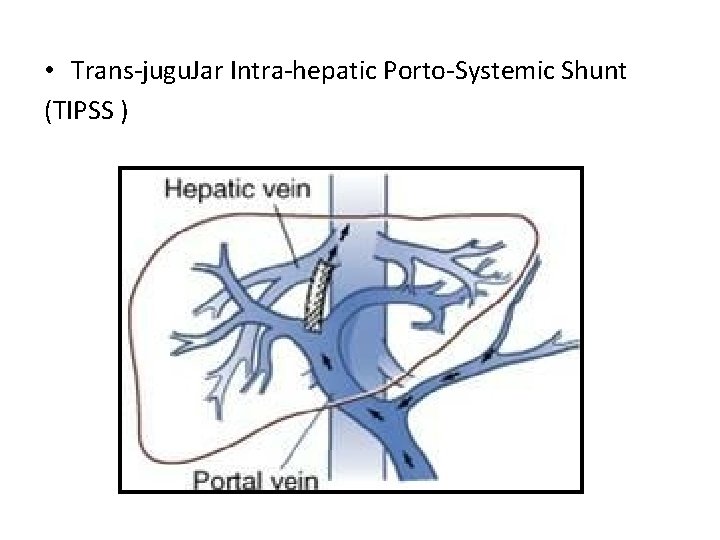
• Trans-jugu. Jar Intra-hepatic Porto-Systemic Shunt (TIPSS )
• Shunt operations – – Portocaval shunt Proximal spleno-renal shunt Mesocaval (Drapanas) shunt Selective shunt (Warren shunt) • Liver Transplant
Chronic Liver Disease
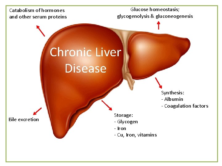
Catabolism of hormones and other serum proteins Glucose homeostasis; glycogenolysis & gluconeogenesis Chronic Liver Disease Synthesis: - Albumin - Coagulation factors Bile excretion Storage: - Glycogen - Iron - Cu, Iron, vitamins
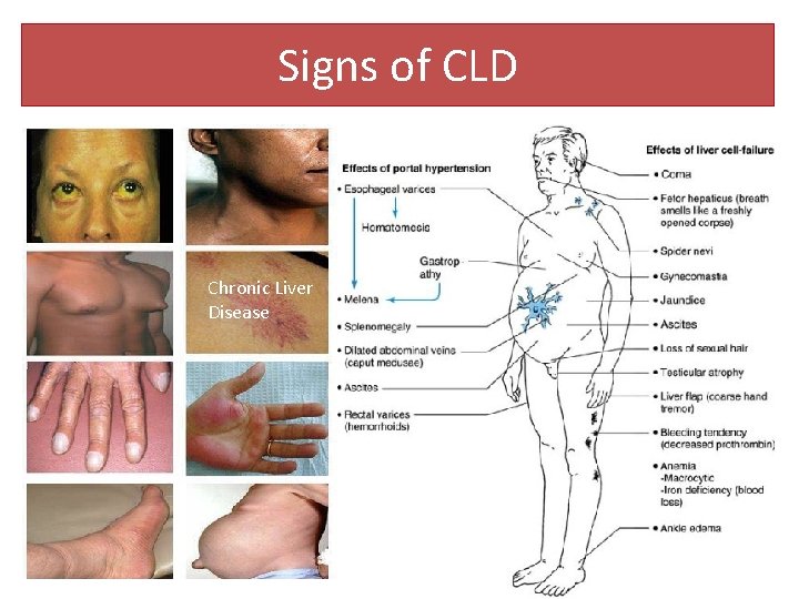
Signs of CLD Chronic Liver Disease
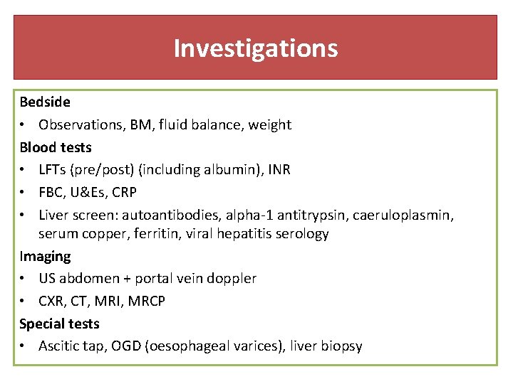
Investigations Bedside • Observations, BM, fluid balance, weight Blood tests • LFTs (pre/post) (including albumin), INR • FBC, U&Es, CRP • Liver screen: autoantibodies, alpha-1 antitrypsin, caeruloplasmin, serum copper, ferritin, viral hepatitis serology Imaging • US abdomen + portal vein doppler • CXR, CT, MRI, MRCP Special tests • Ascitic tap, OGD (oesophageal varices), liver biopsy
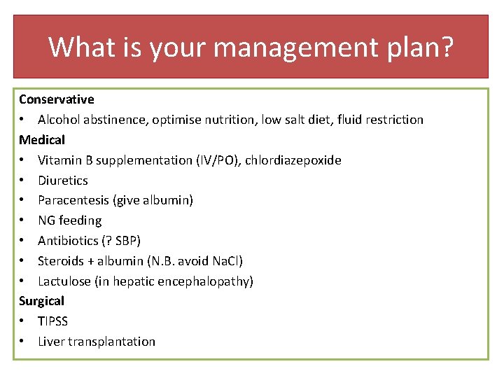
What is your management plan? Conservative • Alcohol abstinence, optimise nutrition, low salt diet, fluid restriction Medical • Vitamin B supplementation (IV/PO), chlordiazepoxide • Diuretics • Paracentesis (give albumin) • NG feeding • Antibiotics (? SBP) • Steroids + albumin (N. B. avoid Na. Cl) • Lactulose (in hepatic encephalopathy) Surgical • TIPSS • Liver transplantation
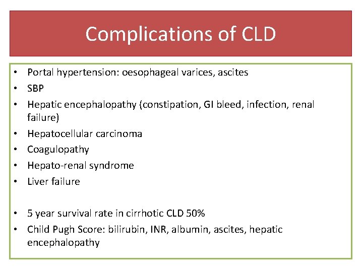
Complications of CLD • Portal hypertension: oesophageal varices, ascites • SBP • Hepatic encephalopathy (constipation, GI bleed, infection, renal failure) • Hepatocellular carcinoma • Coagulopathy • Hepato-renal syndrome • Liver failure • 5 year survival rate in cirrhotic CLD 50% • Child Pugh Score: bilirubin, INR, albumin, ascites, hepatic encephalopathy