
CAPNOGRAPHY By Lafe Bush OBJECTIVES Define Capnography ETCO

CAPNOGRAPHY By Lafe Bush
OBJECTIVES • Define • Capnography, ETCO 2, Capnometry • Review the respiratory cycle • Discuss waveform interpretation • Discuss disease specific waveform presentation
Terminology review • ETCO 2 – Partial pressure of Carbon Dioxide at the end of an exhaled breath • PACO 2 - Partial pressure of CO 2 in the alveoli • Pa. CO 2 - Partial pressure of CO 2 in arterial blood • PETCO 2 – Partial pressure of CO 2 at the end of expiration • (a-ET)PCO 2 – Arterial to end-tidal CO 2 tension/pressure difference or gradient • Pv. CO 2 – Partial pressure of CO 2 in mixed venous blood • V/Q ratio – Ventilation to Perfusion ratio
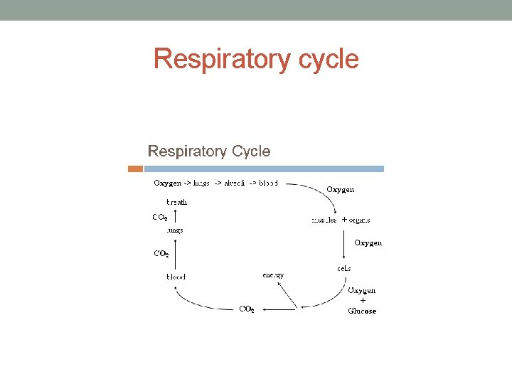
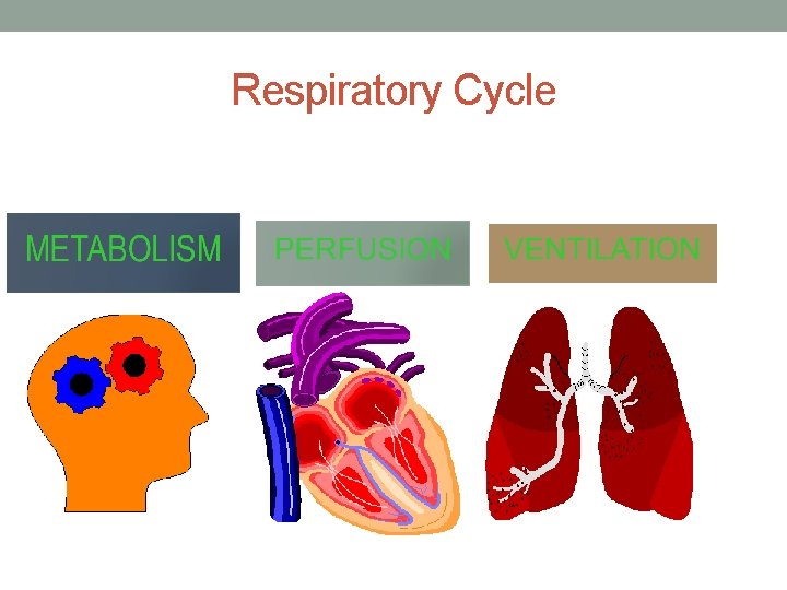
Terminology Review Cont. • Ventilation - moving air into and out of the lungs • Capnography - the continuous analysis and recording of Carbon Dioxide (CO 2) concentrations in respiratory gasses (waveform and number) • Capnometry - the analysis of the gas with no waveform (number only) • Respiration - the process of providing oxygen to the body and removing CO 2 Metabolism - the process of by which an organism obtains energy • Aerobic = Glucose with oxygen • Anaerobic= Glucose without oxygen
Respiratory Cycle • Minute ventilation – total volume of gas entering the lungs per • • minute Tidal Volume (Vt) volume of gas moved in one breath • (approx. 5 -7 ML/KG (500 ML) Dead Space (Vd) – volume of gas that does not reach the alveoli and does not participate in respiration (2 ML/ KG (150 LM) • Anatomical • Alveolar • Physiological Alveolar Ventilation (Va) – volume of gas that reach the alveoli and participates in gas exchange (350 ML) (Vt – Vd = Va) Anything that decreases the Vt only effects the Va not Vd
Respiratory cycle
Respiratory Cycle
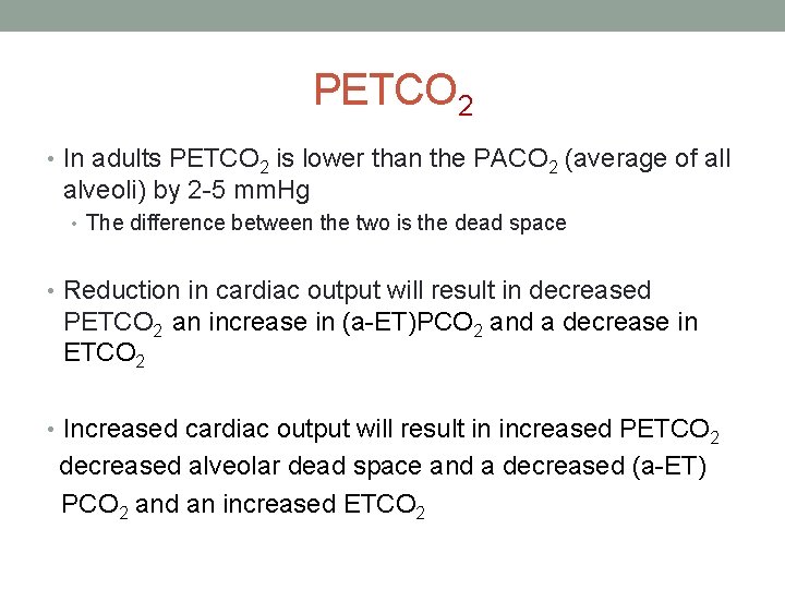
PETCO 2 • In adults PETCO 2 is lower than the PACO 2 (average of all alveoli) by 2 -5 mm. Hg • The difference between the two is the dead space • Reduction in cardiac output will result in decreased PETCO 2 an increase in (a-ET)PCO 2 and a decrease in ETCO 2 • Increased cardiac output will result in increased PETCO 2 decreased alveolar dead space and a decreased (a-ET) PCO 2 and an increased ETCO 2
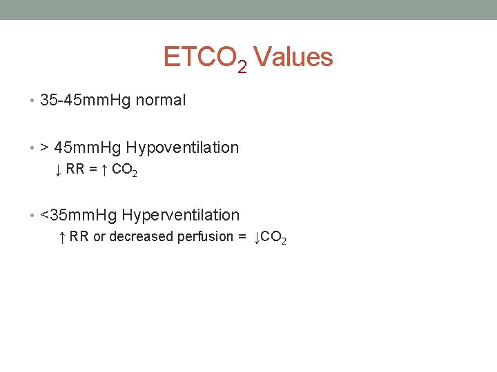
ETCO 2 Values • 35 -45 mm. Hg normal • > 45 mm. Hg Hypoventilation ↓ RR = ↑ CO 2 • <35 mm. Hg Hyperventilation ↑ RR or decreased perfusion = ↓CO 2
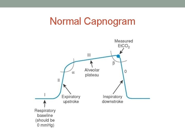
Normal Capnogram
INCREASED ETCO 2 RESPIRATORY • Drug OD • Chocking (partial) • Respiratory failure • Air trapping disease • COPD • Asthma <35 mild, 35 -50 tiring, > 50 tired (better do something)
DECREASED ETCO 2 RESPIRATORY • Hyperventilation (anxiety) • Can be used to train patient • Mucus Plug • Bronchospasm
INCREASED ETCO 2 METABOLISM • Pain • Increased heart rate = increased metabolism • Hyperthermia • Malignant hyperthermia will present with sharp increases in ETCO 2 • Shivering • Increased metabolism
DECREASED ETCO 2 METABOLIC • Diabetic Ketoacidosis (DKA) • ETCO 2 <30 with a >BS = in children • Bradycardia • < ETCO 2 due to decreased metabolism • Tachydysrhythmias • < ETCO 2 due to decreased metabolism • Seizures • <ETCO 2 due to respiratory arrest
DECREASED ETCO 2 PERFUSION • Severe Sepsis • Hypovolemic shock • Cardiogenic shock • Pulmonary embolism
LIMITATIONS OF ETCO 2 • Critically ill patients often have rapidly changing dead space and V/Q mismatch • Higher rates and smaller TV can increase the amount of dead space ventilation • High mean airway pressures and PEEP restrict alveolar perfusion, leading to falsely decreased readings
Pulse Oximetry • Oxygen Saturation • O 2 content/o 2 capacity x 100 • SPO 2 changes lag up to several minutes in patients hypoventilating or apneic • Temperature sensitive • Hemodynamic sensitive
WAVEFORM INTERPITATION • Is there a waveform? • Is baseline flat or moving upward? • Expiratory upstroke? • Alveolar plateau? • Inspiratory downstroke?
FLAT WAVEFORM • Capnograhy equipment not connected • Cardiac arrest/respiratory arrest • Missed intubation / Extubation • Ventilator disconnection
EXPIRATORY UPSTROKE • Air trapping disease = Prolonged or sloped • Alpha angle > 90° should make you consider a V/Q mismatch • Increased ETCO 2 = Rise in upstroke • Relief of air trapping
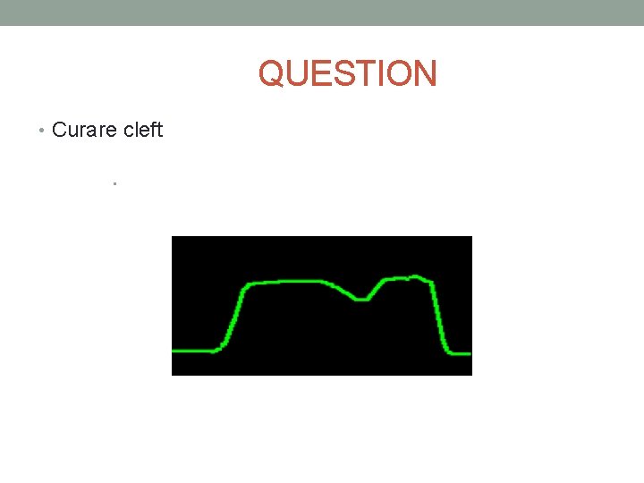
ALVEOLAR PLATEAU • Air trapping = Rising plateau • Curare cleft = small dip • Pneumothorax = Decreasing plateau • Right mainstem intubation = biphasic waveform • ETT cuff leaking = Downward slopping plateau
WHAT CAN WE LEARN FROM ETCO 2 • Asthma, COPD, Anaphylaxis vs. CHF • Hypoventilation due to various medical reasons • Shock • Hyperventilation syndrome
INTUBATED PATIENTS • Verification of ETT • ETT surveillance during transport and movement • Control of hypo/hyperventilation • CPR compression efficacy • Early signs of ROSC • Coming out of paralysis/sedation • ETT size/cuff inflation
TYPES OF DEVICES • Semi – Quantitative capnometry • Only tells you if there is ETCO 2 or not. No number or waveform • Quantitative • Only gives a number no waveform • Waveform • Gives number and waveform
SIDE STREAM VS. MAINSTREAM • Side Stream • Easy to connect • Can be used in awake patients • Patient position is not a factor • Can be used in conjunction with oxygen administration • Mainstream • No sampling tube (dead space) • No problem with water or other fluids in the sensor • No problem with pollutants entering the connector site • No delay in recording • Only utilized with ventilators
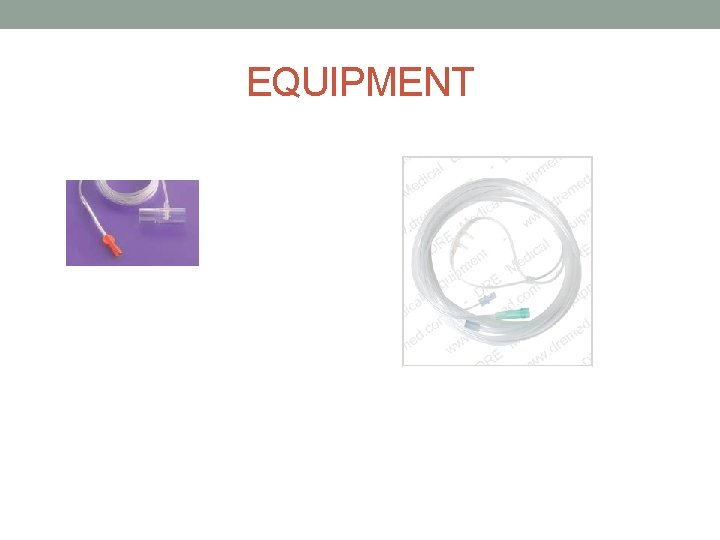
EQUIPMENT
Life Pack Monitor And ETCO 2 • Waveform disappears during charging • LP sweep speed 12. 5 mm/sec print out 25 mm/sec • M sweep speed 6. 25 mm/sec • 4 second screen shot • Shows CO 2 for the last 20 seconds • ETCO 2 must be > 3. 5 to be displayed • RR is an average of the last 8 breaths
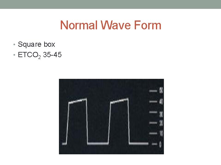
Normal Wave Form • Square box • ETCO 2 35 -45
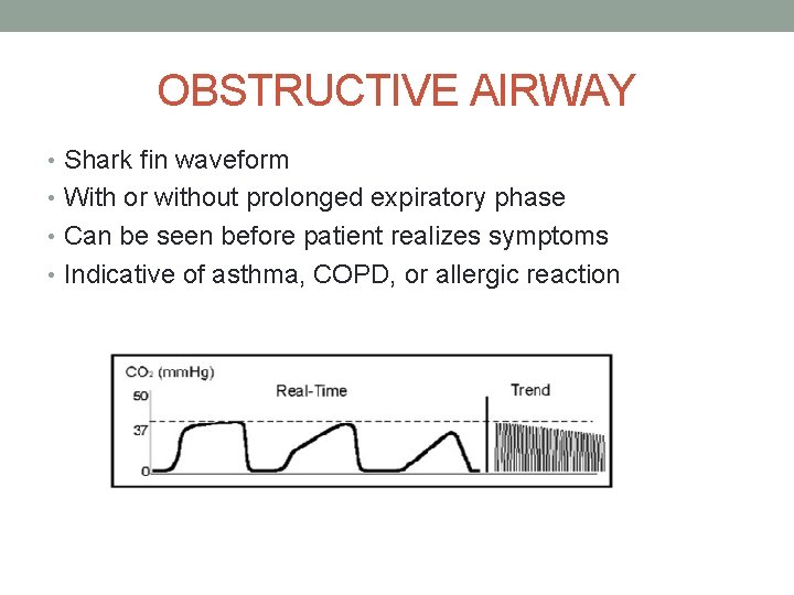
OBSTRUCTIVE AIRWAY • Shark fin waveform • With or without prolonged expiratory phase • Can be seen before patient realizes symptoms • Indicative of asthma, COPD, or allergic reaction
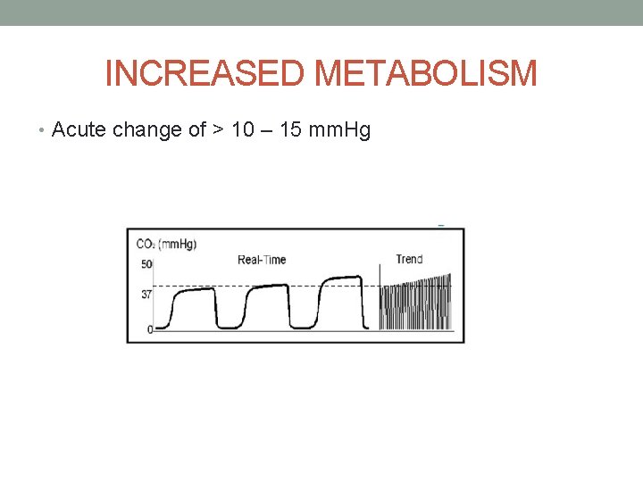
INCREASED METABOLISM • Acute change of > 10 – 15 mm. Hg
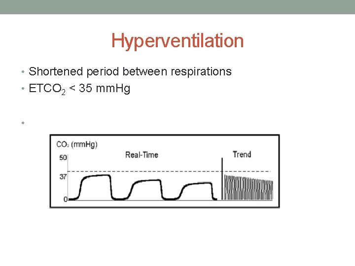
Hyperventilation • Shortened period between respirations • ETCO 2 < 35 mm. Hg •
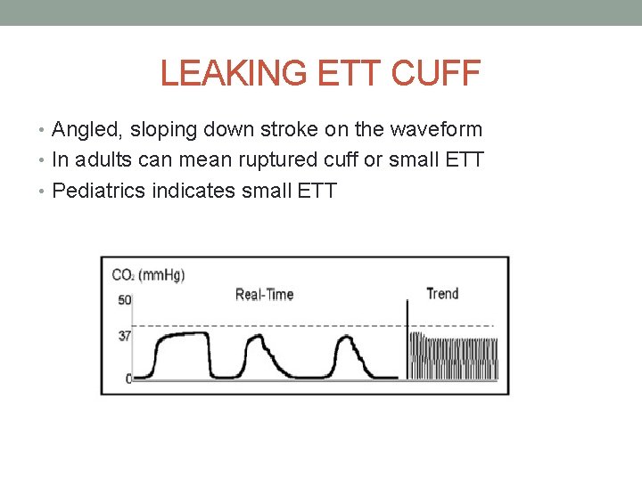
LEAKING ETT CUFF • Angled, sloping down stroke on the waveform • In adults can mean ruptured cuff or small ETT • Pediatrics indicates small ETT
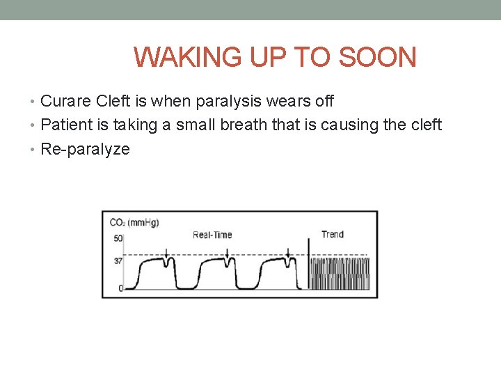
WAKING UP TO SOON • Curare Cleft is when paralysis wears off • Patient is taking a small breath that is causing the cleft • Re-paralyze
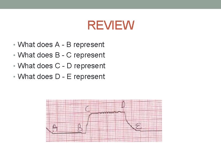
REVIEW • What does A - B represent • What does B - C represent • What does C - D represent • What does D - E represent
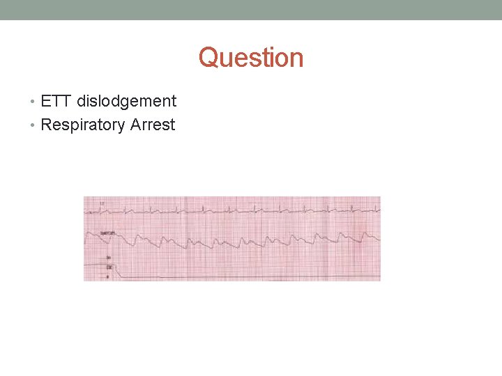
Question • ETT dislodgement • Respiratory Arrest
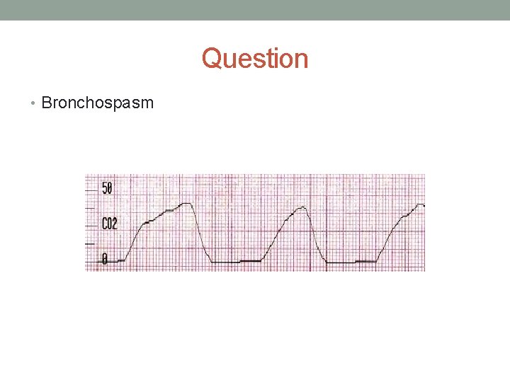
Question • Bronchospasm
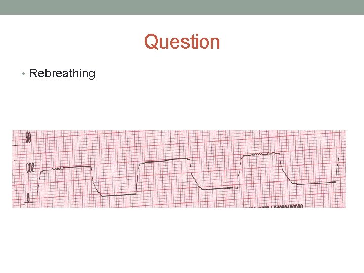
Question • Rebreathing
QUESTION • Curare cleft •
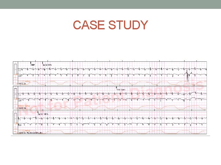
CASE STUDY • Pt awake and alert in exam room at Vanguard Medical. Presents w/ 3 week HX of bronchitis, and asthma. Finished oral steroids. Has been using inhaler. Denies fever, nausea, vomiting, diarrhea. Non productive cough with diffuse wheezing. Spo 2 100% RA. Etco 2 is 12. Carpal-pedal spasms present. Staff gave 1 albuterol Rx prior to ALS arrival. Pt calmed, and encouraged to breathe through her nose. Duo- neb d/c as we are unable to give w/o O 2 and that was making her hyperventilation and finger spasms worse. ALS given per erp order, and PT transported w/o incident. Report to ern.
CASE STUDY
CASE STUDY • 25 yo male complaining of difficulty breathing. Patient has been using his inhaler for the past hour with no improvement. Patient states he has been intubated 1 time before, several years ago. Patient was in obvious distress speaking in 1 -3 word sentences. • HR 120; BP 163/92; SPO 2 94; RR 30; ETCO 2 55
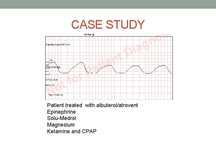
CASE STUDY Patient treated with albuterol/atrovent Epinephrine Solu-Medrol Magnesium Ketamine and CPAP
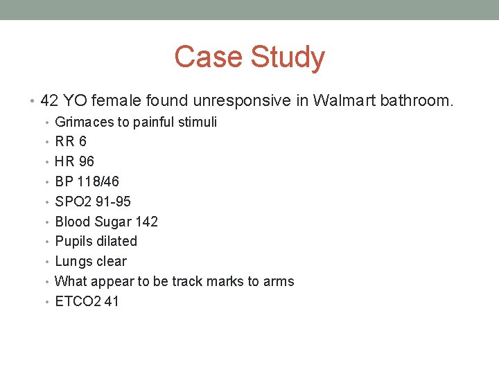
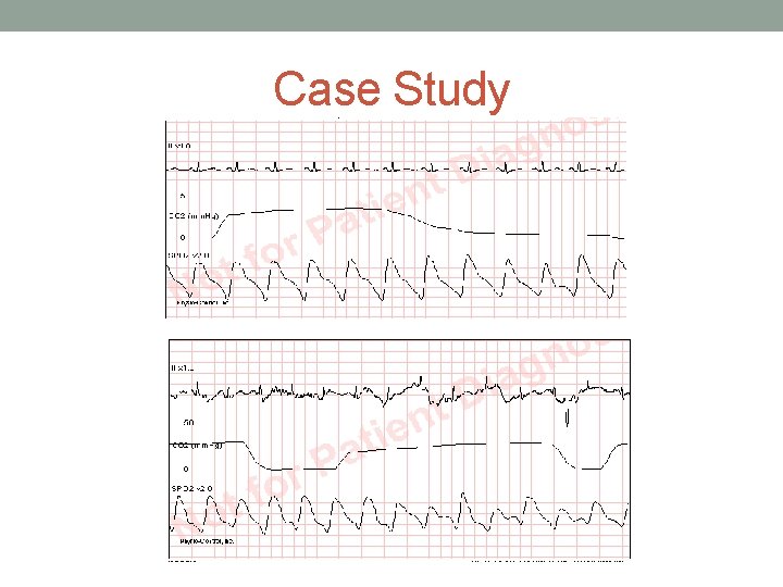
Case Study • 42 YO female found unresponsive in Walmart bathroom. • Grimaces to painful stimuli • RR 6 • HR 96 • BP 118/46 • SPO 2 91 -95 • Blood Sugar 142 • Pupils dilated • Lungs clear • What appear to be track marks to arms • ETCO 2 41
Case Study
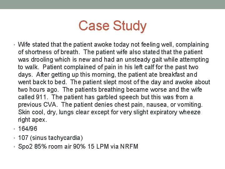
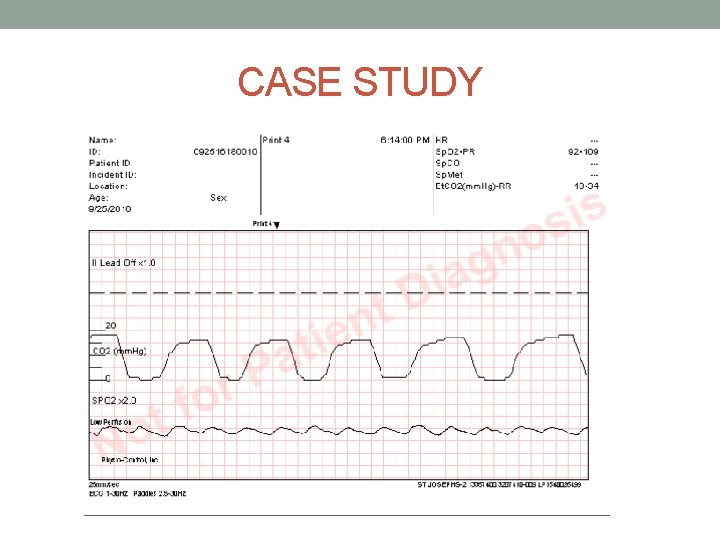
Case Study • Wife stated that the patient awoke today not feeling well, complaining of shortness of breath. The patient wife also stated that the patient was drooling which is new and had an unsteady gait while attempting to walk. Patient complained of pain in his left calf for the past two days. After getting up this morning, the patient ate breakfast and went back to bed. The patient slept most of the day and awoke about two hours ago. The patients breathing became worse and the wife called 911. The patient has garbled speech but this was from a previous CVA. The patient denies chest pain, nausea, or vomiting. Skin cool, dry, lungs clear except for very slight expiratory wheeze right apex. • 164/96 • 107 (sinus tachycardia) • Spo 2 85% room air 90% 15 LPM via NRFM
CASE STUDY
- Slides: 46