Canine Hypoadrenocorticism Addison Disease Unit 3 4 th
 Unit: 3 4 th Professional Dr. Anil Kumar Asst. Professor")


 is administered")
- Slides: 12
Canine Hypoadrenocorticism (Addison Disease) Unit: 3 4 th Professional Dr. Anil Kumar Asst. Professor Dept. of VCC, BVC, Patna
ADDISON DISEASE The adrenal glands are essential for life, being responsible for the minute-to-minute regulation of blood pressure, blood volume, and vascular tone. ADRENAL GLAND Outer Zone (Cortex) Zona fasciculate (70% of the cortex) The zona glomerulosa, Mineralocorticoid (Aldosterone) Ion transport by epithelial cells, resulting in a loss of potassium and retention of sodium. • A lack of secretion of mineralocorticoids (Addison disease) may result in a lethal retention of potassium and loss of sodium. • Inner Zone (Medulla) Glucocorticoid(Cortisol) • • • carbohydrate, protein, and lipid metabolism result in sparing of glucose Decrease lipogenesis and increase lipolysis in adipose tissue suppress inflammatory and immunologic responses Favors the spread of infection. Negative effect on wound healing (fibroblast proliferation and collagen synthesis ) Zona reticularis Sex steroids • Progesterone, estrogens, androgens are adrenal sex hormones
• Adrenal hormones are necessary to control salt, sugar and water balance in the body. • It results from the lack of glucocorticoids, mineralocorticoids, or both • It is seen most commonly in young to middle-aged female dogs and occasionally in horses. Etiology: • Primary—Unknown, but most cases probably result from an autoimmune process and characterized by a lack of glucocorticoids and mineralocorticoids. • Others : üDestruction of the adrenal gland by granulomatous disease ü Metastatic tumor, ü Hemorrhage üInfarction üAdrenolytic agents (mitotane), or üadrenal enzyme inhibitors (trilostane)
FORMS OF HYPOADRENOCORTICISM Primary Hypoadrenocorticism • Characterized by a lack of glucocorticoids and mineralocorticoids • Mostly associated with immune-mediated destruction of adrenal cortical tissue • Mitotane or trilostane for Hyperadrenocorticism treatment • Neoplasia, infection, or infarction of the adrenal glands Atypical Hypoadrenocorticism • Seen in small population of “atypical” dogs • Adrenal destruction is purported to spare the glomerulosa layer, resulting in an isolated glucocorticoid deficiency and so, do not progress to clinically significant mineralocorticoid deficiency • Isolated glucocorticoid insufficiency is more commonly seen in older dogs with vague GI signs, weight loss, normal electrolyte concentrations, hypoalbuminemia and hypocholesterolemia. Secondary Hypoadrenocorticism • It is refers to a central (anterior pituitary) deficiency of ACTH and lead to • isolated glucocorticoid insufficiency • Mineralocorticoids are spared because ACTH does not directly influence their release. • It mainly results from abrupt discontinuation of longterm exogenous administration of corticosteroids or progesterone analog • Rarely, congenital defects of the pituitary gland ( a cystic Rathke’s pouch, neoplasia, or trauma)
Age, Sex and Breed: • Dogs of any sex, age, or breed (including mixed breeds) can develop the disease. • The disease is heritable in certain breeds: Clinical Signs: • The Addisonian crisis is the life-threatening culmination of combined hormone deficiencies that can be fatal if not appropriately treated
DIAGNOSIS: The disease is diagnosed on the basis of a: ü Compatible history üClinical signs ü Laboratory abnormalities üImaging studies and üACTH stimulation test results Note: The combination of Na/K ratio with lymphocyte count is a better screening test for hypoadrenocorticism than either variable alone.
• Patients with atypical hypoadrenocorticism are more likely to exhibit hypoalbuminemia, hypocholesterolemia, and anemia on routine blood work. Diagnostic Imaging: • Thoracic radiographs may reveal a small heart and caudal vena cava, which indicates hypovolemia • On abdominal ultrasonography, atrophy of the adrenal glands with thin and short as compared with normal, but in some cases there may benormal size adrenal glands. ECG: Depending upon the degree of hyperkalemia, the ECG may • Reveal a bradyarrhythmia with absent P waves • Tented T waves • Prolonged QRS complexes, and • decreased R wave amplitude
ACTH Stimulation Test: • The gold standard for diagnosis of all forms of hypoadrenocorticism; it should be performed in any patient suspected of having the disease. • A baseline serum cortisol level > 2 mcg/d. L can be used to rule out hypoadrenocorticism, while a cortisol level ≤ 2 mcg/d. L necessitates an ACTH stimulation test. TREATMENT: The main goal of therapy is to: • Restoration of blood volume • Correction of electrolyte/acid-base disorders • Fluid therapy: is always be instituted prior to the use of adrenal steroid replacement therapy and can be performed while the patient is undergoing the ACTH stimulation test. • Starting with aggressive IV fluid therapy with isotonic crystalloids (0. 9% sodium chloride, Ringer’s lactate solution)
To solve an adrenal crisis is an acute medical emergency: üAn infusion with 0. 9% saline should be started. ü If the dog is hypoglycemic, the saline should include 2. 5%– 5% dextrose. üThe hypovolemia is corrected rapidly by administering 0. 9% saline (60– 70 m. L/kg throughout the first 1– 2 hours). üUrine output should be assessed to determine whether the dog is becoming anuric. ü Fluids should be continued, at a rate appropriate to match ongoing losses, until the clinical signs and laboratory abnormalities have resolved. • If hyperkalemia persists despite fluid resuscitation and mineralocorticoid replacement therapy, suspect renal failure and monitor patient for oliguria or anuria.
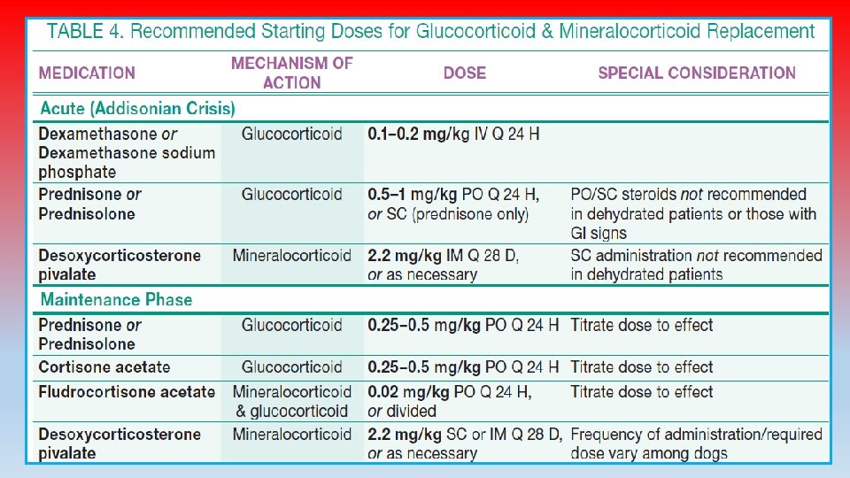
Additionally, to correct life-threatening hyperkalemia: üConsider treatment with dextrose (10% glucose in 0. 9% saline can be given for 30– 60 min to increase potassium movement into the cells) and regular insulin (0. 25– 1 U/Kg, IM, will enhance glucose and potassium uptake), addressing metabolic acidosis, or üTreatment with a beta-adrenergic drug, such as terbutaline or albuterol. • Prednisolone sodium succinate (22– 30 mg/kg) or dexamethasone sodium phosphate (0. 2– 1 mg/kg) may be used in the initial management of shock. • Prednisolone or prednisone should be given at 1 mg/kg, twice a day, for the first few days of therapy and then at 0. 25– 0. 5 mg/kg/day. • Mineralocorticoid replacement therapy is also begun to help with electrolyte imbalances and hypovolemia. • Electrolytes, renal function, and glucose should be monitored regularly to assess response to therapy.
For long term maintenance therapy : • The mineralocorticoid desoxycorticosterone pivalate (DOCP) is administered at 2. 2 mg/kg, IM or SC, every 25– 28 days. • Electrolytes should be measured at 3 and 4 weeks after the first few injections to determine the duration of action. • Alternatively, fludrocortisone acetate is administered PO at 10– 30 mcg/kg/day. Serum electrolytes should be monitored weekly until the proper dose is determined. • Some dogs (especially dogs on DOCP) also require daily oral glucocorticoid therapy to adequately control clinical signs. • in ~50% of dogs, replacement doses of prednisone (0. 2– 0. 4 mg/kg/day) are required • Dogs with atypical Addison disease require only replacement doses of prednisone, although it is recommended that electrolytes be monitored every 3 months for the first year after diagnosis. • Dogs with chronic hypoadrenocorticism should be re-examined every 3– 6 months