CANDIDA Spp Classification Mycoses are classified according to


![Infectious disease Mucocutaneous Manifestation: a] Oral Candidiasis: v infants, diabetes mellitus, HIV-1 & HIV-2.](https://slidetodoc.com/presentation_image_h2/00c8b4c4018856c8a8b22c87776f48e9/image-12.jpg "Infectious disease Mucocutaneous Manifestation: a] Oral Candidiasis: v infants, diabetes mellitus, HIV-1 & HIV-2.")
![Cont……. b] Angular Cheilitis: v Patients presents with sore, erythematous, fissured lesions affecting the](https://slidetodoc.com/presentation_image_h2/00c8b4c4018856c8a8b22c87776f48e9/image-13.jpg "Cont……. b] Angular Cheilitis: v Patients presents with sore, erythematous, fissured lesions affecting the")
![Cont…. d] Vulvovaginitis, Balanitis, Balanoposthitis: v Affect young and middle aged female v Mainly](https://slidetodoc.com/presentation_image_h2/00c8b4c4018856c8a8b22c87776f48e9/image-14.jpg "Cont…. d] Vulvovaginitis, Balanitis, Balanoposthitis: v Affect young and middle aged female v Mainly")
![Cont…. . f] Ocular candidiasis: v There is conjuctival edema, cheesy discharge in conjuctival](https://slidetodoc.com/presentation_image_h2/00c8b4c4018856c8a8b22c87776f48e9/image-15.jpg "Cont…. . f] Ocular candidiasis: v There is conjuctival edema, cheesy discharge in conjuctival")
 – Onychomycosis (nails)")


![Systemic Manifestations 1. 2. 3. 4. 5. 6. Urinary tract candidiasis [UTI] Candiduria [infection](https://slidetodoc.com/presentation_image_h2/00c8b4c4018856c8a8b22c87776f48e9/image-23.jpg "Systemic Manifestations 1. 2. 3. 4. 5. 6. Urinary tract candidiasis [UTI] Candiduria [infection")

, sterile fluid, urine,")


![Cont…. v Histopathology (tissues) – Gomori’s methenamine silver stain [GMS] Yeast-like cells and septate](https://slidetodoc.com/presentation_image_h2/00c8b4c4018856c8a8b22c87776f48e9/image-28.jpg "Cont…. v Histopathology (tissues) – Gomori’s methenamine silver stain [GMS] Yeast-like cells and septate")

")


- Slides: 46
CANDIDA Spp.
Classification Mycoses are classified according to the tissue levels initially colonized. I. III. IV. Superficial mycoses Cutaneous mycoses Subcutaneous mycoses Systemic mycoses A. Opportunistic mycoses B. Deep mycosis
INTRODUCTION - Candida- commonest fungi causing candidiasis -Mucosa, skin, nails and organs of the body. -Yeast like fungi -Acute or chronic, superficial or deep. -Ovoid, round shaped with hyphae -Gram positive
ü These are ovoid shaped with pdeudohyphae 4 -8 micro meter in dm with budding and pseudohyphae ü Contain ergosterol
Candida albicans Ultrastructure
Classification 20 species are considered to be significant pathogens causing various infection in human. Candida albicans Candida Non- albicans C. krusei C. guilliermondii C. kefyr C. dubliniensis C. lipolytica C. rugosa C. tropicalis C. glabrata C. parapsilosis C. lusitaniae C. viswananthi C. zeylanoides C. stellatoidea
Virulence factors of candida species are a. toxins b. enzymes c. adhesin d. complement receptor e. Phenotype switching
Clinical features Candida spp. Are found on mucosal surface It is found mainly as secondary infection in individuals with some immunocompromised condition and rarely as a primary disease. The clinical manifestation of candidiasis can be divided into two broad categories. 1. Infectious disease 2. Allergic disesase
CANDIDIASIS INFECTIONS • Mucocutaneous- Oral Thrush -GIT- Gastritis - Ocular • Cutaneous – Intertriginous, paronychia, diaper dermatitis, granuloma • Systemic – UT, Endocarditis, Pulmonary, Meningitis , Candidemia, Arthritis, Osteomyelitis
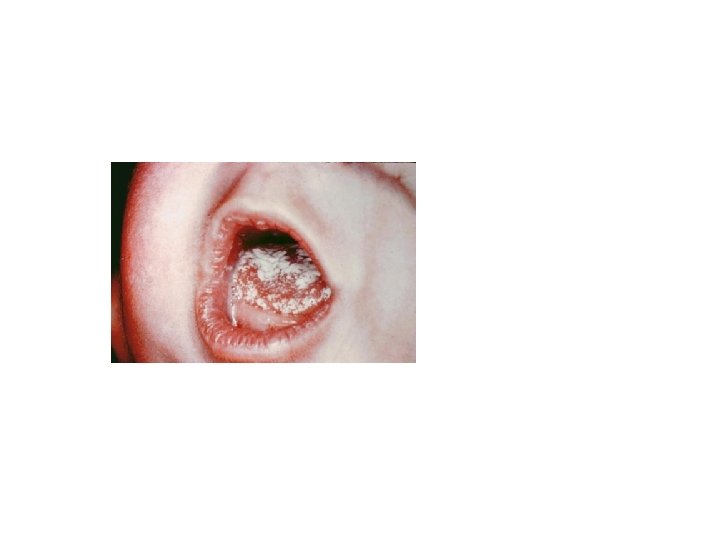
Infectious disease Mucocutaneous Manifestation: a] Oral Candidiasis: v infants, diabetes mellitus, HIV-1 & HIV-2. v Infection begins with congestive reddening of the mucous membrane. v Candida albicans -main cause of oral candidiasis in patient with HIV
Cont……. b] Angular Cheilitis: v Patients presents with sore, erythematous, fissured lesions affecting the angles of the mouth. v Associated with iron deficiency anemia or vitamin B 12 defficiency. v It may also seen in AIDS patients. c] Alimentary: Oesophageal Candidiasis v It may cause burning pain in the substernal area, epigastrium or throat. v It also seen in acute cases as well as in advanced HIV infection.
Cont…. d] Vulvovaginitis, Balanitis, Balanoposthitis: v Affect young and middle aged female v Mainly due to C. albicans and C. glabrata v In male affect non-circumcised and diabetic men v It is a sexually transmitted disease e] Chronic mucocutaneous candidiasis: v Common in patients with deficient cell mediated immunity v Affected sites are mouth, skin, and fingernails
Cont…. . f] Ocular candidiasis: v There is conjuctival edema, cheesy discharge in conjuctival sac, and progressive corneal ulceration. v Most common cause is C. albicans
Cont…. . • Cutaneous infections – Paronychia (skin around nail bed) – Onychomycosis (nails) – Diaper rash – Chronic mucotaneous candidiasis • children with T-cell abnormality 18
Mucosal candidiasis Oral thrush 19
Cutaneous candidiasis Diaper dermatitis 21
chronic superficial candidiasis
Cutaneous candidiasis Onychomycosis and paronychia Chronic mucocutaneous candidiasis 23
Paronychia of nails
Systemic Manifestations 1. 2. 3. 4. 5. 6. Urinary tract candidiasis [UTI] Candiduria [infection of lower urinary tract] Endocarditis Pulmanory candidiasis Meningitis [low birth weight] Candidemia and Septicemia [C. albicans, C. tropicalis, C. krusei] 7. Disseminated Candidiasis 8. Arthritis 9. Osteomyelitis 10. Endopthalmitis
ALLERGIC DISEASE 1. Candidids – This is an allergic manifestation to the metabolites of candida. 2. Eczema 3. Asthma 4. Gastritis
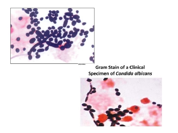
Laboratory Diagnosis v Specimens – Exudate, Blood, tissue (biopsy or autopsy), sterile fluid, urine, CSF, skin scrapping, respiratory secretions v Microscopy (direct on specimen - except blood and urine) – Gram stain [ Presence of yeast and pseudohyphae of candida spp. ] – Calcofluor white stain [ highlight the fungal element] 27
Skin scraping mounted in 10%KOH from superficial candidiasis showing clusters of budding yeast cells and branching pseudohyphae.
Candida species Top: Calcofluor White x 400: Yeast and pseudohyphae Bottom: Gram stain x 1000: Yeast and pseudohyphae 29
Cont…. v Histopathology (tissues) – Gomori’s methenamine silver stain [GMS] Yeast-like cells and septate hyphae GMS
Cont… • Periodic acid-Schiff stain are also done for the demonstration of fungal elements in tissue. Esophagus, vascular invasion, blastoconidia and pseudohyphae, PAS
Fungal culture v The clinical specimen can be cultured on Sabouraud dextrose agar (SDA) with antibacterial antibiotics. v And incubated at 25°C to 37°C for 3 -4 days v Colonies appear white, cream colored, smooth and pasty. v LCB mount prepared from the colonies to examine yeast cells and pseudohyphae.
Candida albicans v The colonies of this yeast are cream colored, pasty, and smooth. v Rate of growth is rapid and they mature in three day. Germ tube is positive. Cornmeal agar at 25°C pseudohyphae are seen in clusters.
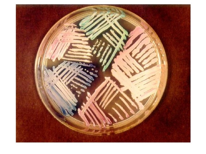
Candida albicans: Sabouraud Agar Morphology: Creamy white yeast, may be dull, dry irregular and heaped up, glabrous and tough Chromagar producing green pigmented colonies on specially designed medium to speciate certain yeasts based on color they produce 34
Chromgar • • C. dubliniensis - Green C. glabrata – Dark violet C. guilliermondii - Pink to lavender C. kefyr - Pink to lavender C. parapsilosis Ivory to pink C. albicans - Green C. krusei -Pink, pale borders C. tropicalis - Steel blue, purple diffusion
Germ tube: v Inoculation of yeast in sheep or human serum incubated at 370 C for 2 to 3 hours Germ Tube: Positive v Long tube like projections extending from the yeast cells. v germinating from the yeast cell without constriction at the point of attachment. e. g. C. albicans, C. dubliniensis Germ Tube: Negative v Shows constriction at the attachment site e. g. other Candida species, esp. C. tropicalis 38
Germ tube • Germ tube positive Germ tube negative
Candida albicans: Cornmeal Agar clusters of blastospores along pseudohyphae at regular Intervals.
Sugar fermentation Candida species Glucose C. albicans AG C. tropicalis AG C. kefyer AG C. guilliermondii AG C. parapsilosis AG C. krusei AG C. glabrata AG Maltose AG AG AG Sucrose Lactose – – AG AG AG – – – –
Sugar Assimilation Reaction Spp. Mal Suc Lac Cel Gal Tre Raff Mel Xyl Ino Dul C. albicans + + + – – – + – – C. tropicalis + + + – – + – + – – C. guillierm + ondii + + – – C. parapsilo + sis + + – + – – – – + – – C. kefyer C. krusei Glu +
Candida antigen, antibody detection 1. Detection of Antibody: v v v Slide agglutination Immunodiffusion Phytohaemagglutination Coelectosynersis Immunopreciptation A and B immunofluorescence 43
Detection of Antigen v Latex agglutination v Immunoblotting
Candida antifungal susceptibility testing • Testing methodology – Microbroth dilution – E-test – Disk diffusion – Vitek 2 45
Candida antifungal susceptibility testing 46
Candida antifungal susceptibility testing 47
Candida - Treatment • Remove infected intravenous lines • Antifungal therapy for systemic infection – Amphotericin B IV – Azoles (fluconazole, itraconazole, voriconazole, posaconazole) – Flucytosine (only with Ampho B because of resistance) – Echinocandins (caspofungin, micafungin) 49
End Of Lecture………. .