CANCER SCREENING TUMOR MARKERS MOHAMED GABER PROFESSOR OF


CANCER SCREENING & TUMOR MARKERS MOHAMED GABER PROFESSOR OF SURGICAL ONCOLOGY ALEXANDERIA UNIVERSITY 2017 -2018

HISTORICAL REVIEW


Breast Screening Program

Data reported in Egypt indicates that breast cancer ranked as a number one malignancy (27. 3%) among females. At present, the mortality from breast cancer is about 15000 annually.

Breast cancer worldwide Estimated age-standardized incidence rates of breast cancer world-wide in 2000. From Ferlay et al. , (2001).

The reduction in breast cancer incidence based upon the present knowledge of risk factors is limited.

Never lactate Getting older Old primigravida Nullipara 1 st degree relative with BC Irradiation Previous Bx atypical changes Risk factors for BC Early Menarche (<12 y). • HRT • Contraceptive pills(very slight) Polyunsat -urated diet Alcohol drinking Obesity Inherited mutation in BC genes (BRCA 1, 2) Late menopause (> 55 y)

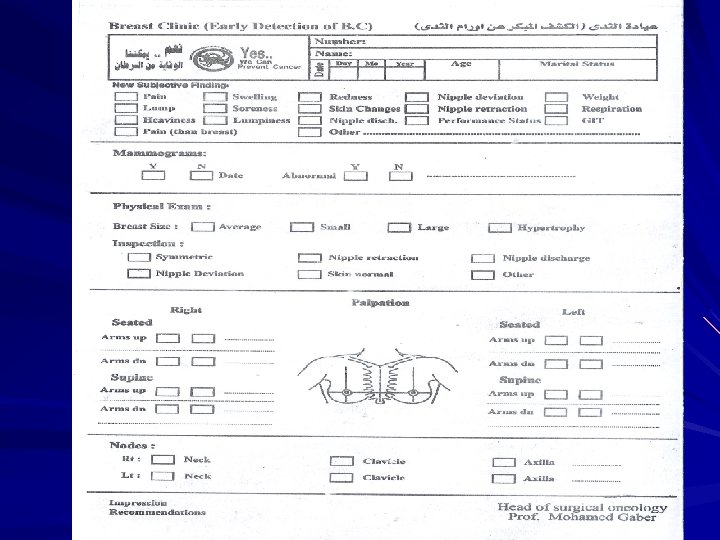
So early detection appears to be the best way, by having properly organized assessment teams who carry out a screening program in a breast clinic.

The expert assessment team working in the specialized breast clinic, should include, surgeons, radiologists, and pathologists with a high quality and special interest for conducting: Ø Clinical examination. Ø Ultrasonography. Ø Mammography. Ø Fine needle aspiration cytology Ø True cut and open biopsies.

What is screening for breast cancer? n A trial to detect BC at a very early stage, in an apparently healthy woman. n Does not prevent cancer. n Concept: Early detection will possibly reduce mortality

What are Breast cancer screening methods?

Breast Self Clinical breast examination Examination CBE BSE Mammography Others : • Ultrasonography • Electrical impedance imaging • Magnetic resonance imaging • Positron emission tomography (PET) • Scinti-mammography • Digital mammography


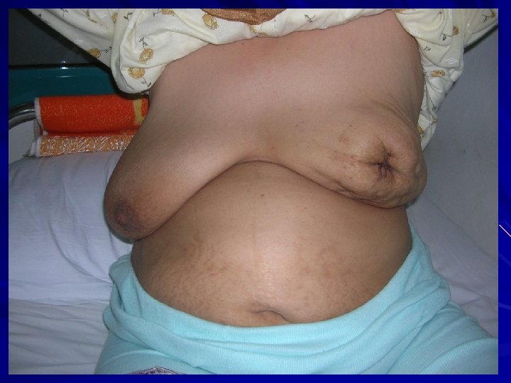
How? 2 steps 1. Initially inspection while standing 2. Next, Supine position,

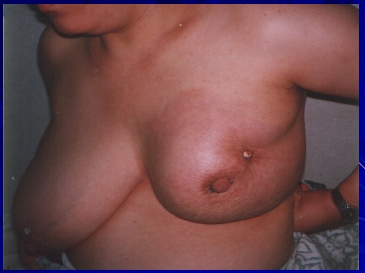
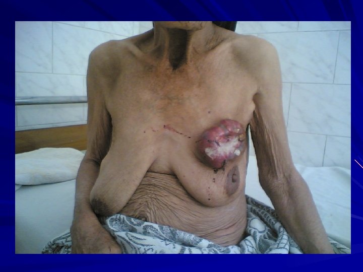
Warning Signs That Should Not Be Ignored 1. Lump, hard knot or thickening 2. Unusual swelling, warmth, redness or darkening 3. Changes size or shape. 4. Dimpling or puckering of the skin of your breast 5. Finding an itchy, scaly sore or rash on the nipple 6. Pulling in of the nipple or other parts of the breast 7. Nipple discharge that starts suddenly 8. Pain in one spot that does not vary with cycle
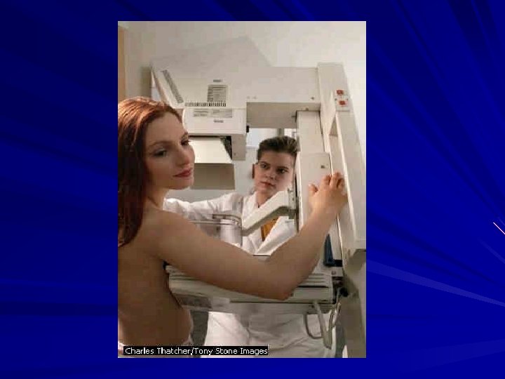

Breast mammography Some radiological findings of mammography image: Breast caner Normal

Questions to be answered 1. How the breast is examined? 2. Look for what during examination ? 3. Diagnostic modalities ( Mammogram, 4. 5. 6. U/S ) What are the important risk factors / What are the types of biopsy? Is fibrocystic disease a premalignant?

SHOULD WE DO BREAST SCREENING? Benefits Vs Obstacles

Benefits Breast screening finds cancers early Breast screening saves lives Breast conserving surgery BCT is possible

Obstacles Breast screening cannot prevent cancer Cultural and social barriers may be a considerable cause of program failure, Mammography is Not 100% perfect, although the most effective& reliable way of detecting BC early, Cancer may occur even in women having regular breast screening “interval cancer”. Relativly high cost,


TUMOR MARKERS

What are tumor markers? A Tumor marker is a biological substance synthesized and released by The tumor or by the host in response to tumor tissue. It may be used to: – Detect the presence of a tumor – Monitor the progress of disease – Monitor the response to treatment

Tumor markers in Cancer Management Screening Diagnosis Staging & Prognosis Therapy Monitoring Detecting recurrence

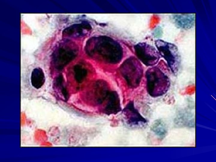
How to identify tumor markers ? On cell: – Immunocytochemistry, Flow cytometry On tissue: – Immunohistochemistry In body fluids: – Blood, urine, CSF, Amniotic fluid
 – cancer")
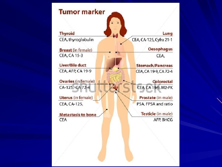
There are many different types of tumour markers, including: – alpha-fetoprotein (AFP) – cancer antigen 125 (CA 125) – cancer antigen 15 -3 (CA 15 -3) – carbohydrate antigen 19 -9 (CA 19 -9) – carcinoembryonic antigen (CEA) – human chorionic gonadotropin (HCG) – prostate-specific antigen (PSA)

Different Types of Tumor Markers Fetal antigens Proteins Hormones Enzymes Steroid Receptors

Oncofetal antigens expressed transiently during normal fetal development and are then turned on again in the formation tumors
 Oncofetal antigen Best tumor marker for monitoring of colon cancer")
CEA (Carcinoembryonic Antigen ) Oncofetal antigen Best tumor marker for monitoring of colon cancer Also elevated in Smokers, Alcoholic cirrhosis & Ulcerative colitis. Good correlation with treatment and prognosis of colon cancer. ↓ CEA after surgical removal of tumor ↑ CEA if malignancy re-occurs or metastasizes. Assay: ELISA and RIA
 Oncofetal antigen ↑ in pregnancy and certain malignancies Useful tumor")
AFP (Alpha –Feto. Protein) Oncofetal antigen ↑ in pregnancy and certain malignancies Useful tumor marker for monitoring Liver cancer, Gonadal malignancies. Serial measurements of AFP used to monitor treatment and post-surgery in patients with HCC. Assay: ELISA and RIA
 PSA is a tissue specific protein secreted by malignant")
PSA ( Prostate Specific Antigen) PSA is a tissue specific protein secreted by malignant prostate cells. PSA is present in low concentrations of normal males, but elevated in malignancies and other conditions. Good screening test for the detection of prostate cancer in older males and the monitoring of previously diagnosed tumors. Complete removal of prostate should result in an undetectable total PSA in serum, Any measurable t. PSA would indicate residual prostatic tissue or metastasis. Assay: Immunoassays (ELISA)

CA 125 is a protein that is associated with normal ductal tissue. Utilized to evaluate gonadal ovarian cancers

Hormones as tumor markers h. CG is a hormone produced by the normal placenta (pregnancy) The presence of h. CG in the plasma at other times indicates the presence of abnormal trophoblastic tissue tumors of the ovary and testis

Enzymes as Tumor Markers Plasma enzyme activities are often increased in patients with cancer. Examples include the increase in alkaline phosphatase activity seen in patients with bone cancer.


THANKZZZZ…. . ZZZZZZ…. . ZZ ZZ

Prof. Mohamed Gaber E-MAIL: profmohdgaber@hotmail. com
- Slides: 47