CAMPBELL BIOLOGY IN FOCUS Urry Cain Wasserman Minorsky

CAMPBELL BIOLOGY IN FOCUS Urry • Cain • Wasserman • Minorsky • Jackson • Reece 5 Membrane Transport and Cell Signaling Lecture Presentations by Kathleen Fitzpatrick and Nicole Tunbridge © 2014 Pearson Education, Inc.

Overview: Life at the Edge § Plasma membrane exhibits selective permeability, some substances cross it more easily than others Video: Membrane and Aquaporin © 2014 Pearson Education, Inc.

§Aquaporins: molecules embedded within the lipid bilayer § Allow billions of water molecules to pass every second

CONCEPT 5. 1: Cellular membranes are fluid mosaics of lipids and proteins § Phospholipids are amphipathic, containing hydrophobic and hydrophilic regions © 2014 Pearson Education, Inc.

Figure 5. 3 Hydrophilic head WATER Hydrophobic tail © 2014 Pearson Education, Inc.

§ fluid mosaic model -membrane is a mosaic of protein molecules bobbing in phospholipid bilayer § Membranes are not static sheets of molecules; fluid © 2014 Pearson Education, Inc.
 Glycoprotein Carbohydrate Glycolipid EXTRACELLULAR SIDE OF")
Figure 5. 2 Fibers of extracellular matrix (ECM) Glycoprotein Carbohydrate Glycolipid EXTRACELLULAR SIDE OF MEMBRANE Cholesterol Microfilaments of cytoskeleton © 2014 Pearson Education, Inc. Peripheral proteins Integral protein CYTOPLASMIC SIDE OF MEMBRANE
 cytoskeletal proteins just")
Fluid Mosaic Model passive transporter adhesion protein active transporter (calcium pump) cytoskeletal proteins just beneath the plasma membrane recognition protein active transporter (ATPase pump) phospholipid receptor cholesterol Lipid bilayer Cytoplasm Plasma Membrane Fig. 5. 4, pg. 77

Do Membrane Proteins Move? §Researchers labeled cell membrane proteins of a mouse cell and a human cell with different markers and fused them. §Observed the hybrid cell under a microscope. §Results?

Figure 5. 4 -1 Results Membrane proteins Mouse cell © 2014 Pearson Education, Inc. Human cell

Figure 5. 4 -2 Results Membrane proteins Mouse cell © 2014 Pearson Education, Inc. Human cell Hybrid cell

Figure 5. 4 -3 Results Membrane proteins Mouse cell © 2014 Pearson Education, Inc. Mixed proteins after 1 hour Human cell Hybrid cell

§ Temperature can affect fluidity §Evolutionary adaptation §Cholesterol reduces fluidity
")
Figure 5. 5 Fluid Unsaturated tails prevent packing. Viscous Saturated tails pack together. (a) Unsaturated versus saturated hydrocarbon tails (b) Cholesterol reduces membrane fluidity at moderate temperatures, but at low temperatures hinders solidification. Cholesterol © 2014 Pearson Education, Inc.

Two Major Populations of Membrane Proteins: §Integral Proteins: transmembrane proteins that go all the way through the membrane §Peripheral Proteins: not embedded in the bilayer; loosely bound to the membrane surface

Stucture of a Transmembrane Protein

§ Six major functions of membrane proteins: § Transport § Enzymatic activity § Attachment to the cytoskeleton and extracellular matrix (ECM) § Cell-cell recognition § Intercellular joining § Signal transduction © 2014 Pearson Education, Inc.

Six Functions of Membrane Proteins §Transport: integral proteins may provide a channel or carrier for specific substances

§ Enzymatic activity: act as an enzyme

§ Attachment to the cytoskeleton or ECM: helps maintain cell shape and stabilizes the location of certain proteins

§ Cell-cell recognition: Some glycoproteins act as identification tags for other cells § Short term cell to cell binding § Ex. White blood cells

Figure 5. 8 Transmembrane glycoproteins Secretory protein Golgi apparatus Vesicle ER ER lumen Glycolipid Plasma membrane: Cytoplasmic face Extracellular face Transmembrane glycoprotein Secreted protein Membrane glycolipid © 2014 Pearson Education, Inc.

§ Intercellular joining: membrane proteins of adjacent cells hook together at different junctions § Long term § Gap junctions and tight junctions
 may have a specific binding site that fits a")
§ Signal transduction: protein (receptor) may have a specific binding site that fits a chemical messenger § Relays messages to the inside of the cell
 Transport ©")
major functions of membrane proteins Figure 5. 7 a Enzymes ATP (a) Transport © 2014 Pearson Education, Inc. (b) Enzymatic activity (c) Attachment to the cytoskeleton and extracellular matrix (ECM)
")
major functions of membrane proteins Figure 5. 7 a Signaling molecule Receptor Glycoprotein (d) Cell-cell recognition © 2014 Pearson Education, Inc. (e) Intercellular joining (f) Signal transduction
 molecules dissolve in lipid bilayer")
The Permeability of the Lipid Bilayer § Hydrophobic (nonpolar) molecules dissolve in lipid bilayer and cross it easily § Polar molecules, such as sugars, do not © 2014 Pearson Education, Inc.
 substances § Some -channel")
Transport Proteins: § Transport proteins allow passage of hydrophilic (polar) substances § Some -channel proteins- have a channel for certain substances © 2014 Pearson Education, Inc.

§Aquaporins: channel proteins for passage of water

§ Carrier proteins: transport proteins that bind to molecules and change shape to transport them

§Diffusion: movement of particles of any substance to spread out into the available space § Responsible for direction from high concentration to low concentration § Does not require energy

Diffusion Stepped Art Fig. 5. 7 a, p. 80

Diffusion Stepped Art Fig. 5. 7 b, p. 80

CONCEPT 5. 3: Passive transport is diffusion of a substance across a membrane with no energy investment § Dynamic equilibrium: as many molecules cross the membrane in one direction as in the other © 2014 Pearson Education, Inc.
 WATER Net diffusion Equilibrium (a)")
Figure 5. 9 Molecules of dye Membrane (cross section) WATER Net diffusion Equilibrium (a) Diffusion of one solute Net diffusion Equilibrium (b) Diffusion of two solutes © 2014 Pearson Education, Inc.

Diffusion Rate Depends on the Following: §Steepness of concentration gradient § Concentration gradient: the region along which the density of substance will increase or decrease) §Molecular size §Temperature §Electrical or pressure gradients

§ Osmosis: the diffusion of water molecules across a membrane
 Higher concentration of solute Sugar molecule")
Figure 5. 10 Lower concentration of solute (sugar) Higher concentration of solute Sugar molecule H 2 O Selectively permeable membrane Osmosis © 2014 Pearson Education, Inc. More similar concentrations of solute

Recap: Forms of Passive Transport: § Diffusion movement of substance across a biological membrane from high conc to low conc §Facilitated Diffusion: Diffusion through a membrane channel or carrier §Osmosis: Diffusion of water §**None of these require energy**

§Tonicity ability of a solution to cause a cell to gain or lose water

§Isotonic : Solute concentration is equal on both sides of membrane

§Hypertonic solution: Solute concentration is greater in the solution than the inside of cell membrane

§Hypotonic solution: Solute concentration of the solution is less than the inside of cell membrane

Figure 5. 11 Isotonic Hypotonic Animal cell H 2 O Lysed Cell wall H 2 O Shriveled Normal H 2 O Plant cell H 2 O Hypertonic Turgid (normal) © 2014 Pearson Education, Inc. Flaccid Plasmolyzed

§https: //www. youtube. com/watch? v=7 -QJ-UUX 0 i. Y §Think of it this way, the water always follows the solute

§ Osmoregulation, the control of solute concentrations and water balance § The protist Paramecium caudatum, which is hypertonic to its pondwater environment, has a contractile vacuole that can pump excess water out of the cell Video: Paramecium Vacuole © 2014 Pearson Education, Inc.

Figure 5. 12 Contractile vacuole © 2014 Pearson Education, Inc. 50 m

Osmoregulation §https: //www. youtube. com/watch? v=qf. Wx 8 msg. Hq. M §https: //www. youtube. com/watch? v=pah. Ut 0 RCKYc

Water Balance of Cells with Walls §Cells immersed in a hypotonic solution § Causes cell membrane to press against the cell wall creating pressure: Turgor Pressure § Makes the cell turgid: firm § When plant cell loses water it becomes flaccid: limp § https: //www. youtube. com/watch? v=BLTc. VNy. Oh. Uc

§ Facilitated Diffusion: Diffusion diffusion through a transmembrane protein § Ex. channel proteins and carrier proteins § Ex. Gated channels: ion channels that open and close in response to a stimulus

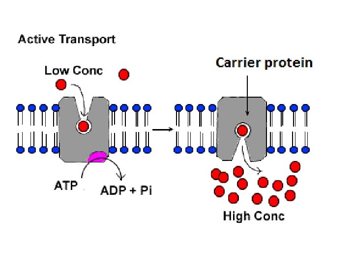
5. 4 The Need for Energy in Active Transport § Active transport moves substances against their gradients § requires energy © 2014 Pearson Education, Inc.

§Active transport allows cells to maintain different internal/external conditions §Ex. Animal cells are more negative inside than outside § Transport proteins support this gradient § How? § ATP transfers a phosphate group to the transport protein to change the protein’s shape allowing solute to enter/exit

Ion Transport: §With membrane transport, energy is required to move ions against their electrochemical gradient §Combination of two gradients: §The ion’s concentration gradient §The ion’s membrane potential
 separation of charge across the cell membrane §Inside of a cell")
§Membrane potential: (voltage) separation of charge across the cell membrane §Inside of a cell is more negative than the outside § Due to an unequal distribution of cations and anions on each side
")
§Ions diffuse down their electrochemical gradient (not just concentration gradient)

Electrogenic pump: transport protein that generates a voltage across a membrane §If electrical forces due to membrane potential oppose diffusion of an ion down its concentration gradient, active transport is needed

Cotransport: Coupled Transport by a Membrane Protein Figure 5. 17 Proton pump Sucrose-H cotransporter Sucrose Diffusion of H Sucrose

Ex. Sodium Potassium Pump §Actively transports ions §Pumps 3 Na+ out for every 2 K+ in § Provides a 1+ from cytoplasm to extracellular fluid to maintain gradient

Sodium. Potassium Pump EXTRACELLULAR FLUID 1 Figure 5. 14 © 2014 Pearson Education, Inc. CYTOPLASM [Na ] high [K ] low [Na ] low [K ] high 2 6 3 5 4 ADP

§https: //www. youtube. com/watch? v=7 Ae. Vnx. GWEpw

CONCEPT 5. 5: Bulk transport Exocytosis and endocytosis § Exocytosis: transport vesicles migrate to membrane, fuse with it, and release their contents § Endocytosis: reversal of exocytosis- cell takes in matter by forming new vesicles § 3 types § Phagocytosis (“cellular eating”) § Pinocytosis (“cellular drinking”) § Receptor-mediated endocytosis © 2014 Pearson Education, Inc.

Figure 5. 18 Phagocytosis Pinocytosis Receptor-Mediated Endocytosis EXTRACELLULAR FLUID Solutes Pseudopodium Plasma membrane Coat protein “Food” or other particle Food vacuole CYTOPLASM © 2014 Pearson Education, Inc. Coated pit Coated vesicle Receptor

Figure 5. 18 a Phagocytosis Pseudopodium of amoeba EXTRACELLULAR FLUID Solutes Bacterium Food vacuole 1 m Pseudopodium “Food” or other particle An amoeba engulfing a bacterium via phagocytosis (TEM) Food vacuole CYTOPLASM © 2014 Pearson Education, Inc.
")
Pinocytosis Plasma membrane 0. 25 m Figure 5. 18 b Pinocytotic vesicles forming (TEMs) Coat protein Coated pit Coated vesicle © 2014 Pearson Education, Inc.

Figure 5. 18 c Receptor-Mediated Endocytosis Coat protein 0. 25 m Plasma membrane Top: A coated pit Bottom: A coated vesicle forming during receptormediated endocytosis (TEMs) © 2014 Pearson Education, Inc. Receptor

5. 6 Cell Communication §Adjacent cells can communicate through molecules traveling through junction and cell to cell recognition

5. 6 Plasma Membrane Plays a Key Role in Cell Signaling §Adjacent cells can communicate through molecules traveling through junction and cell to cell recognition §Types of Signaling Molecules: §Local regulators: messenger molecules that travel short distances § Ex. growth factors, synaptic signaling

§Hormones: messenger molecules that travel long distances § Through circulatory system § Ex. insulin

Figure 5. 19 Long-distance signaling Local signaling Target cell Secreting cell Local regulator diffuses through extracellular fluid. (a) Paracrine signaling Secretory vesicle Electrical signal along nerve cell triggers release of neurotransmitter. Endocrine cell Neurotransmitter diffuses across synapse. Target cell is stimulated. Blood vessel Hormone travels in bloodstream. Target cell specifically binds hormone. (b) Synaptic signaling (c) Endocrine (hormonal) signaling © 2014 Pearson Education, Inc.

The Three Stages of Cell Signaling: A Preview § cells receiving signals undergo the following stages: § Reception: signaling molecule binds to a receptor protein in the membrane or inside the cell § Transduction: a series of steps convert the signal to form a specific response in the cell § Response: resulting cellular activity § https: //www. youtube. com/watch? v=-db. Rterut. HY Animation: Signaling Overview © 2014 Pearson Education, Inc.

Figure 5. 20 -1 EXTRACELLULAR FLUID Reception Receptor Signaling molecule © 2014 Pearson Education, Inc. CYTOPLASM Plasma membrane

Figure 5. 20 -2 EXTRACELLULAR FLUID Reception CYTOPLASM Plasma membrane Transduction Receptor Relay molecules Signaling molecule © 2014 Pearson Education, Inc.

Figure 5. 20 -3 EXTRACELLULAR FLUID Reception CYTOPLASM Plasma membrane Transduction Response Receptor Activation Relay molecules Signaling molecule © 2014 Pearson Education, Inc.

Reception §Signaling molecule is “fits” the receptor on the cell membrane protein §Binds to receptor §Behaves as a ligand (molecule that binds to another molecule): causes receptor protein to change shape
: activates response with")
Two Types of Transmembrane Receptors: § 1. G Protein-Coupled Receptor (GPCR): activates response with the help of a G protein § G Protein: molecule that binds to GTP (energy molecule)
 G protein Adenylyl cyclase")
Figure 5. 25 First messenger (signaling molecule such as epinephrine) G protein Adenylyl cyclase G protein-coupled receptor Second messenger Protein kinase A Cellular responses © 2014 Pearson Education, Inc.

§ 2. Ligand-Gated Ion Channel: membrane receptor that acts as gate for ion with its shape change

Transduction Stage §Usually a series of steps: cascade § One molecule activates another, etc. § At each activation, a protein changes shape § Caused by phosphorylation: addition of phosphate groups to a protein

§ Protein kinase: enzyme that transfers phosphate groups from ATP to a protein § (you can think of it as a relay molecule) § Newly added phosphate group causes protein to change shape due its interaction with amino acids (polar, charged, etc) § Now activated, it can hydrolyze another ATP and attach the phosphate to another protein kinase §Phosphates removed by protein phosphatases

§Phosphates removed by protein phosphatases § Deactivates protein §https: //www. youtube. com/watch? v=9 s 9 GJx 6 XJZM Cascade phosp

Figure 5. 26 Growth factor Reception Receptor Phosphorylation cascade Transduction CYTOPLASM Inactive transcription factor Active transcription factor Response DNA Gene NUCLEUS © 2014 Pearson Education, Inc. m. RNA

Small Molecules and Ions as Second Messengers § Second messengers: nonprotein, water-soluble molecules or ions that spread through cell by diffusion § Released by receptor protein § Often influences cascade effect § Cyclic AMP and calcium ions are examples § c. AMP rapidly actives phosphorylation of many proteins © 2014 Pearson Education, Inc.

https: //www. youtube. com/watch? v=Vatd. TJka 3_M

Response § Many signaling pathways regulate protein synthesis § Turn on or off specific genes
- Slides: 86