Calcium Metabolism Primary Hyperparathyroidism Calcium metabolism 99 of

Calcium Metabolism Primary Hyperparathyroidism
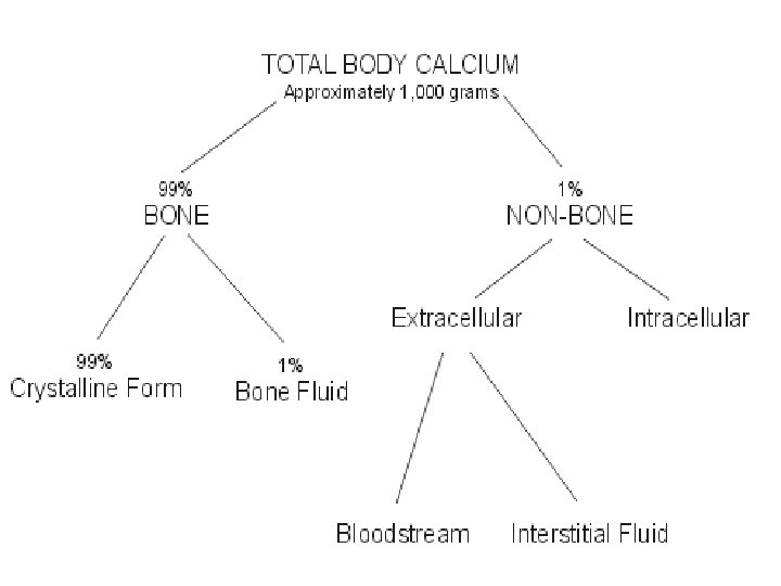

Calcium metabolism • • • 99% of total body calcium in the bone 1% in ICF , ECF , cell membranes * Calcium content 400 mg/kg in infant 950 mg/kg in adult * 50% ionized 40% bound to albumin. 10% citrate, phosphate

NORMAL PTH SECRETION/ACTIONS • PTH 1 -84 • secreted within seconds after hypocalcemia • Intact PTH half life of 2 -4 min (renal and hepatic clearence)

• Calcitriol: inhibits PTH synthesis • Mg: binds to Calcium Sensing Receptor • low Mg causes: - PTH resistance - reduced PTH secretion • Low Ca, high PO 4 stimulate PTH secretion

ACTIONS OF PTH A. Skeletal actions of PTH : • release of calcium by activation of bone resorption

B. Renal actions of PTH : 1. Ca Reabsorption: on distal tubule to stimulate ca reabsorption.

• 2. inhibit tubular reabsorption of phosphate • 3. Synthesis of calcitriol : 1 -alpha hydroxylase in proximal tubules

Primary hyperparathyroidism Mostly asymptomatic Mild hypercalcemia • Mostly > 45 years. • F: M (2: 1)

Pathogenesis and etiology • The routine measurement led to a marked rise in the incidence

ETIOLOGY 1. Radiation exposure: to the head and neck, 20 - 40 yrs earlier

Etiology • 2. Genetic or chromosomal defects : a. Cyclin D 1/PRAD 1 gene: parathyroid adenoma 1, proto-oncogene 20 -40 % of sporadic parathyroid adenomas
: sporadic nonfamilial parathyroid adenomas 16 % with sporadic")
b. MEN 1 (tumor suppressor gene): sporadic nonfamilial parathyroid adenomas 16 % with sporadic tumors.

• C. HRPT 2 gene : inactivating mutations in parathyroid ca • D. RET gene : rarley found in sporadic primary hyperparathyroidism

PATHOLOGY IN PRIMARY HYPERPARATHYROIDISM 1. Adenoma: Single adenoma: 89 % double adenomas: 5 % rarely in the thymus.

2. Glandular hyperplasia: 6 % of cases 3. Carcinoma: 1 -2% of cases at least one: - local invasion of contiguous structures - lymph node - distant metastases

Clinical manifestations – Primary. Hyperparathyroidism • asymptomatic hypercalcemia Biochemical screening detects 80 % of pts

ASYMPTOMATIC PRIMARY HYPERPARATHYROIDISM mild and sometimes only intermittent hypercalcemia. The mean s ca is < 1. 0 mg/d. L above nl • nonspecific symptoms: fatigue, weakness, anorexia, mild depression

CLASSICAL MANIFESTATIONS Anorexia, nausea, constipation, Polydipsia, and polyuria. • Osteitis fibrosa cystica: is rare (<5 % of pts) • Nephrolithiasis: most common complication, 15 -20 % of newly diagnosed pts.

Osteitis fibrosa cystica bone pain X-ray

• Subperiosteal bone resorption Tapering of distal clavicles

Before after surgery
")
• Brown tumors of pelvis ( ? PTH ca)

Neuromuscular/Psych symptoms • Neuromuscular: weakness and fatigue. Myopath is rare • Neuropsychiatric: depressed mood, psychosis,
 > cancellous")
Bone mineral density • Decreased BMD : cortical sites (forearm and hip) > cancellous sites (spine)

Cardiovascular • HTN is common • Mean carotid intima-media thickness was significantly higher compared with controls

Mortality • increased mortality: primarily due to CVS disease

DIAGNOSIS Serum calcium : confirmed PTH

Measurement of PTH -80 -90 %: high PTH -10 -20 %: minimally elevated or high normal

DIFFERENTIAL DIAGNOSIS 1. Malignancy: often evident clinically Intact PTH: very low in hypercalcemia of malignancy mediated by PTHr. P

• 2. FHH: an inactivating mutation in the Ca-SR in the parathyroid glands and the kidneys - A f/h of hypercalcemia (young children) - absence of symptoms and signs of hypercalcemia

• 15 - 20 % of pts with FHH: mildly elevated PTH • • FHH vs primary hyperparathyroidism: low urine ca excretion and Ca/Cr clearance ratio

3. Drugs : thiazide diuretics and lithium. reduce u ca excretion and can cause mild hypercalcemia (up to 11. 5 mg/d. L ) • thiazide should be stopped and calcium and PTH assessed 2 -3 months later

Lithium • in bipolar disorder • s/e: nephrogenic DI, hypothyroidism and hyperparathyroidism. • PTH less inhibited by calcium • It decreases renal Ca excretion (independent of PTH)

4. Secondary hyperparathyroidism: High PTH and nl or low ca

causes: - renal failure and impaired calcitriol production - inadequate calcium intake or absorption: - vitamin D deficiency - malabsorption.

TESTS TO CONFIRM PRIMARY HYPERPARATHYROIDISM 1. Urinary Ca excretion : distinguishes PHPT from FHH • If low ( < 200 mg/day): FHH vs hyperparathyroidism with vit D deficiency
 before management decisions")
2. 25 Vitamin D: replete low levels ( ≤ 50 nmol/L) before management decisions 3. Serum phosphorus: decreased or low nl

• Renal imaging: renal U/S should be performed if kidney stones are suspected.

• Localization studies: U/S, technetium-99 m sestamibi CT, or MRI intraoperative PTH monitoring

Management of primary hyperparathyroidism - Parathyroidectomy: first line therapy - indicated in asymptomatic patients if : • S Ca >= 1. 0 mg/d. L above the upper nl • Cr clearance <60 m. L/min • BMD at hip, lumbar spine, or distal radius (T score <-2. 5) and/or previous fragility fracture • Age < 50 years

ALTERNATIVES TO SURGERY • Avoid: thiazide lithium volume depletion prolonged bed rest / inactivity high calcium diet (>1000 mg/day) • Encourage physical activity • Adequate hydration to minimize the risk of nephrolithiasis.
. A low calcium diet may lead")
• Moderate calcium intake (800 -1000 mg/day). A low calcium diet may lead to further increases in PTH secretion and bone disease • Maintain moderate vitamin D intake (400 -600 IU/ d)

• Monitoring: S Ca and Cr annually and bone density (hip, spine, and forearm) every 1 -2 years is sufficient.

Drug therapy • Bisphonates: Drug of choice for pts who do not meet surgical criteria or who prefer to avoid surgery

• Calcimimetics: activate the calcium-sensing receptor in the parathyroid gland, thereby inhibiting PTH secretion - cinacalcet: - Rx of 2 hyperparathyroidism associated with renal failure - hypercalcemia in parathyroid cancer

HYPERCALCEMIA TREATMENT OF HYPERCALCEMIA regardless of etiology
 do")
INDICATIONS FOR TREATMENT 1. Asymptomatic or mildly symptomatic hypercalcemia (Ca <12 mg/d. L) do not require immediate treatment. 2. S Ca of 12 -14 mg/d. L may be well-tolerated chronically, but acutely requires more aggressive measures 3. S Ca >14 mg/d. L requires treatment, regardless of symptoms.

SALINE HYDRATION isotonic saline at 200 -300 m. L/h initially • if edema: loop diuretic may be used
 IM or sc q 12 hrs. Nasal calcitonin")
CALCITONIN • Salmon calcitonin (4 IU/kg) IM or sc q 12 hrs. Nasal calcitonin is not effective • lowers ca by 1 -2 mg/d. L within 4 -6 hours • Efficacy limited to the first 48 hrs, even with repeated doses (tachyphylaxis)

BISPHONATES • Pamidronate: malignancy acute primary hyperparathyroidism immobilization hypervitaminosis D sarcoidosis • (60 mg over 2 -4 hrs): NL ca for >2 wks • Serum ca decreases in 1 -2 days

• Zoledronic acid: drug of choice for malignancy-associated hypercalcemia • 4 mg/15 min • a single dose normalized calcium in 88 % of pts the median duration control 32 -43 days
 • prednisone 20")
GLUCOCORTICOIDS • high Ca with overproduction of calcitriol (sarcoidosis and lymphoma) • prednisone 20 to 40 mg/day reduces serum calcium within 2 -5 days

• HYPOCALCEMIA

Tetany
: 1. Destruction of the parathyroid glands")
Etiology • A. HYPOCALCEMIA WITH LOW PTH (HYPOPARATHYROIDISM): 1. Destruction of the parathyroid glands - Surgical : most common cause Transient: 20 % permanent: 0. 8 -3 % after total thyroidectomy

2. Autoimmune : polyglandular autoimmune syndrome type I presents in childhood - candidiasis - hypoparathyroidism - adrenal insufficiency during adolescence.

• 3. Abnormal parathyroid gland development: Genetic defects: - X-linked, autosomal recessive - Di. George syndrome
: - childhood - low Ca,")
B. HYPOCALCEMIA WITH HIGH PTH 1. PTH resistance: (Pseudohypoparathyroidism): - childhood - low Ca, high phosphate, and elevated PTH 2. Vitamin D deficiency or resistance 3. Extravascular deposition: deposition in tissues or binding within the vascular space

4. Hyperphosphatemia : CKD / ARF/ rhabdomyolysis/ tumor lysis 5. Osteoblastic metastases: breast or prostate cancer 6. Acute pancreatitis : precipitation of calcium soaps in the abdominal cavity 7. Sepsis or severe illness: hypocalcemia in critically ill or postsurgical pts 80 -90 %.

• C. DISORDERS OF MAGNESIUM METABOLISM: 1. magnesium depletion: PTH resistance (Mg < 1 mg/d. L) or by decreasing PTH secretion - Malabsorption - Chronic alcoholism - Cisplatin therapy - diuretic therapy - aminoglycosides 2. Severe hypermagnesemia: suppressing the secretion of PTH ( mg > 6 mg/d. L)

Treatment of hypocalcemia • IV calcium : - acute symptomatic pts - asymptomatic hypocalcemia with acute decreases to ≤ 7. 5 mg/d. L • 1 - 2 g of calcium gluconate ( 90 - 180 mg elemental calcium) in 50 m. L of 5 % D over 10 - 20 minutes • iv solution: 100 m. L (10 amp) of 10 % calcium gluconate in 1000 m. L of 5 % dextrose in water, at 50 m. L/h

• Iv calcium continued until the patient is recieving oral calcium and Calcitriol • Oral calcium: mild acute hypocalcemia (7. 5 8. 0 mg/d. L) or for chronic hypocalcemia. • 1500 - 2000 mg of elemental calcium • Vitamin D supplement is also needed.

• Thank you


Clinical manifestations and diagnosis of MEN 1

INTRODUCTION • MEN 1 : autosomal dominant: - parathyroid glands - anterior pituitary - pancreatic islet cells Also: - duodenum: gastrinomas, and carcinoid tumors - adrenal adenomas, and lipomas

DEFINITION OF MEN 1 • presence of two of the three main MEN 1 tumor types (parathyroid, entero-pancreatic endocrine adenomas, and pituitary adenomas) • Familial MEN 1 : index MEN 1 case with at least 1 relative with 1 of 3 main MEN 1 tumors

PRIMARY HYPERPARATHYROIDISM in MEN 1 • most common : almost 100 % by age 40 -50 yrs • 1 -2 % of primary hyperparathyroidism are due to MEN 1 • different features from the sporadic form : • The M: F ratio is even in MEN 1 in contrast to the female predominance in sporadic form • in MEN 1 presents in the 2 nd - 4 th decade of life (2 decades earlier than in sporadic form)

• Multiple gland involvement in MEN 1. 80 -85 % of pts with sporadic disease have single parathyroid adenomas.

• A high rate of recurrent hyperparathyroidism after subtotal parathyroidectomy. recurrence rate > 50 % at 12 yrs.

Diagnosis • hypercalcemia with inappropriately high PTH

PITUITARY ADENOMAS Clinically apparent in 15 - 20 % by CT or MRI ; Pathological prevalence > 60 %. The most common type is a prolactinoma, GH-producing, ACTH-producing, gonadotroph and clinically non-functioning tumors can also occur. • Multiple tumors are rare • •

macroadenomas - 85 % had macroadenomas versus 42 % in non-MEN 1 patients - MEN 1 patients had tumors that were larger and more aggressive than those in non-MEN patients

• The approach to diagnosis and therapy of pituitary adenomas in patients with MEN 1 is similar to that in patients with sporadic adenomas.

PANCREATIC ISLET CELL/GASTROINTESTINAL ADENOMAS • 1/3 of pts with MEN 1 • The most common cause of symptomatic disease is the Zollinger-Ellison (gastrinoma) syndrome, leading to multiple peptic ulcers. • 60 % of pts with MEN 1 have ZES or asymptomatic elevation in serum gastrin

• MEN 1 is present in 20 - 60 % of pts with ZES • Symptomatic insulinomas also occur • VIPomas and glucagonomas are rare

• The prevalence of nonfunctioning tumors is 30 - 80 %. • clinically nonfunctioning Pancreatic Endocrine Tumors may be malignant causing liver metastases

Zollinger-Ellison syndrome • the gastrinomas in MEN 1 are multifocal, often very small • the duodenum is a common site of gastrinomas in MEN 1 and sporadic form • Tumors in the pancreas do not usually secrete gastrin

• The risk of death from malignant MEN 1 gastrinoma is < sporadic

• ZES : initial manifestation of MEN 1 in 40 % • The onset of ZES symptoms preceded the dx of hyperparathyroidism in 45 % of pts • Pituitary disease occurred in 60 % of patients.

Diagnosis • Suspected clinically by the presence of multiple peptic ulcers or symptoms like diarrhea similar to sporadic form

• nonfamilial gastrinoma: the usual cause is ectopic ACTH release from the islet-cell tumor. • Patients with MEN 1 and ZES who develop Cushing's syndrome usually have a corticotroph adenoma of the pituitary

Insulinoma • In MEN 1 are often small, may be multiple,

Nonfunctioning pancreatic Endocrine tumors • Detected as early as ages 12 -14 in asymptomatic children • The risks of metastasis and death are low for tumors ≤ 20 mm
,")
Carcinoid tumors • Thymic carcinoid tumors occur in MEN 1 (2. 6 5 %), - mostly in men - heavy smoking ? a risk factor Thymic carcinoids : are typically nonfunctional (in contrast to the substantial incidence of ectopic Cushing's syndrome in pts with sporadic thymic carcinoid), and tend to be aggressive

Thymic carcinoids in MEN 1 • CT and MRI were sensitive for detecting the tumors • All patients underwent surgical resection with high rate of recurrence

Cutaneous tumors • Angiofibromas and collagenomas were more common in MEN 1 patients • lipomas were present in 17 %

others - adrenal tumors, especially nonfunctional adrenocortical adenomas, - gastric carcinoids, - pheochromocytoma (very rarely) - angiomyolipomas - spinal cord ependymomas.

SCREENING OF FAMILY MEMBERS IN MEN 1 KINDREDS • little evidence that early, presymptomatic detection actually reduces overall morbidity or mortality in MEN 1 • one low-cost option for screening is measurement of serum calcium

DNA TESTING IN DIAGNOSIS OR SCREENING FOR MEN 1 • Direct DNA testing for MEN 1 mutations is available • DNA testing in specific individuals can be helpful when the diagnosis of MEN 1 is unable to be clearly established on clinical grounds and would alter management

current evidence does not support routine use of intensive laboratory evaluation and/or imaging studies in the absence of relevant signs or symptoms
 : - bone")
PTH receptors 1. PTH / PTHr. P receptor (type 1 R) : - bone and kidney - recognizes PTH and PTH-related protein
: - binds PTH but not")
• 2. PTH 2 receptor (type 2 R): - binds PTH but not PTHr. P
- Slides: 96