Caenorhabditis elegans THE GENETICS OF CAENORHABDITIS ELEGANS S

Caenorhabditis elegans

THE GENETICS OF CAENORHABDITIS ELEGANS S. BRENNER Medical Research Council Laboratory of Molecular Biology, Hills Road, Cambridge, CB 2 2 QH, England Manuscript received December 10, 1973 ABSTRACT Methods are described for the isolation, complementation and mapping of mutants of Caenorhabditis elegans, a small free-living nematode. About 300 EMS-induced mutants affecting behavior and morphology have been characterized and about one hundred genes have been defined. Mutations in 77 of these alter the movement of the animal. Estimates of the induced mutation frequency of both the visible mutants and X chromosome lethals suggests that, just as in Drosophila, the genetic units in C. elegans
 Mutants published by")
C. elegans timeline. • • • Developed by Sydney Brenner (1963) Mutants published by Brenner (1974) 1976 postembryonic cell lineages determined (Sulson and Horwitz) 1982 "Programmed cell death" (Horwitz et al. ) (Nobel Prize Brenner, Sulson, Horwitz 2002) 1983 complete embryonic cell lineages determined (Deppe et al. , Sulson et al. ) 1986 Complete connectivity of nervous system established (White et al. ) "The mind of a worm” 1991 -98 RNAi and mi. RNA discovered in worms. (Nobel Prize: Fire, Mello 2006) 1994 - First use of GFP in animals (Nobel Prize: Chalfie, 2008) 1998 First animal genome sequenced (97 Mb, now 100. 3 Mb) Ø This is about 1/30 the size of the human genome (3 Gb). C. elegans have about 20, 000 coding sequence genes, more than half that of humans (30, 000 -40, 000 genes). First animal to be sequenced! Knowing the sequence allows genes of interest to be easily cloned. Also it opened opportunity for reverse genetic approach.

Figure 2 | A simple F 2 screen. a | A population of wild-type hermaphrodites is exposed to a mutagen and genes are randomly mutated in the germ cells (mutated germ cells are indicated in red). For example, one sperm could be mutated for the gene unc-32, which is required for the correct functioning of the nervous system. Fertilization of an egg by this sperm will result in a heterozygous F 1 individual. Because this animal is a self-fertilizing hermaphrodite, it will produce eggs and sperm that bear this mutated gene; one quarter of its F 2 progeny will be homozygous for the mutation and result in a coiled phenotype b | An unc-32(f 131) mutant. unc-32 encodes the a subunit of the V 0 complex of the vacuolar ATPase 119 — a proton pump that generates the energy that is required for transporting neurotransmitter into synaptic vesicles. Animals that lack the proton pump do not release normal levels of neurotransmitter at the synapse and therefore have uncoordinated movement.
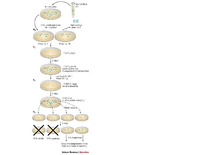

Mutant Isolation n n EMS induces high forward mutation rate Hermaphrodite heterozygotes for mutation will produce ¼ homozygous mutant progeny by self-fertilization Severe effect mutations recovered because of self-fertilization (e. g. , complete paralysis of body wall muscles) 1600 genes on genetic map from mutant screens n Examples n n n Dumpy (Dy) – abnormal body shape Long (Lon) – abnormal body shape Unc – uncoordinated Phenotypes capitalized and not italicized Genes and alleles are in lower-case italics Protein products are all in upper case (e. g. , DPY-10)

Mutants http: //130. 15. 90. 245/photos. htm Bli Muv wt Bmd trp-4 Unc

WHY C. elegans? • Short generation time: between 3 days-1 week depending on temperature. • Stocks can be frozen: the eggs survive the freezing process so stocks don’t have to be continuously propagated. • Very easy to propagate and recover: a square of agar can be cut of starved plates and placed upside down on new agar plates, recovering the strain. This method can also be used to propagate strains. • Very cheap to maintain: essentially all you need for C. elegans research is a microscope and an incubator, no expensive animal house costs. Temperature sensitive: the worms grow at different speeds depending on the temperature. • Present no biohazard: not much paperwork and no IRB necessary. Agar plates do not require special disposal. • The first complex organism to have its genome sequenced: therefore it’s the most annotated. • The fate of all cells are known: mostly all cells have been traced through development, making C. elegans a great developmental model.

• • Simple organisms with complex structures: Even though these worms are a relatively simple organism, they contain many complex structures including, a complete nervous system, gut, and an extensive germline. Very easy to do forward genetic screens. RNAi was discovered in C. elegans and can be incorporated by feeding. The Arliger lab has created an extensive library of RNAi constructs in bacteria. These can be grown up and fed to the worms. Embryos and adults are translucent. Easy to GFP labeled structures and other phenotypes. Balancers for each chromosome exist. Recessive sterile and lethal mutations can be stably propagated and easily detected. Also many balancers are marked via GFP. Hermaphroditic, self mating allows for efficient generation of recessive mutants. Rare males (0. 05%) Large broad size: for easy screening and large N value. Single Hermaphrodite has about 300 progeny.

Drawbacks • No site directed mutagenesis, due to only one crossover per chromosome. There a couple ways around this. Forward genetics screens are very easy. Also transgenic constructs can be either injected or bombarded into worms. • No cell lines available • dissection of specific tissues is unrealistic

Anatomy of the worm 959 somatic cells XX karyotype become hermaphr • With one less chromosome, males develop 79 more neurons and 25 more muscle cells than hermaphrodites, used for mating. X 0 karyotype become males 1031 cells

Males arise spontaneously via nondisjunction of X during hermaphrodite gametogenesis

Dosage compensation in. C. elegans

Germline and Mating Hermaphrodite Image from Hansen lab First 40 germ cells that enter meiosis in each arm of gonad develop into ~150 sperm deposited into spermatheca Sexual fate switches and rest of germ cells are oocytes

Life cycle of Caenorhabditis elegans a spheroid of cells: ectoderm - hypodermis and neurons, mesoderm - pharynx and muscle, endoderm - germline and intestine. Organogenesis terminal differentiation http: //www. wormatlas. org

C. elegans cell lineage • One amazing advantage of worms is that every worm has the exact same number of somatic cells! Ø Ø Ø Hatching larva=558 cells Males (XO)=1031 somatic cells+ ~1000 sperm Hermaphrodite (XX)= 959 somatic cells+ ~2000 eggs and sperm. • Somatic cells arise by an INVARIANT cell lineage. The divisions are invariant in pattern, timing, and orientation of each division.

Embryonic Development in C. elegans gastrulation C - 17 Figure C. 5 a wormlike shape before hatching • 3 days • Embryonic development begins in uterus • Hermaphrodite lays her eggs 2 hours after fertilization • Blastoderms – large cells produced by early cleavage 1. Unequal cleavages in early development 2. Until 28 cell stage all blastoderms contact surface of embryo • Gastrulation - At 28 cell stage, two gut precursor cells begin to move to embryo interior 1. Three germ layers produced (endoderm, mesoderm, ectoderm) 2. Later, cell division ceases while cells differentiate 3. Contractile proteins squeeze embryo into wormlike shape 4. 14 hours after fertilization, egg hatches 5. L 1 larvae is 250 mm long and has 558 cells • Next 50 hours – L 2, L 3, L 4 stages of development separated by molts

Lineage Diagram of Early Cleavages Give Rise to Six Founder Cells Which Each Give Rise to Various Features of Adult A more general method of examining the roles of individual cells during all stages of C. elegans development is laser ablation C - 18

Germline formation in C. elegans 6 Founder Cells +98 die +14 die Small posterior cell goes germline +1 dies

Where do specific tissues come from? Lineages do not strictly produce single tissue types. AB MS E C D P 4 While intestine & germ-line come from single founder cells, the muscle, nervous system & skin cells each arise from multiple lineages. This means, for example, that muscles do not have one “founder” cell early in development.

Cell death can be a genetically programmed cell fate. Indicate death event The Nematode Caenorhabditis Elegans. 1988. Editor William B. Wood , Cold Spring Harbor Laboratory. § Specific cells with diverse developmental origins undergo programmed cell death at specific times during development. § Programmed cell death is characterized by a series of specific morphological changes. § There must be genes that control both the decision to express that fate and the execution.

APOPTOSI Stimoli fisiologici e non fisiologici • Restringimento cellulare • Organelli non danneggiati • Condensazione della cromatina • Formazione di corpi apoptotici • Mancanza di infiammazione NECROSI • Danni meccanici • Esposzione ad agenti chimici • Rigonfiamento cellulare • Organelli danneggiati • Alterazione della cromatina • Lisi cellulare • Risposta infiammatoria

Nell’uomo processi apoptotici sono coinvolti in: • Sviluppo embrionale • Sviluppo del sistema nervoso centrale • Turn-over cellulare • Selezione timica • Spegnimento delle risposte immunitarie In base agli studi condotti su C. Elegans si è stabilito di suddividere il processo apoptotico in 4 fasi: • induzione • esecuzione • riconoscimento e fagocitosi del corpo cellulare • degradazione dello stesso da parte della cellula fagocitica

APOPTOSI Attualmente la maggior parte delle nostre conoscenze sull’apoptosi provengono da studi compiuti sul nematode Caenorhabditis Elegans. Tre geni sembrano svolgere un ruolo nella morte cellulare programmata di questo nematode: ced-3, ced-4 e ced-9. Mentre i prodotti genici di ced-3 e ced-4 promuovono la morte cellulare, il prodotto di ced-9 svolge un ruolo protettivo. Il gene ced-9 codifica una proteina analoga a Bcl-2, un inibitore della morte cellulare in altri sistemi cellulari.

Un ermafrodita di C. Elegans genera 1000 nuclei di cellule somatiche durante lo svilppo, ma 131 muoiono Ced-3 e ced-4 sono necessari perché avvengano le 131 morti cellulari normali Se ced-3 e ced-4 vengono inattivati in seguito a mutazione le cellule destinate a morire sopravvivono differenziandosi in tipi cellulari riconoscibili L’espressione eccessiva di ced-3 e ced-4 fa morire molte cellule che normalmente sopravviverebbero

Quando il gene bcl-2 umano è trasferito in C. Elegans inibisce la morte cellulare normale del verme. E’ in grado di controbilanciare gli effetti della mutazione in ced-9 che causerebbe la morte prematura nel corso dello sviluppo. Il meccanismo di morte cellulare programmata e la sua regolazione sono fenomeni conservati nel corso dell’evoluzione dai vermi all’uomo.

Programmed Cell Death - Apoptosis C - 27 Fig. C. 15

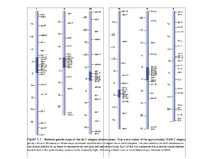
Nuclear Genome of C. elegans • • • 97 Mb – very small (2/3 size of Drosophila) Six chromosomes (I-VI) – about same size No defined centromere – holocentric Detailed physical map of cosmids and YACs Recombination rates vary considerably 1 gene every 5 kb 20, 000 genes Proteins match homologous sequences of other organisms 20% proteins carry out core biological functions Rest involved in processes required only in multicellular organisms Repetitive sequences – e. g. , 7 kinds of TEs

Assembly of kinetochores on monocentric and holocentric chromosomes. Dernburg A F J Cell Biol 2001; 153: F 33 -F 38
 Schematic illustrating the hierarchy for mitotic kinetochore assembly Dotted lines indicate groups of")
(B) Schematic illustrating the hierarchy for mitotic kinetochore assembly Dotted lines indicate groups of proteins that have been shown to co-purify from C. elegans extracts in immunoprecipitations. The colored ovals group proteins together whose individual depletions result in a similar phenotype. (Red) “Kinetochore Null”/KNL proteins whose depletion is characterized by a failure to assemble kinetochores. Consequently, segregation is severely defective and spindle poles separate prematurely. (Blue) proteins whose depletion results in relatively subtle chromosome segregation defects. In MIS embryos, kinetochore assembly occurs but at a slower rate and to a reduced extent relative to wild-type. (Yellow) proteins whose depletion results in a chromosome alignment and segregation defects of intermediate severity. Spindle poles separate prematurely. (Green) proteins, whose depletion causes sister chromatids to co-segregate to the same spindle pole. This defect likely arises from an inability to polymerize microtubules at kinetochores.

Holocentric chromosomes

Why the nematode centromeres are holocentric? The existence of mechanisms to avoid the problems connected with this type of centromere structure in different cell types suggests that there are selective evolutionary pressures favouring the maintenance of a diffuse centromere in nematodes. The holocentric organization of chromosomes could be related to the mosaic development of nematodes: each cell (with few exceptions) passes through a predetermined number of mitotic divisions and differentiates into a predetermined terminal state. So, one cell cannot be substituted by, nor substitute for, another cells. An important consequence is that a single cell death can be lethal to the embryo. Since chromosome breakages in monocentric chromosomes are cause of cell lethality due to the loss of acentric fragments, the holocentric organization of chromosomes should increase the resistence to breakages because chromosome fragments are maintained through cell division. The holocentric chromosomes could have been selected in nematodes as a strategy to avoid the potentially lethal effects of unrepaired chromosome breakages.

Gene Expression • 70% of m. RNAs have one of two splice-leader sequences trans-spliced onto the 5 end of the message • Trans-splicing with splice-leader sequences produces single-gene m. RNAs • Conserved among all nematode genera examined • 25% of adjacent genes are transcribed as operons C - 35

Trans-splicinig 36 Fig. C -C. 3
 Reporter-gene fusion")
Gene expression 3 approaches to study gene expression in C. elegans: 1) Reporter-gene fusion with transformation (GFP, Lac. Z) 2) In situ hybridization using m. RNA 3) Immunofluorescence with specific antibody

Transformation ØDNA transformation by microinjection is a very fast, simple, inexpensive, and efficient way to generate transgenic C. elegans strains. Injection requires little to no preparation and only few worms need to be injected to obtain transgenic lines. ØThe C. elegans hermaphrodite germline is a syncytium containing hundreds of mitotically active oocytes at different stages of maturation and almost all nuclei share a common cytoplasm. Ø Different DNA species can be co-injected (plasmids, PCR products, cosmids, fosmids, BACs, YACs) with selectable injection markers (fluorescent reporter genes, dominant or recessive phenotypic markers). This DNA mix will be processed by DNA end ligation and homologous recombination into large, heritable DNA concatemers called extrachromosomal arrays. Ø These episomes are recognized by the replication machinery and usually segregate like chromosomes during cytokinesis and meiosis 40 X DIC http: //130. 15. 90. 245/photos. htm

Purposes of transformation: • identification of genes by rescuing a mutant phenotype using a WT copy of the gene or the gene homolog from another species • expression pattern using the gene of interest with reporter (gene_X_promoter: : GFP) • interference of a biological process by overexpression of WT or mutated gene

Stable Integration of C. elegans Transgenes transgene integration is generally achieved by irradiating extrachromosomal lines with γ or UV radiation. Transgene sequences are randomly integrated into the genome, most likely, following erroneous repair of a chromosomal break. The resulting stable lines need to be extensively outcrossed to remove background mutations. Moreover, it is recommended to obtain independent integrated lines because some variability in expression can be observed, probably due to positional effects and the amount of integrated DNA. • Expose a plate with ca 50 L 4 worms bearing the extrachromosomal array, to 3500– 4800 rads of x- or gamma rays • Look for worms positive for marker - in case of GFP fluorescent worms - who give a 100% fluorescent progeny Øpick a few hundred F 1 s to individual plates Øpick 4 -5 F 2 s from each F 1

Analysis of the C. elegans genome sequence indicates that approximately 12 % of the C. elegans genome is derived from transposable elements Figure 1. Active transposons in C. elegans. The structure of the transposons that proved to be mobile are depicted in the left panel. They all are class II DNA tranposons flanked by Terminal Inverted Repeats (boxes) and containing one open reading frame (arrows) encoding the transposase enzyme. Distribution of the different transposons in the genome of the C. elegans Bristol N 2 strain is represented in the right panel using the same color code as on the right (Tc 7 are in light blue; however, as represented on the left panel, 36 of the 38 outer bp of Tc 7 are identical to the one of Tc 1) The sizes of the chromosomes are in Mb. Reprinted from Fischer et al. (2003). Copyright © 2003 the Genetics Society of America.

Tc 1 and Tc 3 are the most active and best characterized transposons in C. elegans. They are part of the superfamily TC 1/mariner Tc 1 is 1, 610 bp long and contains two 54 bp terminal inverted repeats (TIRs Analysis of spontaneous and reversible mutations of the myosin heavy-chain unc-54 demonstrated the mobile nature of Tc 1. The subsequent characterization of additional spontaneous unc-22 mutations lead to the identification of Tc 3 (Collins et al. , 1989). Tc 1/mariner transposases all contain a triad of acidic residues (DDE or DDD) with a characteristic spacing which is shared by a superfamily of endonucleases. Primary sequence conservation of the Tc 1/mariner transposases is relatively low (about 15 % identity among the superfamily) but phylogenetic analysis suggests that all Tc 1/mariner were derived from a common ancestor and might all transpose through similar mechanisms. Tc 3 is an element of 2, 335 bp with 462 bp TIRs. The genome of the Bristol N 2 strain contains 31 and 22 copies of Tc 1 and Tc 3, respectively. These numbers are strain dependent. In some strain isolates such as Bergerac, Tc 1 transposition is active in the germ line and each haploid genome contains up to 300 -500 Tc 1 copies.

Other active transposons The Tc 2 element was initially identified as a polymorphism marker (Levitt and Emmons, 1989). It is 2, 074 bp in length and has perfect terminal inverted repeats of 24 bp (Ruvolo et al. , 1992). There are 4 full-length Tc 2 copies in the N 2 genome, each flanked by a TA dinucleotide at either end. In addition, up to 300 copies of remnant Tc 2 elements have been detected in the genome The first Tc 4 element was identified as a mutagenic insertion in the gene unc-86 It is a foldback element of 1. 6 kb which contains almost perfect terminal inverted repeats of 774 bp with a 57 -bp unique internal sequence. No open reading frame can be detected within Tc 4. A variant class of Tc 4 (Tc 4 v, 5 copies in the N 2 genome) contains a 2, 343 bp sequence which replaces 477 bp in one of the inverted repeats. A transcript from Tc 4 v has been detected. It may encode a 537 -aa protein which resembles transposases of the DDE superfamily. Tc 4 v might provide in trans the transposase required to mobilize all Tc 4 elements

The Tc 5 element is present in four copies per haploid genome. Tc 5 encodes a putative 532 amino acid transposase which is overall 33 % identical to the Tc 4 v transposase Tc 7 is a 921 bp element that uses the Tc 1 A transposase for transposition. It is made up of two 345 bp inverted repeats separated by a unique sequence that does not contain an ORF.

The genome of C. elegans also contains class I retrotransposons. These elements are subclassified into Long Terminal Repeat (LTR) retrotransposons that resemble retroviruses but usually lack the gene encoding the envelop protein and non-LTR retrotransposons. They can be grouped in 19 families related either to the gypsy or Bel clades of retrotransposons These elements only constitute 0. 2 % of the C. elegans genome (Zagrobelny et al. , 2004) suggesting that retrotransposons have been altogether strongly counterselected in this compact genome as compared to other species such as Homo sapiens in which more than 40 % of the genome is composed of retroelement sequences.

Mechanisms of Tc 1 and Tc 3 transposition Tc 1/mariner transposons move via a "cut-and-paste" mechanism: transposase binds the TIRs, catalyses excision and subsequent reinsertion into target DNA in a TA dinucleotide and leaves behind a double-strand DNA break which is repaired by the cellular machinery.

Transposon-based tools for C. elegans research Gene identification in forward genetic screens Insertional mutagenesis with transposons generates mutant alleles that are tagged by the presence of a transposon. This molecular tag can subsequently be used to identify the mutated gene. Identification of Mutations Caused by Insertion of a Transposable Element

However, using Tc elements as mutagens has some major drawbacks. • • • First, the mobilization of Tc transposons is not restricted to a single class of elements in mutator strains. Second, there are several copies of each transposons in the genome which complicate the identification of the mutagenic insertion. Third, in the mutator strains that are used, transposition is not controlled. Some Tc insertions are poorly mutagenic either because they are in introns, or because they are removed from the mature m. RNA by aberrant splicing Spontaneous re-excision can generate mutagenic footprints that generate a stronger phenotype but can no longer be detected in a transposon tagging strategy. These limitations have been circumvented by mobilizing the Mos 1 transposon in the germ line of C. elegans (Bessereau et al. , 2001). Mos 1 is a member of the Tc 1/mariner family and was isolated from Drosophila mauritiana (Jacobson et al. , 1986). The Mos 1 element is absent from the C. elegans genome and controlled mobilization of Mos 1 is achieved by conditional expression of the Mos 1 transposase. Mos 1 mutagenesis is 10 times less efficient than chemical mutagens but the cloning of mutated genes is extremely fast since Mos 1 insertions represent rare tags that are easy to localize in the genome

Mos 1 Transposon Mutagenesis Mos 1 mutagenesis is based on a binary system. Two extrachromosomal transgenes are used: one that contains multiple copies of the Mos 1 transposon (transposon array), and another that provides the transposase under the control of a heat-shock inducible promoter. When the transposase activity is induced in doubly transgenic worms, Mos 1 can jump from the ‘transposon array’ into the genome. The extrachromosomal arrays are lost in subsequent generations, eliminating the risk for Mos 1 re-excision. Mos 1 is approximately 10 times less efficient than EMS as a mutagen under optimal conditions. This low efficiency has to be contrasted with the ease, speed, and precision with which Mos 1 can be located.

Transposon-based tools for C. elegans research Site-directed mutagenesis Gene targeting techniques based on homologous recombination, such as those used in mice and yeast, are difficult in C. elegans. Transposons might represent an interesting alternative to engineer specific mutations in the genome. In 1992, Plasterk and Groenen (Plasterk and Groenen, 1992) demonstrated that a transgene containing a fragment of the unc-22 gene could be used as a template to repair a double-strand break caused by Tc 1 excision out of the unc-22 locus in a mut-6 background. Point mutations contained in the transgene were copied in the genome during the repair process. Therefore, transgene-instructed gene conversion provides a strategy to engineer mutations or introduce exogenous sequence in the genome. However, such events were rare (2. 105 event per meiosis), thus preventing this technique from being widely utilized in C. elegans.

Mos 1 Excision Induced Transgene-Instructed Gene Conversion Mos. DEL: Mos 1 -Mediated Deletion
 The goal of")
Schematic presentation of Mos 1 mediated Single Copy Insertion (Mos. SCI) The goal of Mos. SCI (Single copy insertion of transgene is to engineer single-copy transgenes at a defined location in the genome. A DSB induced by Mos 1 excision is repaired by homologous recombination with an invitro-engineered template which contains the desired transgene in combination with the unc-119(+) minigene. twk-18(gf) and fluorescent m. Cherry markers are negative selection markers. twk-18(gf) is a temperature-sensitive dominant mutation in a potassium channel, which paralyzes the animals at 25°C but not at 15°C. m. Cherry markers are expressed in the pharynx, body muscle, and nervous system for visual identification of array-carrying animals. Following loss of the array, single-copy transgenic animals are isolated. L and R homologous regions = 1. 4 kb.

Manipulating Gene Activity by RNAi was first discovered in C. elegans, rapidly generalized to all other model systems from plants to vertebrates and recognized as a revolutionary approach to modify gene expression. RNAi immediately became an attractive gene knock-down technology in C. elegans. With RNAi and the complete genome sequence, it was theoretically possible for the first time to easily and rapidly test the function of a large number of genes in a systematic fashion. This led to remarkable large-scale screens for cytokinesis, essential genes and cell polarity, lifespan, fat regulation, and gene function at the whole-genome level. However, it also became clear that not all genes were affected by RNAi and affected genes often did not show phenotypes as strong as corresponding genetic null mutants. Intriguingly, RNAi seemed to be much more efficient in certain tissues, with neurons being often refractory to RNAi. This surprising result led researchers to investigate the basis for this tissue specificity and identify sensitized mutant backgrounds which display increased RNAi sensitivity.

RNAi can be applied at any stage during development, which makes it easier to study essential genes or to study a gene's function at different stages of development. ds. RNA can be efficiently delivered in worms by four protocols. • Worms can either be (1) injected with in vitro-transcribed ds. RNA 7; • soaked in buffer containing concentrated in vitro-transcribed ds. RNA; • fed bacteria expressing ds. RNA from an engineered plasmid (clones for ∼ 94% of all C. elegans genes are available from RNAi-feeding libraries generated by the Ahringer and Vidal labs and can be procured from commercial suppliers (Source Bio. Science Life. Sciences and Open Biosystems, respectively). ); • trans formed with a vector expressing ds. RNA in a given tissue.

Microinjection Soaking in ds. RNA Feeding bacteria expressing ds. RNA
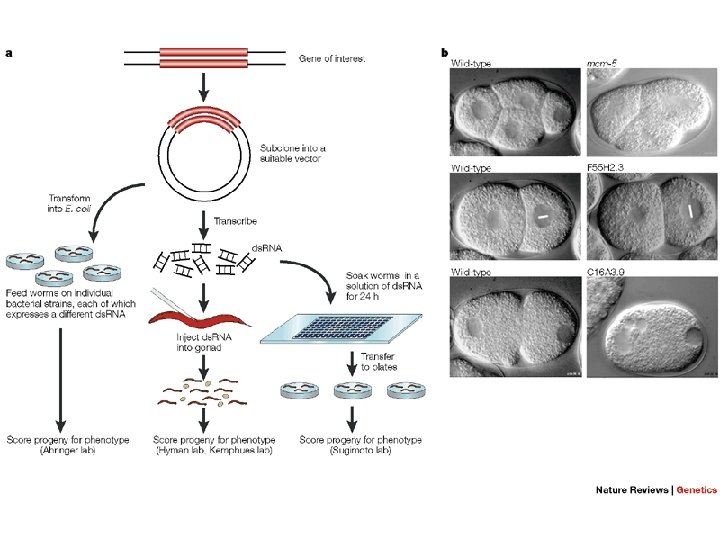

Plasmids for feeding Timmins L. & Fire A. 1998. Nature, 395: 854. Segments were cloned between flanking copies of bacteriophage T 7 promoter into a bacterial plasmid vector. A bacterial strain BL 21/DE 3 expressing T 7 polymerase Gene from an inducible Lac promoter was used as a host. Large-scale screens for RNAi deficient mutants. Production of genome wide collections of RNAi feeding constructs
 A")
However, a few limitations of RNAi have to be kept in mind. (1) A major disadvantage of RNAi compared to genetic mutation is the variable efficiency and potency of RNAi depending on the experimental conditions. (2) It is not possible to assess unambiguously the degree to which a gene's function is knocked down by RNAi. 3) As discussed previously, some tissue types are insensitive to RNAi in the wild-type context but sometimes also in sensitized backgrounds. (4) The efficiency of RNAi decreases rapidly when multiple genes are targeted at once, limiting genetic interactions tests based solely on RNAi. (5) A given RNAi clone can sometimes target more than one gene, based on sequence similarity. If no mutant allele is available to confirm an RNAi result, it is important to conduct RNAi with distinct non-overlapping ds. RNA sequences derived from the target gene to avoid off-target effects
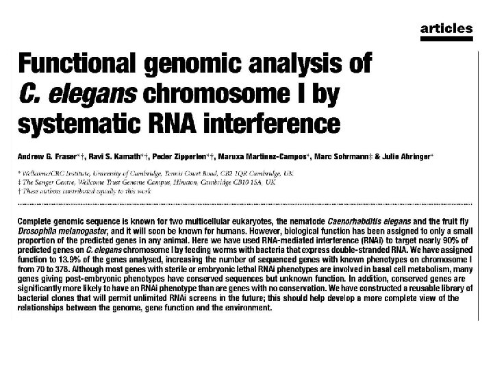

The authors have constructed a library of bacteria expressing ds. RNA corresponding to genes on chromosome I. Chromosome I is the second smallest chromosome, has few duplicated gene clusters and has no striking unusual features. Each individual bacterial clone is able to synthesize ds. RNA designed to target a single gene; In total, the resulting library contains 2, 445 independent clones, corresponding to 2, 416 predicted genes, a total of 87. 3% of the 2, 769 currently predicted genes of chromosome I.
, Ste (sterile), Unc (uncoordinated) and Pep (viable postembryonic")
Functional classes of Emb (embryonic lethal), Ste (sterile), Unc (uncoordinated) and Pep (viable postembryonic phenotype) genes a, Predicted products of genes that gave Ste, Emb, Unc or viable post-embryonic (Pep) RNAi phenotypes were placed into functional classes as described in Methods. Genes whose products could not be accurately classified into any of the eight functional classes were placed into the unknown category (white). Numbers denote the percentage of genes in each functional class; pie charts illustrate these numbers graphically. b, Pie charts show distributions of predicted gene products grouped as follows: basal metabolic category (red) comprises the classes of DNA, RNA, protein and intermediate metabolism; specialized functions (blue) comprises cell-cycle and chromosome dynamics, cell biology and cellular structure, gene-specific transcription factors and signal transduction. Worms show the tissue affected in each phenotypic class shaded in grey. c, Distribution of genes giving rise to non-viable RNAi phenotypes in C. elegans (worm) or to non-viable phenotypes following disruption in S. cerevisiae (yeast).


- Slides: 66