C N S DEVELOPMENT LECT1 DR SHAHAB Assoc

C. N. S DEVELOPMENT LECT#1 DR SHAHAB Assoc Professor Anatomy KGMC

Learning objectives • Neural tube formation, neural crest cells origin; • Brain development • Primary brain vesicles • Secondary brain vesicles

Stages of Embryonic development

Neurulation Neural Tube • The nervous system develops when the notochord induces its overlying ectoderm to become neuroectoderm and • This now develop into the neural plate. • The neural plate folds along its central axis to form a neural groove • lined on each side by a neural fold

• The two neural folds fuse together and pinch off to become the neural tube. • Fusion of the neural folds begins in the middle of the embryo and moves cranially and caudally. • The cranial open end of the tube is the anterior (rostral) neuropore, and • the caudal open end of the tube is the posterior (caudal) neuropore. • The anterior neuropore closes on or before day 26 and • the caudal neuropore closes before the end of the fourth week.

Neural Crest • Some cells from the neural folds give rise to pleuripotent neural crest cells, • they migrate widely in the embryo to give rise to many nervous structures: • Spinal ganglia (dorsal root ganglia) • Ganglia of the autonomic nervous system • Ganglia of some cranial nerves • Sheaths of peripheral nerves • Meninges of brain and spinal cord • Pigment cells • Suprarenal medulla • Skeletal and muscular components in the HEAD

Summary: Embryonic Development • Nervous system develops from ectoderm – by 3 rd week, neural plate becomes a groove with neural folds along each side – by 4 th week, neural folds join to form neural tube – lumen of the neural tube develops into central canal of spinal cord & ventricles of the brain – cells along the margin of the neural groove is called the neural crest • develop into sensory and sympathetic neurons & schwann cells – NB: By 4 th week, neural tube exhibits 3 anterior dilations

Stages of Embryonic development

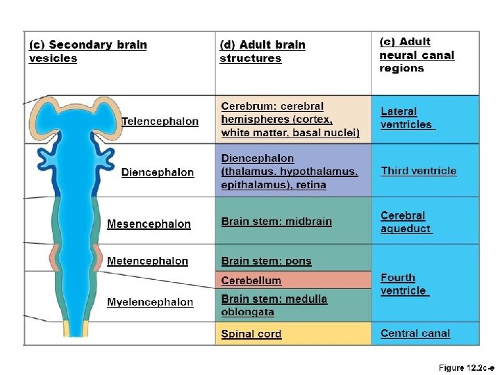
Brain development • By wk 4, the neural tube forms three Primary Brain Vesicles. • The primary brain vesicles give rise to five Secondary Brain Vesicles, • which give rise to the various adult structures.

Brain Development • 4 th week – forebrain – midbrain – hindbrain • 5 th week – telencephalon – diencephalon – mesencephalon – metencephalon – myelencephalon

Brain Development

Brain-5 vesicles/adult derivatives

Week 5 brain stage development
 Secondary vesicles Telencephalon Diencephalon Adult structures Cerebral hemispheres, consisting")
Primary vesicles Forebrain vesicle (prosencephalon) Secondary vesicles Telencephalon Diencephalon Adult structures Cerebral hemispheres, consisting of the cortex and medullary center, basal ganglia, lamina terminalis, hippocampus, the corpus striatum, and the olfactory system Thalamus, epithalamus, hypothalamus, subthalamus, neurohypophysis, pineal gland, retina, optic nerve, mamillary bodies Midbrain vesicle (mesencephalon) Mesencephalon Hindbrain vesicle (rhombencephalon) Metencephalon Pons and cerebellum Myelencephalon Medulla Midbrain
 • Fluid filled cavities found within the developing brain")
Ventricular System (Cerebrospinal Fluid System) • Fluid filled cavities found within the developing brain system originate from lumen of the developing neural tube • 5 cavities- lateral vent-1 pair • -3 rd vent • -4 th vent • Lined by choroid plexus made of ependymal cells-BBB

Ventricles in forebrain • Lateral ventricles-one pair • Ventricles in brainstem: • Mesencephalon 3 rd/cerebral aqueduct • Metencephalon 4 thventricle • Mylencephalon 4 thventricle

Ventricular communication in the brain • Lateral-3 rd vent foramen of Monro • 3 rd to 4 thvent cerebral aqueduct of sylvius • 4 thto lateral aperture foramen of luschka • 4 thto median aperture foramen of mengedie

Lateral view

Superior view

Anterior view

Occipital Lobe y pyramid

Medial view
- Slides: 23