
by Zach London Goals Identify axial sagittal and
by Zach London
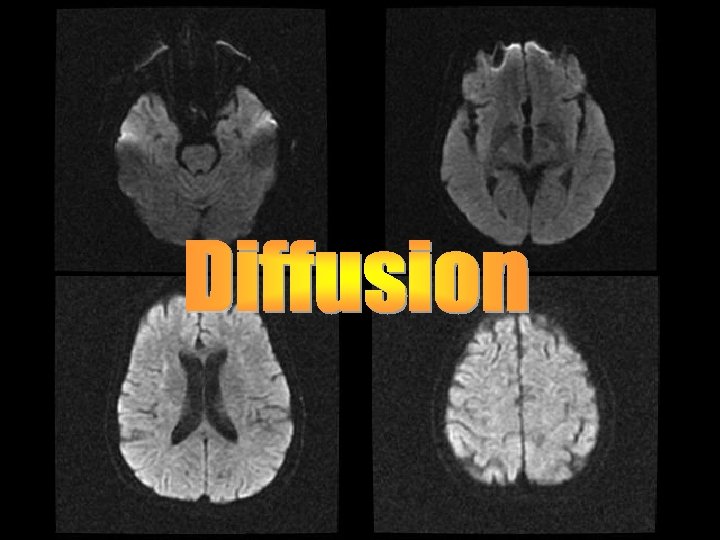
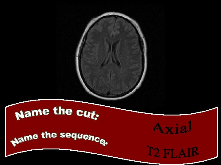
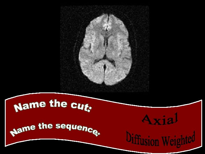
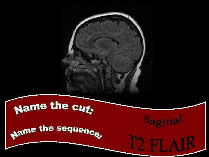
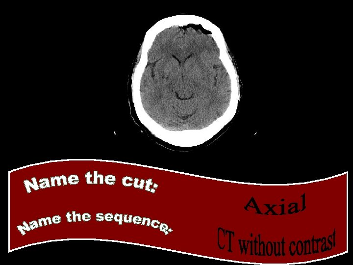
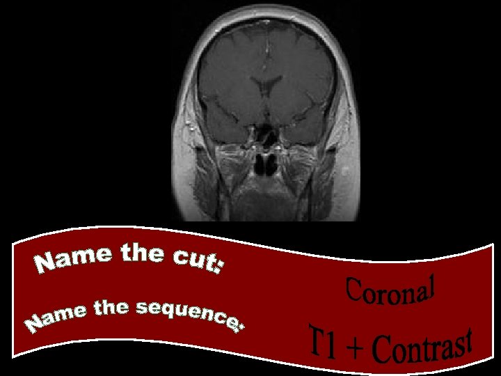
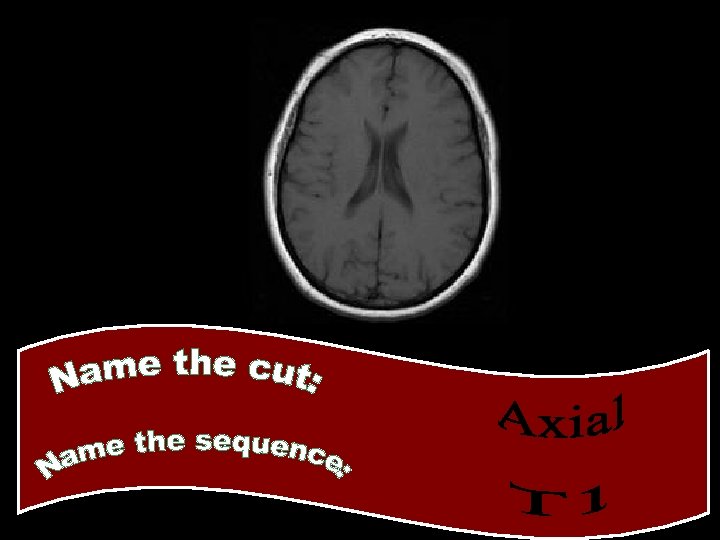
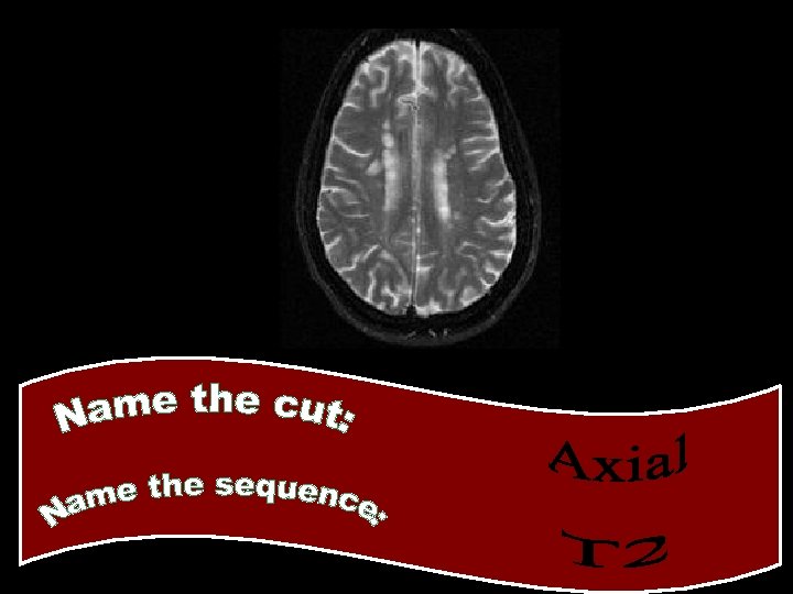
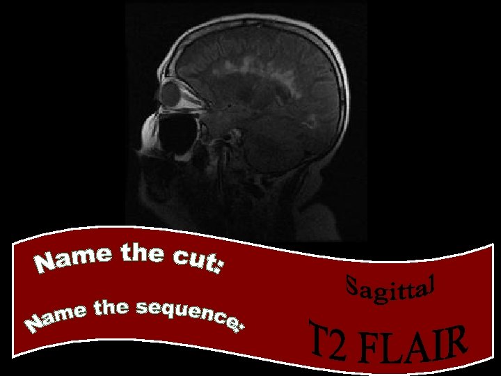
Goals • Identify axial, sagittal, and coronal images • Identify the following: – CT head without contrast – CT head with contrast – MRI brain • Identify the following MRI sequences – – – T 1 T 2 FLAIR T 1 + contrast Diffusion Weighted Images
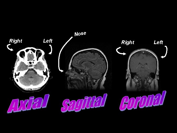
Nose Right Left

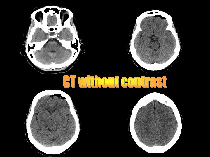
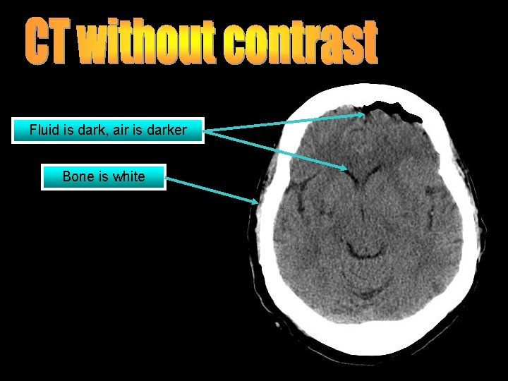
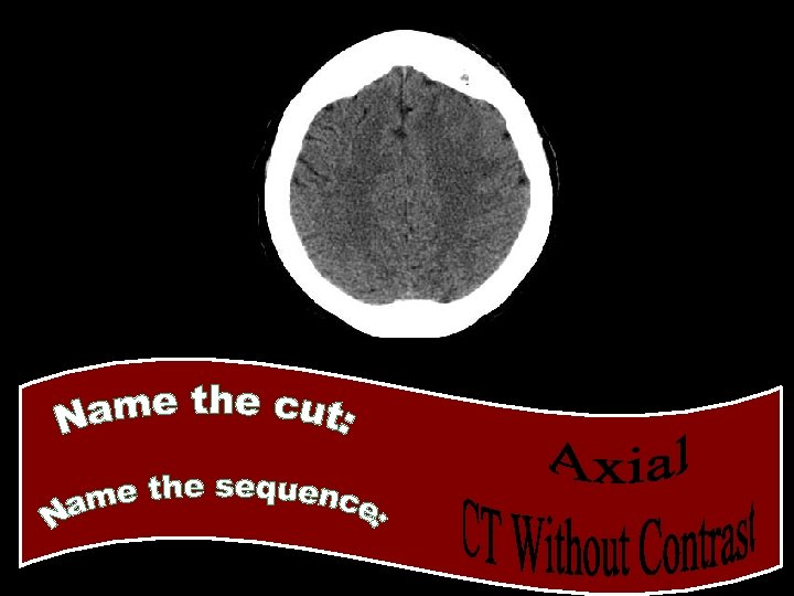
Fluid is dark, air is darker Bone is white
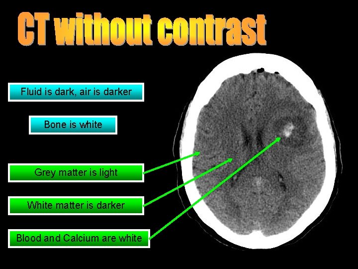
Fluid is dark, air is darker Bone is white Grey matter is light White matter is darker Blood and Calcium are white
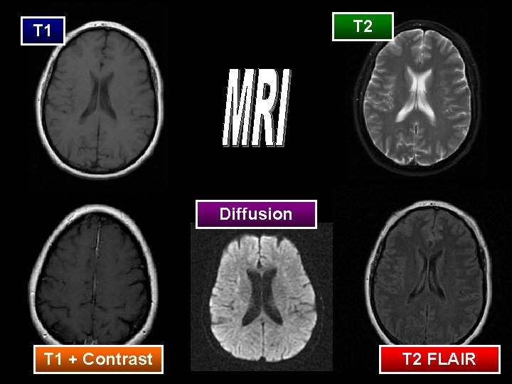
T 2 T 1 Diffusion T 1 + Contrast T 2 FLAIR
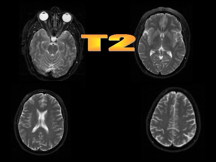
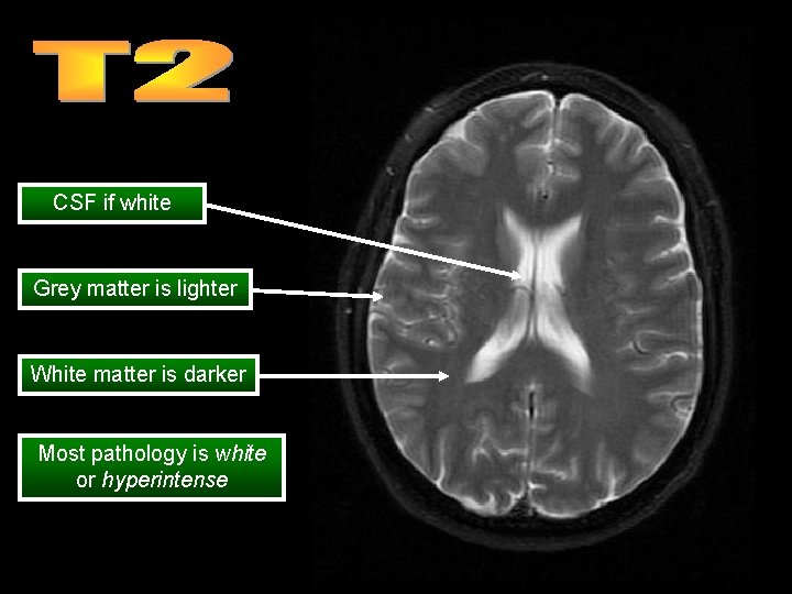
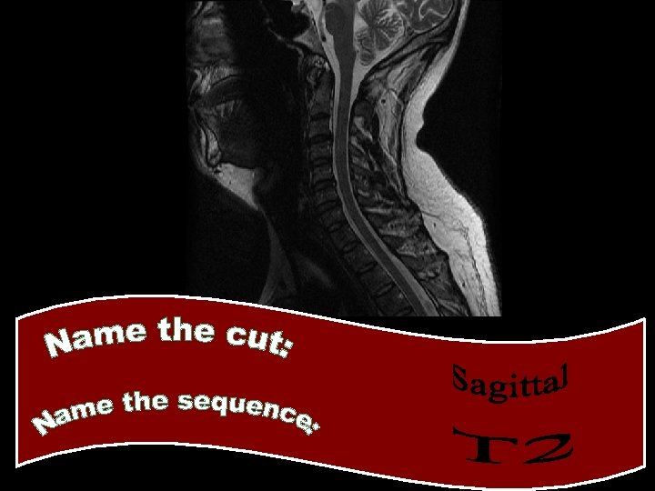
CSF if white Grey matter is lighter White matter is darker Most pathology is white or hyperintense
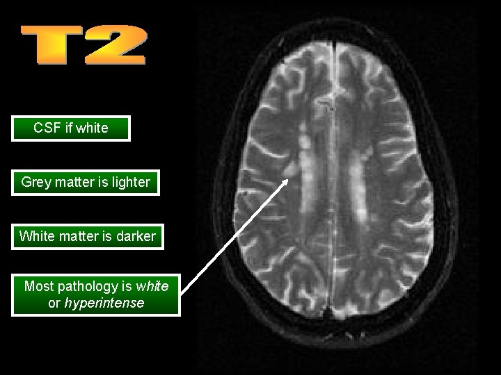
CSF if white Grey matter is lighter White matter is darker Most pathology is white or hyperintense
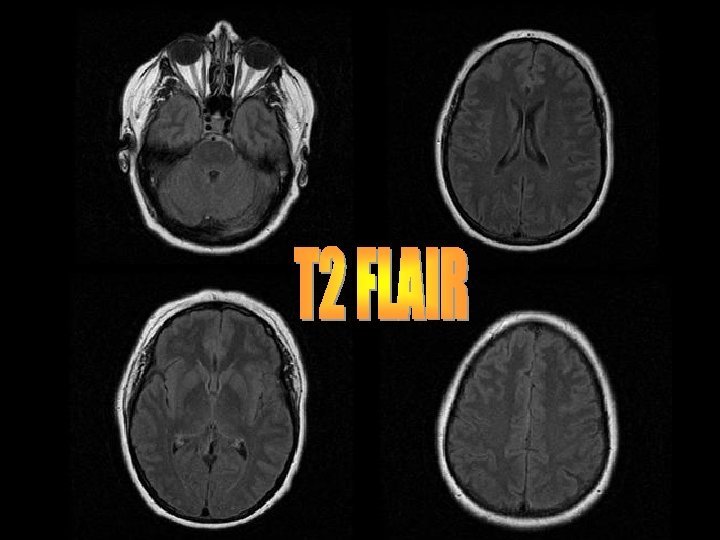
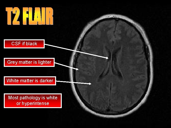
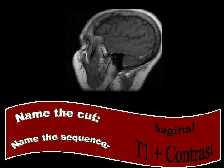
CSF if black Grey matter is lighter White matter is darker Most pathology is white or hyperintense
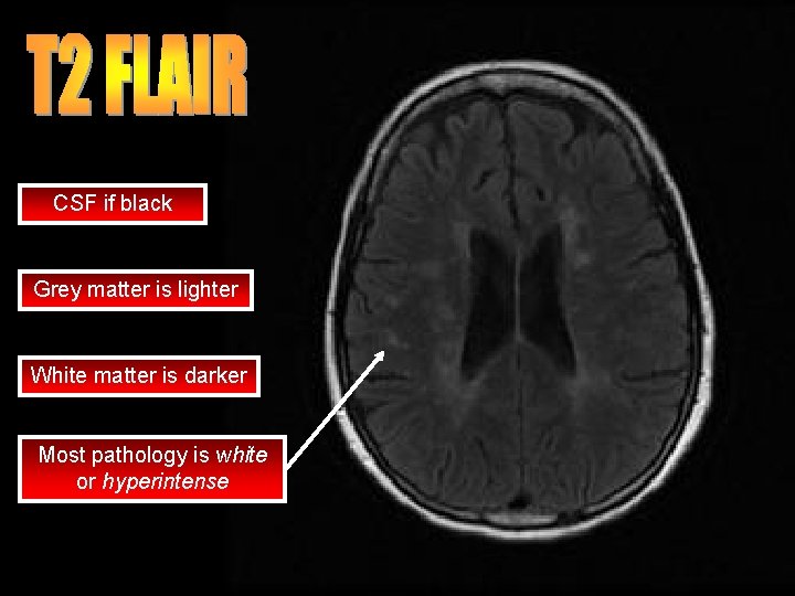
CSF if black Grey matter is lighter White matter is darker Most pathology is white or hyperintense
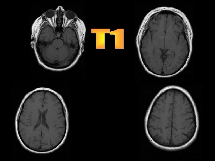
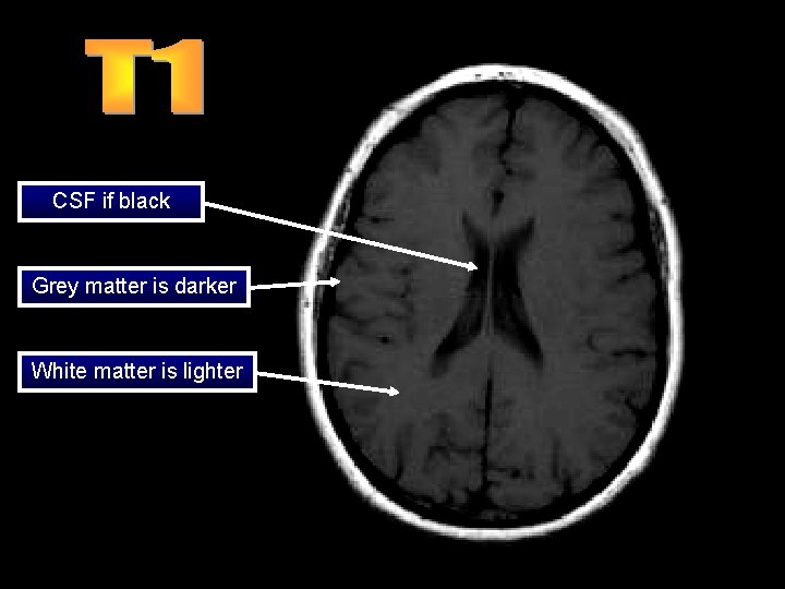
CSF if black Grey matter is darker White matter is lighter
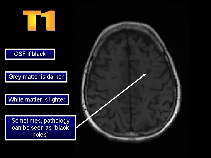
CSF if black Grey matter is darker White matter is lighter Sometimes, pathology can be seen as “black holes”
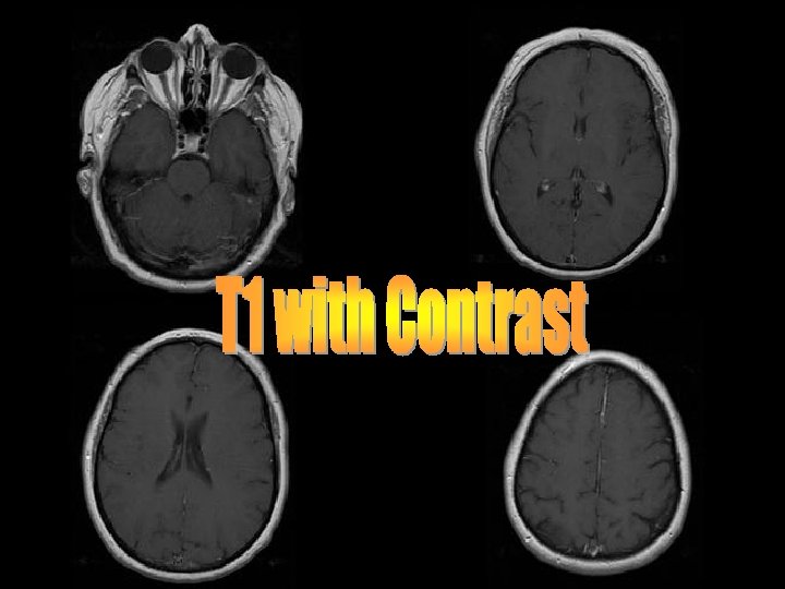
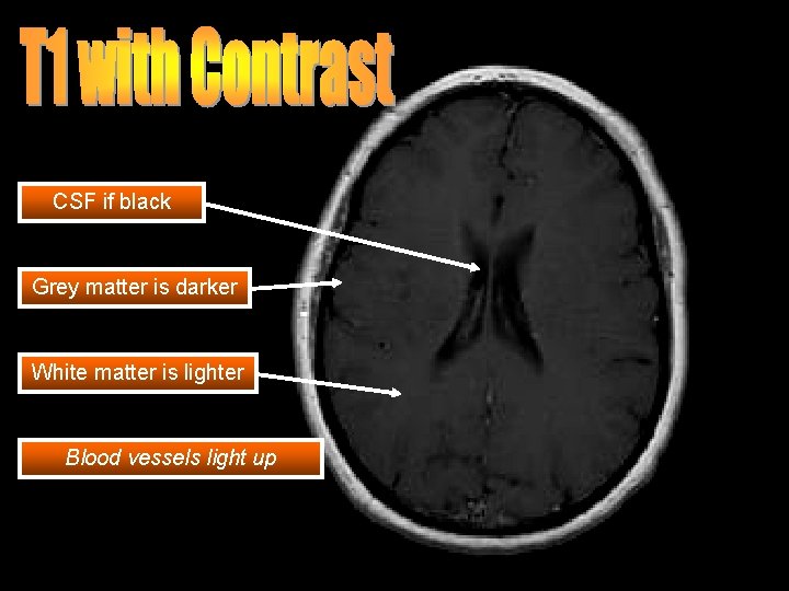
CSF if black Grey matter is darker White matter is lighter Blood vessels light up
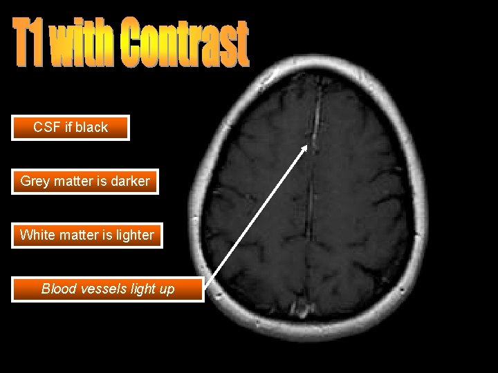
CSF if black Grey matter is darker White matter is lighter Blood vessels light up
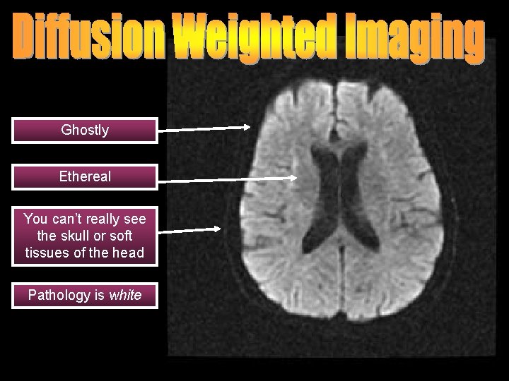
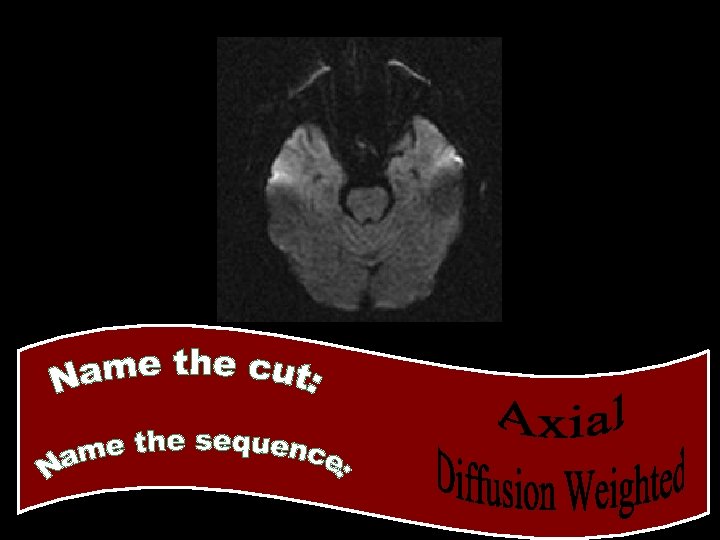
Ghostly Ethereal You can’t really see the skull or soft tissues of the head Pathology is white
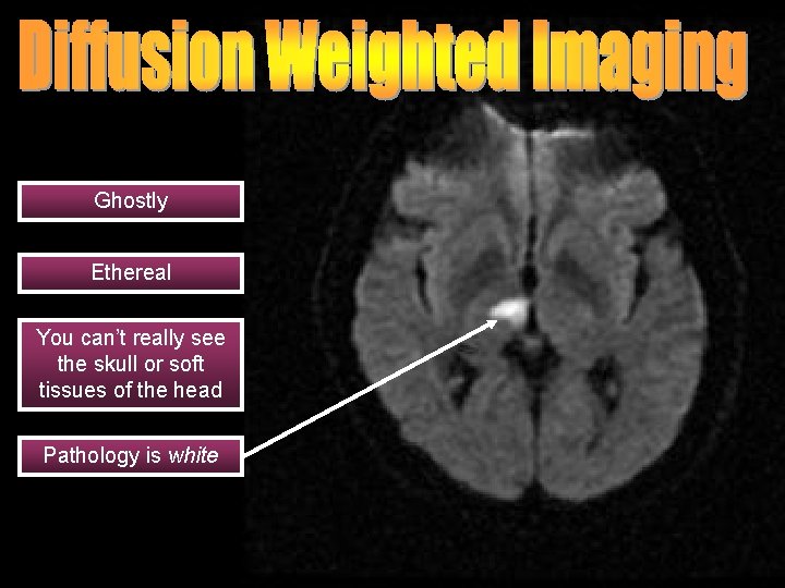
Ghostly Ethereal You can’t really see the skull or soft tissues of the head Pathology is white
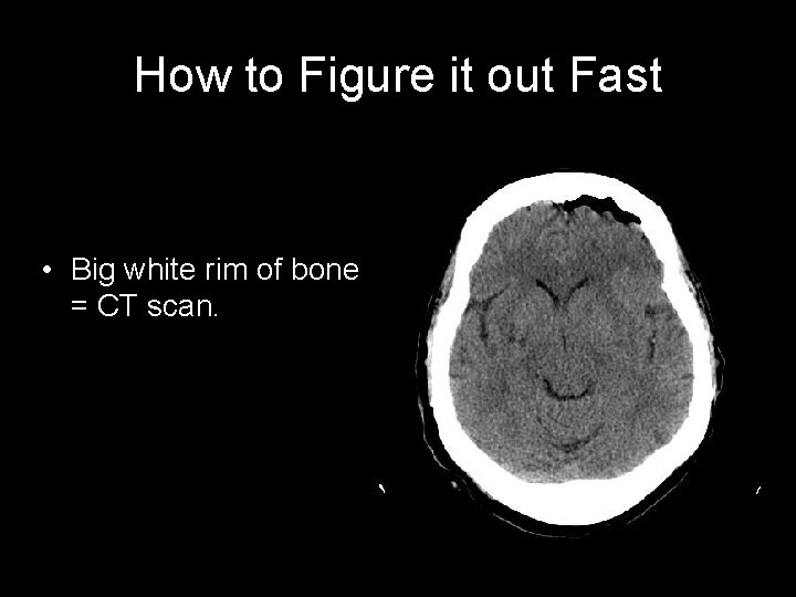
How to Figure it out Fast • Big white rim of bone = CT scan.
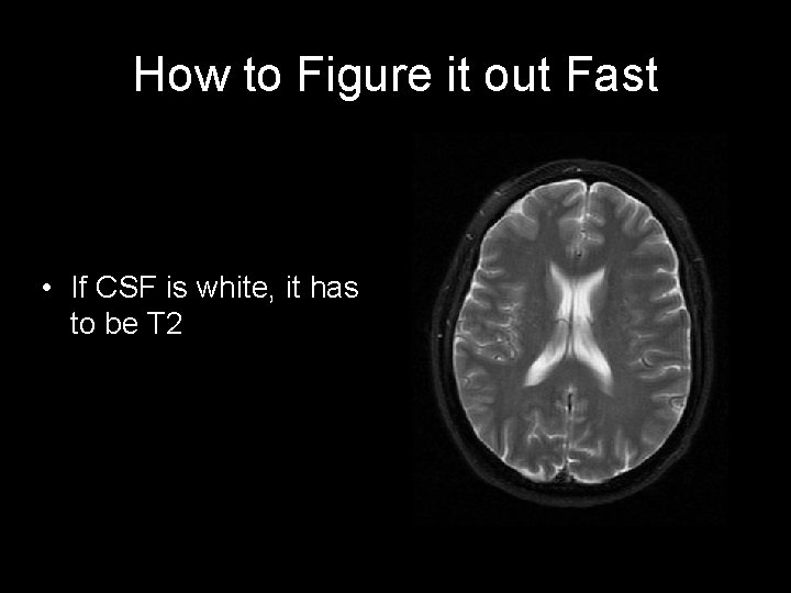
How to Figure it out Fast • If CSF is white, it has to be T 2
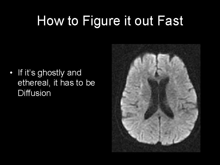
How to Figure it out Fast • If it’s ghostly and ethereal, it has to be Diffusion
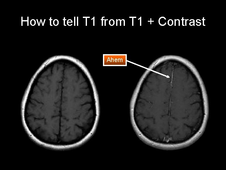
How to tell T 1 from T 1 + Contrast Ahem
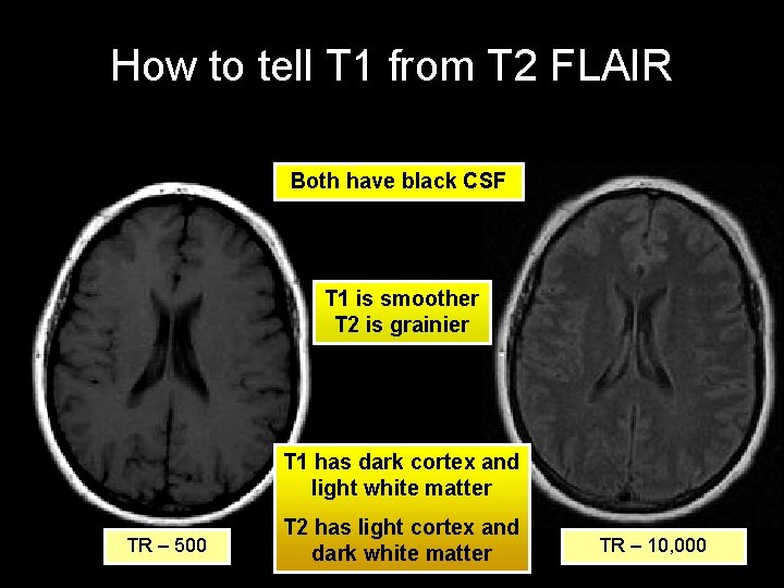
How to tell T 1 from T 2 FLAIR Both have black CSF T 1 is smoother T 2 is grainier T 1 has dark cortex and light white matter TR – 500 T 2 has light cortex and dark white matter TR – 10, 000

We Hope You Enjoyed

- Slides: 44