
By Tiffany Shaw MBCh B II 2002 Anaemia
By Tiffany Shaw MBCh. B II 2002
Anaemia – Definition w Reduced Hb concentration in blood w Compared to normal range for the particular gender + age w NOT reduced red cell count (unlike leucopenia or thrombocytopenia, which is defined by low counts)
Symptoms + Signs Symptoms: Signs: w w w w w SOB on exertion Tiredness Headache Depends on Angina cause of anaemia Pallor Tachycardia +/- Jaundice +/- Koilonychia +/- Splenomegaly +/- Sore corners of mouth
Symptom + Signs w w w Koilonychia = nail change, usually seen in Fe deficiency. Nails become brittle, spoon shaped, and ridged. Sore mouth corners = angular cheilosis, seen in Fe, B 12, or folate deficiency. Jaundice = due to increased bilirubin production as a result of increased haemolysis (haemolytic anaemia)
Investigation for Suspected Anaemia Full Blood Count 1. 2. 3. 4. 5. 6. 7. 8. Hb concentration Haematocrit Red cell count MCV (mean cell volume) MCH (mean cell haemoglobin) White cell count Differential white cell count (neutrophils, lymphocytes, monocytes, eosinophils, basophils) Platelet count
Investigation for Suspected Anaemia 1. 2. Hb concentration: confirms anaemia + assesses severity MCV: is this microcytic, normocytic, or macrocytic? Microcytic = MCV < normal (76) Normocytic = MCV normal (76 – 96) Macrocytic = MCV > normal (> 96) 3. 4. MCH: is this normochromic or hypochromic? Other cell counts: is this isolated anaemia or pancytopenia?
Classification Based on MCV Microcytic ~ Normocytic ~ Macrocytic ~ • Fe deficiency • Chronic disease • B 12 deficiency • Thalasaemia • Haemolysis • Folate deficiency • Sideroblastic • Marrow disease • Liver disease • Chronic disease • Marrow disease • Haemolysis (MCV pushed up by reticulocytosis)
Investigating Microcytic Anaemia Clue One --- Iron Studies w Serun Iron (free Fe) w Ferritin (stored Fe) w Iron Binding Capacity IBC (transferrin) NB: Transferrin is the iron binding protein in blood and amount of transferrin reflects capacity to bind Fe.
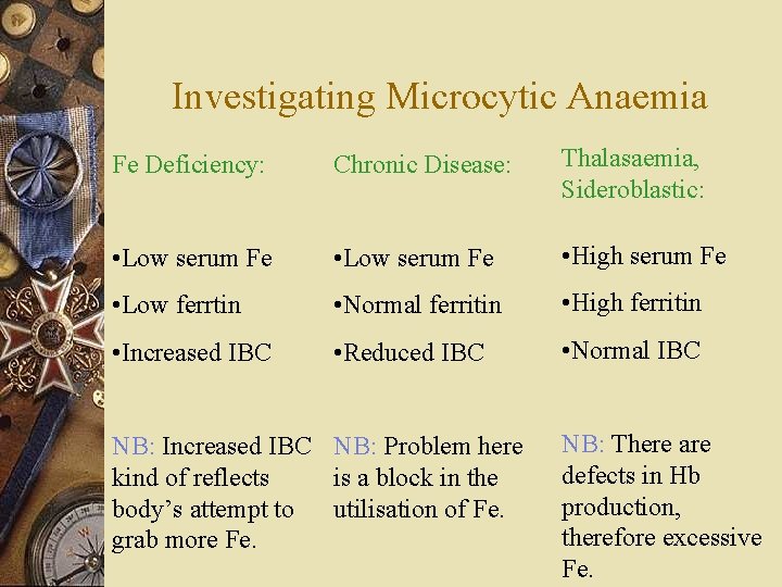
Investigating Microcytic Anaemia Fe Deficiency: Chronic Disease: Thalasaemia, Sideroblastic: • Low serum Fe • High serum Fe • Low ferrtin • Normal ferritin • High ferritin • Increased IBC • Reduced IBC • Normal IBC NB: Increased IBC NB: Problem here kind of reflects is a block in the body’s attempt to utilisation of Fe. grab more Fe. NB: There are defects in Hb production, therefore excessive Fe.
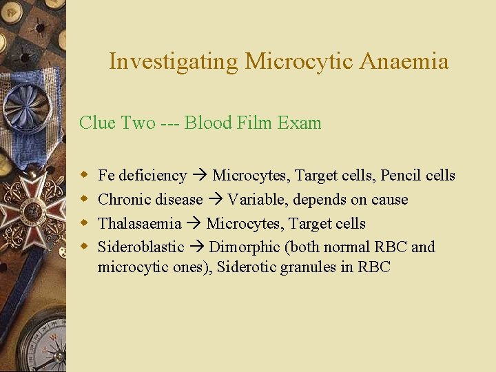
Investigating Microcytic Anaemia Clue Two --- Blood Film Exam w w Fe deficiency Microcytes, Target cells, Pencil cells Chronic disease Variable, depends on cause Thalasaemia Microcytes, Target cells Sideroblastic Dimorphic (both normal RBC and microcytic ones), Siderotic granules in RBC
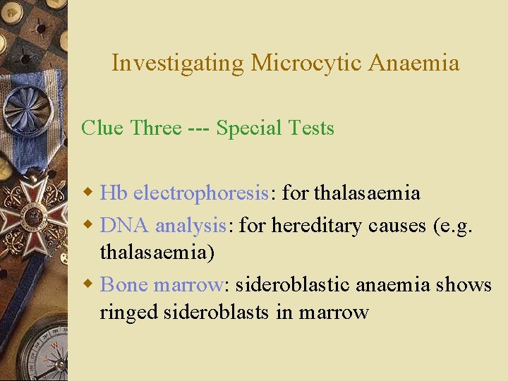
Investigating Microcytic Anaemia Clue Three --- Special Tests w Hb electrophoresis: for thalasaemia w DNA analysis: for hereditary causes (e. g. thalasaemia) w Bone marrow: sideroblastic anaemia shows ringed sideroblasts in marrow
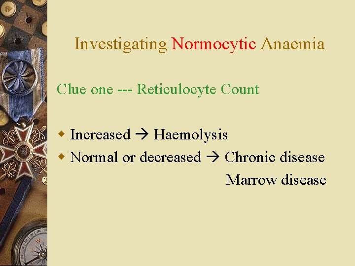
Investigating Normocytic Anaemia Clue one --- Reticulocyte Count w Increased Haemolysis w Normal or decreased Chronic disease Marrow disease
Investigating Normocytic Anaemia Clue Two --- Other Cell Counts w Normal Chronic disease probably w Abnormal Marrow disease likely
Investigating Normocytic Anaemia Clue Three --- Special Tests w Bone marrow biopsy for ? marrow diseases w Other tests for ? chronic diseases 1. U + E 2. Creatinine 3. LFT 4. Thyroid function …etc.
Investigating Macrocytic Anaemia Clue One --- Reticulocyte Count w Increased Haemolysis w Normal or decreased B 12 deficiency Folate deficiency Liver disease Marrow disease
Investigating Macrocytic Anaemia Clue Two --- B 12 / Folate Level w Low Deficiency w Normal Liver disease Marrow disease NB: For B 12, folate, and Fe deficiency, further investigations must be made to determine the cause of deficiency, so that appropriate therapy can be instituted.
Investigating Macrocytic Anaemia Clue Three --- Other Cell Counts w Normal liver disease probably (confirm with LFT, liver u/s…etc. ) w Abnormal probably marrow disease (confirm with marrow biopsy…etc. )
Fe Deficiency Physiology: w Source of Fe = both in animal + vegetable products w Animal Fe more easily absorbed w Site of absorption = proximal jejunum w Vit C enhances absorption w Daily loss = urine, skin, faeces, hair, menstrual blood loss
Fe Deficiency Findings associated with Fe deficiency: w w w Anaemia (microcytic) Koilonychia Angular cheilosis Glossitis Hair thinning Abnormal Fe studies (see before)
Fe Deficiency Causes: 1. Blood loss • Menorrhagia • GI bleed • Haematuria …etc. . 2. Increased demand • Pregnancy • Growth 3. 4. Inadequate intake Malabsorption • Gastrectomy • Coeliac disease
Fe Deficiency Mx: w Treat underlying cause if possible (e. g. bleed) w Oral Fe replacement – Ferrous sulphate best, gluconate second w Taken before meals TDS w Continue for 4 -6/12 w Reticulocytosis typically begins 7 days after Rx w Fe replacement IV or IM if oral not possible
Thalasaemia w Hereditary anaemia w Impaired production of components of the globin w Classified into a and b depending on which chain is deficient w Lack of a chain = a thalasaemia w Lack of b chain = b thalasaema
Thalasaemia a b w Impaired production of a chain in the globin. w Severity depends on number of genes deleted w 4/4 = hydrops fetalis (intrauterine death) w 3/4 = haemoglobin H disease (severe anaemia) w 1 - 2/4 = a trait (mild anaemia) w Impaired production of b chain in the globin w Major = complete failure of production (severe anaemia) w Intermedia = partial failure (moderate anaemia) w Minor = asymptomatic or mild anaemia
Thalasaemia Diagnosis: Associated findings: w Microcytic anaemia w Blood film examination w Hb electrophoresis w DNA analysis w Usual signs of anaemia w Failure to thrive w Hepatosplenomegaly w Expansion of the skull w High serum + storage Fe
Thalasaemia Mx: w w w Transfusion (packed red cells) prn Iron chelation therapy with DFX Splenectomy prn Bone marrow transplantation (definitive Rx) Genetic counseling
Sideroblastic Anaemia w Defect of haem synthesis w Increased Fe present as granules arranged in a ring around nucleus of developing RBC in marrow (ringed sideroblasts) w Peripheral blood shows microcytic hypochromic RBC w May be hereditary or acquired
Sideroblastic Anaemia Causes: 1. 2. 3. 4. Hereditary type (X-linked) Alcohol Radiation therapy Lead poisoning
Sideroblastic Anaemia Mx: w Transfusion prn w Iron chelation therapy w Some benefit from Vit B 6 (esp the hereditary type)
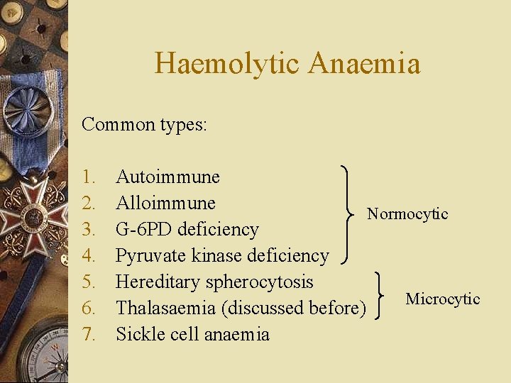
Haemolytic Anaemia Common types: 1. 2. 3. 4. 5. 6. 7. Autoimmune Alloimmune Normocytic G-6 PD deficiency Pyruvate kinase deficiency Hereditary spherocytosis Microcytic Thalasaemia (discussed before) Sickle cell anaemia
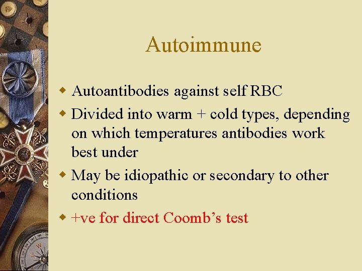
Autoimmune w Autoantibodies against self RBC w Divided into warm + cold types, depending on which temperatures antibodies work best under w May be idiopathic or secondary to other conditions w +ve for direct Coomb’s test
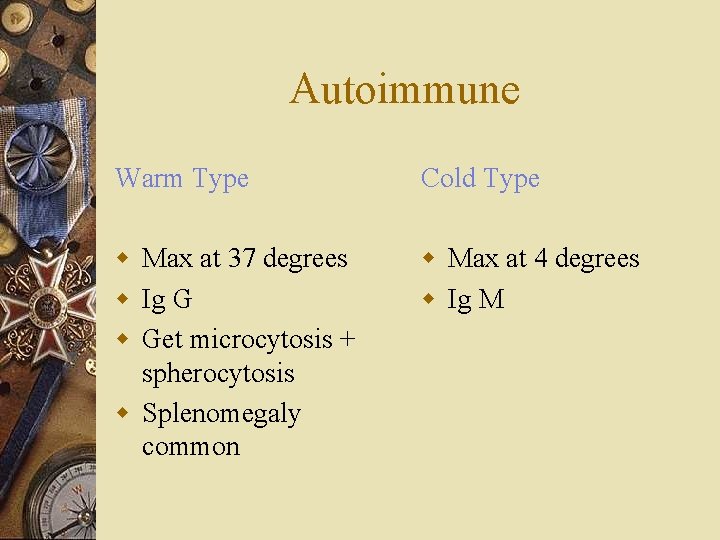
Autoimmune Warm Type Cold Type w Max at 37 degrees w Ig G w Get microcytosis + spherocytosis w Splenomegaly common w Max at 4 degrees w Ig M
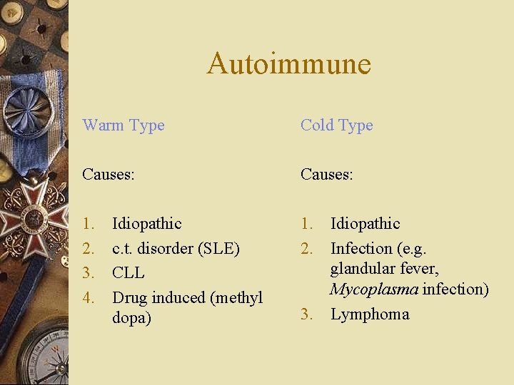
Autoimmune Warm Type Cold Type Causes: 1. 2. 3. 4. 1. Idiopathic 2. Infection (e. g. glandular fever, Mycoplasma infection) 3. Lymphoma Idiopathic c. t. disorder (SLE) CLL Drug induced (methyl dopa)
Autoimmune Warm type Cold type Mx: 1. Steroids 2. Transfusion prn 3. Splenectomy if steroids fail 4. Other immunosuppressives 1. Avoid cold environment 2. Immunosuppressives (chlorambucil, cyclophosphamide) 3. Transfusion prn
Alloimmune w Mismatched blood transfusion w Haemolytic disease of the newborn w Post bone marrow transplantation w Post organ transplantation
Hereditary Spherocytosis w Hereditary anaemia w Most common one in Caucasian w Autosomal dominant w Defect in RBC membrane w RBC becomes spherical w Therefore destroyed prematurely in spleen w Splenomegaly common
Hereditary Spherocytosis Special test --- Osmotic Fragility Test w Spherical RBC increased volume : surface area ratio w More prone to lysis by osmosis w Measure degree of lysis of RBC in different concentrations of saline spherical ones lyse more w Therefore confirm spherocytosis
Hereditary Spherocytosis Mx: w Splenectomy to stop destroying RBC prematurely
G-6 PD Deficiency w G-6 PD = enzyme required to generate ATP to maintain shape of RBC w Deficiency RBC more susceptible to oxidation stress, e. g. ingestion of certain drugs / fava beans w Haemolysis occurs with such ingestion w Normal at other times
6 -GPD Deficiency Mx: w Stop offending drugs / fava beans w Transfusion prn during attacks
Vit B 12 Deficiency Physiology: w Source of B 12 = food of animal origin w Storage = in liver, enough for 2 – 4 years w Absorption = in terminal ileum 1. Vit B 12 ingested 2. Binds to “R protein” in saliva, gastric juice 3. IF secreted by parietal cells releases Vit B from R protein 4. Form IF-Vit B 12 complex 5. Complex absorbed in terminal ileum 6. Transcobolamin in plasma strips off Vit B 12 and transport to tissues
Vit B 12 Deficiency Diagnosis: Associated findings: w Macrocytic anaemia w Low B 12 level w w w Features of anaemia Mild jaundice Glossitis Angular cheilosis Neuropathy (often tingling in feet +/- visual or psych disturbance)
Vit B 12 Deficiency Causes: w Inadequate intake (e. g. vegan) w No IF (gastrectomy, pernicious anaemia) w Malabsorption (ileal resection, Crohn’s disease)
Vit B 12 Deficiency Pernicious Anaemia: w Autoandibodies develop against parietal cells or intrinsic factors w Therefore no IF w Can’t absorb Vit B 12 w Peak incidence at 60 years old
Vit B 12 Deficiency Distinguish between no IF & ileal problems: 1. Hx (e. g. gastrectomy) 2. Test for autoantibodies against parietal cells or IF 3. Radioactive Vit B 12 study Give radio-labelled Vit B 12 +/- IF. IF corrects gastric problems, but not ileal problems.
Vit B 12 Deficiency Mx: 1. Correct underlying cause if possible (e. g. adequate intake) 2. IM Vit B 12 injection 3. 6 injections to replace deficit (1 every 2 – 3 days) 4. Maintenance (1 injection 3/12)
Folate Deficiency Physiology: w Source = green vegetables + animal liver w Body store = 4/12 supply w Absorption = through proximal jejunum
Folate Deficiency Diagnosis: Associated findings: w Macrocytic anaemia w Low folate level w w w Features of anaemia Mild jaundice Glossitis Angular cheilosis No neuropathy!!
Folate Deficiency Cause: 1. Inadequate intake 2. Malabsorption (coeliac disease, bowel resection) 3. Drug induced (anti-convulsant)
Folate Deficiency Mx: 1. Treat underlying cause if possible (e. g. coeliac disease) 2. Folate replacement 3. Oral folate 5 mg OD for 4 -6/12
- Slides: 49