By Dr Manjula vastrad Asst Prof Dept of

By Dr. Manjula vastrad Asst Prof Dept of Rachana Shareera SMVVS RKM AMC Vijayapur Email : 1 6/20/2021 manjula. prasad 2010@gmail. com

� Arise from the brain & pass through or into the cranial bones (thus cranial nerves) � Each cranial nerve has both a number, designated by a roman numeral, and a name. . � Numbered I to XII roughly in order from anterior to posterior, in which the nerves arise from the brain. � The names designate a nerve’s distribution or function. . � All the nerves are distributed in the head and neck except the X , which also supplies the structures of thorax & abdomen 6/20/2021 2

6/20/2021 3

OLFACTORY NERVE OPTIC NERVE OCCULOMOTOR NERVE TROCHLEAR NERVE TRIGEMINAL NERVE ABDUCENS NERVE FACIAL NERVE VESTIBULOCOCHLEAR NERVE GLOSSOPHARYNGEAL NERVE VAGUS NERVE ACCESSARY NERVE HYPOGLOSSAL NERVE 6/20/2021 4

6/20/2021 5
 carry axons of sensory neurons and thus")
� Three cranial nerves (I, II &VIII) carry axons of sensory neurons and thus called special sensory nerves and are associated with the special senses of smelling, seeing and hearing. . � Five cranial nerves(III, IV, VI, XI &XII) are classified as motor nerves because they contain only axons of motor neurons. . � The remaining four cranial nerves(V, VII, IX & X)are mixed nerves-they contain axons of both sensory neurons entering the brain stem and motor neurons leaving the brainstem. 6/20/2021 6

CRANIAL NERVES: CLASSIFICATION SENSORY/ MOTOR/ AFFERENT EFFERENT OLFACTORY OCCULOMOTOR TROCHLEAR MIXED TRIGEMINAL FACIAL OPTIC ABDUCENS GLOSSOPHARYNGEAL ACCESSARY VAGUS VESTIBULO COCHLEAR HYPOGLOSSAL 6/20/2021 7

6/20/2021 8

v Component: Sensory v Function: Smell v Origin: Olfactory receptor nerve cells v Opening to the Skull: Openings in cribriform plate of ethmoid bone. 6/20/2021 9

6/20/2021 10

6/20/2021 11

6/20/2021 12
 Ø Cerebrospinal")
CLINICAL ANATOMY Ø Anosmia -Loss of smell (injury to anterior cranial fossa) Ø Cerebrospinal fluid rhinorrhoea 6/20/2021 13

6/20/2021 14

q Component: Sensory q Function: Vision q Origin: Back of the eyeball q Opening to the Skull: Optic Canal 6/20/2021 15

6/20/2021 16

6/20/2021 17

6/20/2021 18

§ Component: Motor § Origin: Anterior surface of the midbrain § Opening to the Skull: Superior orbital fissure § Function: § Raises upper eyelid § Turns eyeball upward, downward and medially § Constricts pupil § Accommodates the eye 6/20/2021 19

6/20/2021 20
Superior, Inferior & Medial Recti Muscles 2)Inferior oblique")
Innervates : - Extra-Occular Muscles : 1)Superior, Inferior & Medial Recti Muscles 2)Inferior oblique Muscle - Also Levator Palpabre Superioris

It has an accessory nucleus situated medial to the main nucleus, it is called as EW nucleus/Edinger Westphal nucleus, it is parasympathetic nucleus for sphincter pupillae. Constrict the pupil and accommodate the lens. The fibers travel in the IIIrd nerve, so damage to that nerve will also produce a dilated pupil.

Lateral strabismus- medial rectus is paralysed & lateral rectus is unopposed. Inability to move the eye medially laterally. Ptosis – as ipsilateral levator palpabre superioris is paralysed. Mydriasis (dilated pupil of affected side) and unresponsiveness to light as the sphincter pupillae is non- functional and 6/20/2021 23

PTOSIS MYDRIASIS STRAMISMUS

6/20/2021 25

ü Component: Motor ü Origin: Posterior surface of the midbrain ü Opening to the Skull: Superior orbital fissure ü Supply : Superior oblique muscle ü Function: Assisting in turning eyeball downward and laterally 6/20/2021 26

6/20/2021 27

v UNIQUE FEATURES: ü It is the only cranial nerve attached to the dorsal aspect of the brainstem (exits the brainstem dorsally) ü It is the thinnest & shortest and is particularly vulnerable to traumatic injury.

� Eye points superolaterally. � Attempted movements in these directions (e. g. , reading or walking down stairs) may cause diplopia. � This condition often causes vertical double vision as the weakened muscle prevents the eyes from moving in the same direction together. 6/20/2021 29

V 1. V 2. V 3. Ophthalmic Nerve Maxillary Nerve Mandibular Nerve 6/20/2021 30

It is the largest cranial nerve. Composed of three divisions � Ophthalmic (V 1) � Maxillary (V 2) � Mandibular (V 3) Conveys sensory impulses from various areas of the face (V 1) and (V 2), and supplies motor fibers (V 3) for mastication. Fibers run from the face to the pons via the Superior Orbital Fissure (V 1), Foramen Rotundum (V 2), and the Foramen Ovale (V 3) 6/20/2021 31

6/20/2021 32

v Component: Sensory v Origin: Anterior aspect of the pons v Opening to the Skull: Superior orbital fissure v Function: v Cornea v Skin of forehead v Scalp v Eyelids and nose v Mucuos membranes of paranasal sinuses and nasal cavity 6/20/2021 33

6/20/2021 34

o Component: Sensory o Origin: Anterior aspect of the pons o Opening to the Skull: Foramena rotundum o Function: o Skin of the face over maxilla o Teeth of the upper jaw o Mucous membrane of the nose, the maxillary sinus and palate 6/20/2021 35

o Component: Mixed o Origin: Anterior aspect of the pons o Opening to the Skull: Foramen ovale Motor Function: o Muscles of mastication i. e. Massater $ Temporalis muscles. Sensory Function: • Skin of cheek • Skin over mandible and side of head • Teeth of lower jaw and TMJ • Mucous membrane of mouth and anterior part of tongue 6/20/2021 36

6/20/2021 37

6/20/2021 38

Trigeminal neuralgia: is a sensory disorder of CN V that is characterized by sudden attacks of excruciating, lightening like jabs of facial pain. A paraoxysm (sudden sharp pain) can last for 15 mins or more. � The cause of trigeminal neuralgia is unknown , but some investigators believe that it can be due to a anomalous blood vessel that compresses the nerve. � � Injury leads to inability to bite down and chew, inability to close jaw. � In severe cases, nerve is cut; relieves agony but results in loss of sensation on that side of the face. 6/20/2021 39

6/20/2021 40

6/20/2021 41

ü Component: Motor ü Origin: Anterior Surface of hindbrain between pons and medulla ü Opening to the Skull: Superior orbital fissure ü Function: supply Lateral rectus muscle, turns eyeball laterally 6/20/2021 42

Medial strabismus: the affected eye deviates medially by the unopposed action of the lateral rectus muscle. The individual may be able to move the affected eye to the midline, but no further, by relaxing the medial rectus muscle.

6/20/2021 44

ü Component: Mixed ü Origin: Anterior Surface of hindbrain between pons and medulla üOpening in the skull : Internal acoustic meatus, Facial canal, Stylomastoid foramen ü Motor functions: o Facial expression o Transmit parasympathetic impulses to lacrimal and salivary glands (submandibular and sublingual glands) ü Sensory function is taste from taste buds of anterior twothirds of the tongue 6/20/2021 45
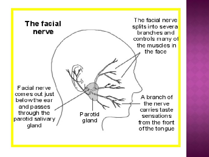
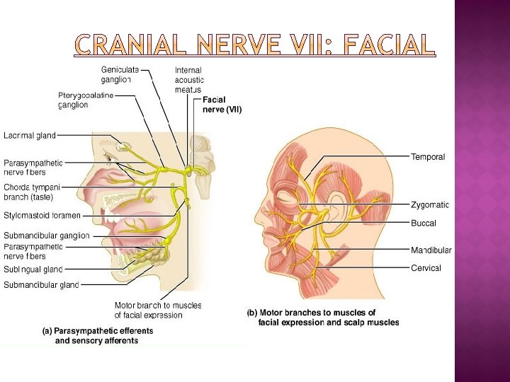

q Bell’s palsy: paralysis of facial muscles on affected side. q loss of taste sensation q Lower eyelid droops q Corner of mouth sags q Tears drip continuously and eye cannot be completely closed 6/20/2021 48

6/20/2021 49

6/20/2021 50

Other Names : Auditory / Acoustic Nerve Component : Sensory Origin o Vestibular – Saculae/saccule/semicircular canals o Cochlear – Organ of Corti Opening to the Skull: Internal acoustic meatus Function: o Vestibular – Saculae/saccule/semicircular canals – Balance position of head o Cochlear – Organ of Corti – Hearing 6/20/2021 51
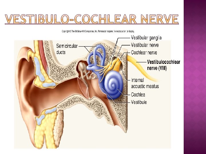

ØDamage produces: Deafness Dizziness Nausea Loss of balance Vertigo

6/20/2021 54

Component : Mixed Origin: Medulla oblongata Opening to the Skull: Jugular foramen Function: • Motor Stylopharyngeus muscle – assists swallowing • Sensory: carries sensory signals from tympanic cavity, auditory tube, posterior third of the tongue, pharynx, including the soft palate, tonsil. • Also supply to Carotid sinus. • Secretomotor: Parotid gland 6/20/2021 55

6/20/2021 56

� Complete lesion of the Glossopharyngeal nerve results in the following : 1. 2. 3. Loss of taste and common sensations over the posterior one third of the tongue. Difficulty in swallowing Loss of salivation from the parotid gland

6/20/2021 58

Vagus comes from the Latin word meaning “wandering. ” Component : Mixed Nerve( Longest Cranial Nerve) Origin: Medulla Oblongata Opening In The Skull: Jugular Foramena 6/20/2021 59

Function: Sensory: ü carries sensory information from skin at the back of the ear and in the external acoustic meatus, part of the external surface of the tympanic membrane, the Pharynx, larnyx, trachea, esophagus, thoracic & abdominal viscera Motor: ü One muscle of the tongue (palatoglossus). ü Muscles of the soft palate. ü Intrinsic muscles of larynx ü Pharynx (except stylopharyngeus) ü Smooth muscles of Esophagus, Stomach and Heart. Parasympathetic: ü To smooth muscle and glands of the pharynx, larynx, and thoracic and abdominal viscera 6/20/2021 60
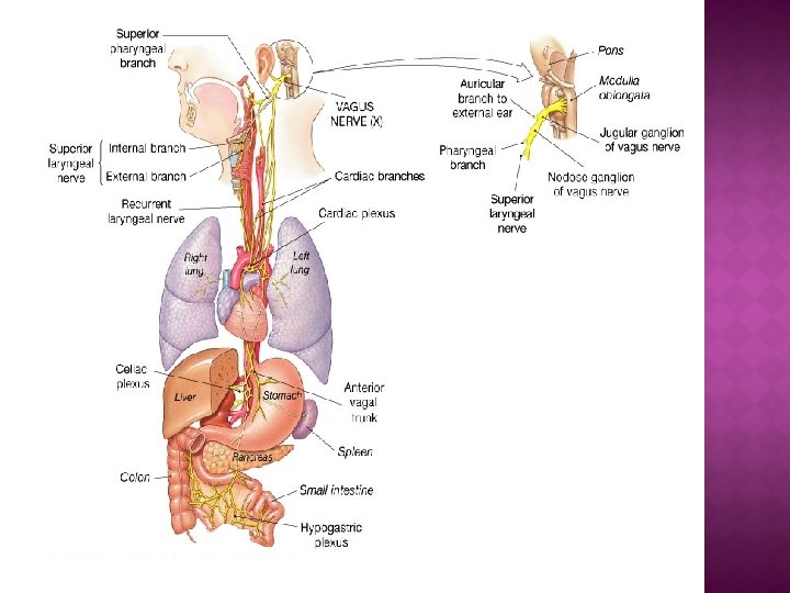

Lesion Of Vagus Nerve Leads To : Ø Dysphagia Ø Total destruction incompatible with life

6/20/2021 63

6/20/2021 64

o Component: Motor o Origin: Cranial root emerging from the medulla and a spinal root arising from the superior region of the spinal cord(C 1 -C 6). Spinal root passes upward into the cranium via the foramen magnum o Opening in the skull: Jugular Foramena q. Function: v. Innervates the Trapezius and Sternocleidomastoid, which move the head and neck 6/20/2021 65
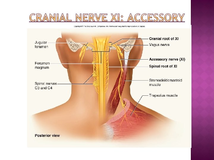

Lesion may result the followings : Ø Shoulder droop Ø Weakness turning head to opposite side. Ø Weakness & difficulty in raising the arm above horizontal

6/20/2021 68

ü Component: Motor ü Origin: Anterior Surface of medulla ü Opening to the Skull: Hypoglossal canal ü Function: Supply Extrinsic and Intrinsic muscles of the tongue, which contribute to swallowing and speech. ( Except Palatoglossus) 6/20/2021 69

6/20/2021 70

If damaged, difficulties in speech and swallowing. Inability to protrude tongue. The hypoglossal nerve accompanies the tonsillar artery on the laterial wall of the pharynx and this wall is vulnerable to injury during tonsillectomy.

6/20/2021 72
- Slides: 72