BVSc AH General Pathology CELL INJURY Dr Sanjiv
 CELL INJURY Dr. Sanjiv Kumar Assistant Professor, Deptt. of")
BVSc & AH (General Pathology) CELL INJURY Dr. Sanjiv Kumar Assistant Professor, Deptt. of Pathology, BVC, Patna

INTRODUCTION • Rudolph Virchow, 'Father of Cellular Pathology', put forward the concept that disease begins at the cellular level. • The normal cell lives in a hostile environment In other words, it exists in a state of striking disequilibrium with its external environment. For example, the concentration of calcium ions outside the cell is 10, 000 times higher than that inside. If all this calcium were to enter the cell, it will prove toxic, and kill the cell. This can happen during cell injury.

• It is the plasma membrane of the cell that constitutes the structural and functional barrier, which separates the intracellular from the hostile extracellular environment.

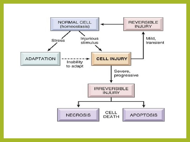
DEFINITIONS • The normal cell has to live within a fairly narrow range of function and structure. • Even then, 1 t is able to handle normal physiological demands, so-called normal homeostasis (L. staying same). • Somewhat more excessive physiological stresses, or some pathological stimuli, bring about adaptation. • That is, the cell modifies its structure and function in response to changing demands and stresses.

• It then acquires a new but steady state and preserves its health. • If the adaptive capability is exceeded, or in certain cases when adaptation is not possible, a sequence of regressive changes occurs, collectively known as cell injury (L. re, retro = back; gress = step, i. e. , step back, go back, fall in health; also called retrogressive or degenerative changes). • Within certain limits, injury is reversible and cells return to a normal state, but with severe and persistent stress, the cell reaches a 'point of no return', suffers irreversible injury and then dies.

Cellular Adaptation

• Adaptation, reversible injury, irreversible injury, and cell death are states of progressive encroachment on the cell's normal function and structure.
 is an extremely")
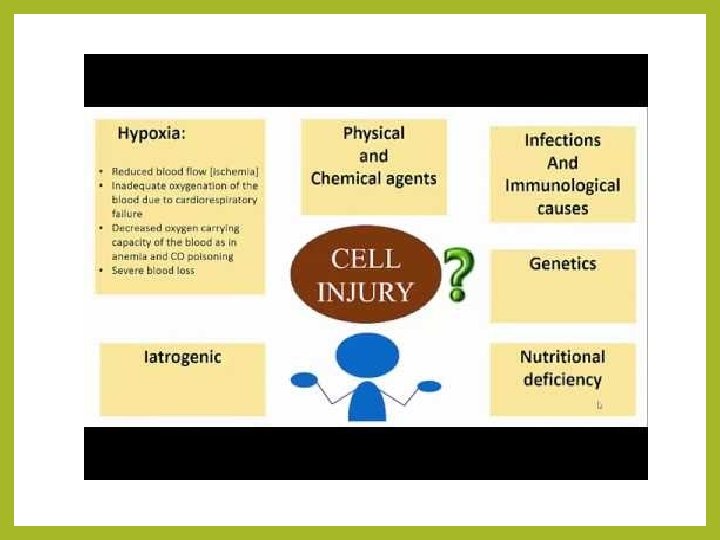
Causes of Cell Injury • Hypoxia: Hypoxia (loss of oxygen supply) is an extremely important and common cause of cell injury and cell death. It affects cells aerobic oxidative respiration. • Physical agents: Physical agents include mechanical trauma, extremes of temperatures (burns and deep cold), radiation, electric shock, and sudden changes in atmospheric pressure. • Chemical agents and drugs: Virtually any chemical substance or drug can cause cell injury.

• Infectious agents: These agents range from the submicroscopic viruses to the large tapeworms. In between are the bacteria, fungi, rickettsiae, chlamydiae, mycoplasma, protozoa, and higher forms of parasites. • Immunological reactions: Although the immune system serves in the defence against infectious agents, immune reactions may also cause cell injury. Examples include anaphylactic reaction to a foreign protein or a drug, and autoimmune diseases. • Nutritional imbalances: Nutritional deficiencies such as avitaminoses and others are important causes of cell injury. Ironically, excesses of nutrition are also important causes of morbidity and mortality. • Genetic defects: Genetic defects may cause cell injury. The genetic injury may result in a defect as gross as congenital malformations, or in as subtle a change as the single amino acid substitution in haemoglobin. S in sickle cell anaemia.

GENERAL CONSIDERATIONS Biochemical mechanisms of cell injury • The biochemical mechanisms responsible for cell injury and cell death are complex. All the same, there are certain principles that are applicable to most forms of cell injury. 1. The morphological changes of cell injury become noticeable only after some critical biochemical system within the cell has been deranged. Thus, the first lesion to develop is biochemical (molecular) in nature. This, in turn, causes structural changes first at an electron microscopical level (ultrastructural lesion), then light microscopic lesions develop, and these, when extensive, produce gross lesions.

2. The cellular response to injurious stimuli depends on the type of injury, its duration, and its severity. 3. The results of an injurious stimulus depend on the type, status, adaptability, and genetic make-up of the injured cell. The same injury has different result depending on the cell type. 4. Although the exact biochemical site of action for many injurious agents is difficult to determine, four intracellular systems are particularly exposed to attack:
 Cell membrane. It is on the maintenance of the integrity of cell membrane")
(1) Cell membrane. It is on the maintenance of the integrity of cell membrane that the ionic and osmotic homeostasis of the cell and its organelles depends, (2) Oxidative phosphorylation and production of adenosine triphosphate (ATP), (3) Synthesis of enzymic and structural proteins, and (4) Preservation of the integrity of genetic apparatus of the cell. The structural and biochemical components of a cell are so closely inter -related that, whatever the exact point of initial attack, injury at one site leads to wide-ranging secondary effects.

Common Biochemical Mechanisms • With many injurious stimuli the exact pathogenic mechanisms that lead to cell death are incompletely understood. In spite of this, there are several common biochemical pathways in the mediation of cell injury and cell death, whatever the causative agent. These include: ATP DEPLETION: High-energy phosphate in the form of ATP is required for many 1. processes within the cell. ATP is produced in two ways: i. The major route is oxidative phosphorylation of ADP, a reaction that requires oxygen, and ii. the second is the glycolytic pathway that can generate ATP in the absence of oxygen using glucose obtained either from body fluids, or from hydrolysis of glycogen. ATP depletion and decreased ATP synthesis are common consequences of both ischaemic and toxic injury.

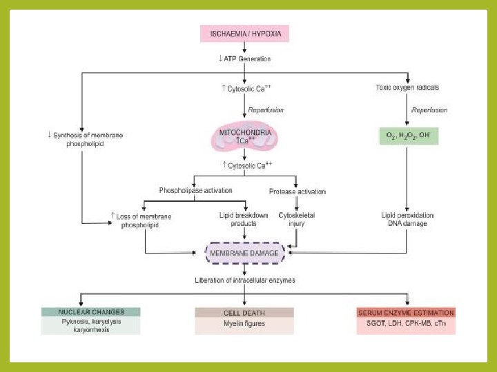
2. Lack of oxygen or generation of oxygen-derived free radicals: Partially reduced activated oxygen species are also important mediators of cell death. They are highly toxic molecules that can damage lipids, proteins, and nucleic acids. These molecules are referred to as activated or reactive oxygen species. 3. Loss of calcium homeostasis: Concentration of calcium in cytosol is up to 10, 000 times lower than the concentration of extracellular calcium, or of calcium sequestered (isolated) within mitochondria and endoplasmic reticulum. Ischaemia and toxins increase cytosolic calcium concentration due to a net influx (entry) of extracellular calcium (Ca++) through the plasma membrane, and also because of the release of Ca++ from mitochondria and endoplasmic reticulum. Increased cytosolic calcium, in turn, activates a number of enzymes, with harmful cellular effects.

• The enzymes activated by calcium include: i. phospholipases, which cause membrane damage, ii. proteases, which break down structural and membrane proteins, iii. ATPases, which accelerate ATP depletion, and iv. endonucleases, which break down nuclear chromatin. 4. Defects in membrane permeability: Changes in membrane permeability may also be secondary to a loss of ATP synthesis, or may result from calcium-mediated activation of phospholipases. 5. Mitochondrial damage: Since all cells depend on oxidative metabolism, mitochondrial integrity is most important for cell survival. Irreparable damage to mitochondria will ultimately kill cells.

Reversible and Irreversible Cell Injury The sequence of events in reversible and irreversible cell injury will be discussed by two model systems: (1) ischaemic and hypoxic injury, and (2) free radical-induced cell injury.

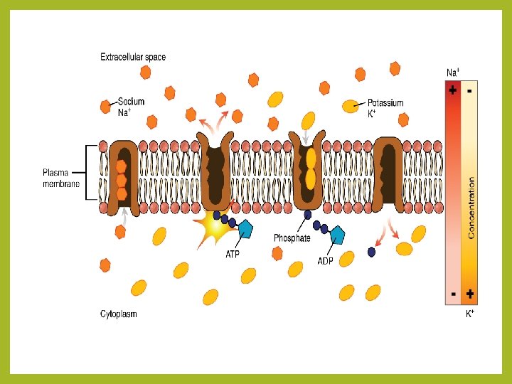
Reversible Cell lnjury Ischaemic and Hypoxic Injury • The first point of attack of hypoxia is the cell's aerobic respiration, that is, oxidative phosphorylation by mitochondria. • As the oxygen tension within the cell decreases, there is loss of oxidative phosphorylation and decreased generation of adenosine triphosphate (ATP). • Sodium is maintained at a higher concentration outside the cell than inside by an energy-dependent (ATP-driven) "sodium pump", that is, by Na+ and K+-adenosine triphosphatase (ATPase) enzyme. • This pump also keeps concentration of potassium significantly higher inside the cell than outside. • The decreased ATP concentration, following acute hypoxia, reduces activity of the plasma membrane "sodium pump“.

Ischaemic and Hypoxic Injury

Nuclear Change

Irreversible cell lnjury • Irreversible injury is associated morphologically with severe swelling of mitochondria, extensive damage to plasma membranes, and swelling of lysosomes. • Extracellular calcium enters into the cell. Large, amorphous, calcium-rich densities accumulate in the mitochondrial matrix. • After this, there is continued loss of proteins, essential coenzymes, and ribonucleic acids from the hyperpermeable plasma membrane. • The cells may also leak metabolites, which are vital for the reconstitution of ATP, thus further depleting net intracellular high-energy phosphates.

• The falling p. H (due to accumulation of lactic acid and inorganic phosphates) causes injury to the lysosomal membranes. • This is followed by leakage of their lysosomal enzymes into the cytoplasm and activation of acid hydrolases. • Lysosomes contain RNases, glucosidases, and cathepsins. DNases, proteases, phosphatases, • Activation of these enzymes leads to enzymatic digestion of cytoplasmic and nuclear components. • Finally, the dead cell may become replaced by large masses composed of phospholipids called myelin figures. • These are then either phagocytosed by other cells, or degraded further into fatty acids. • Calcification of such fatty acid residues may occur with the formation of calcium soaps.
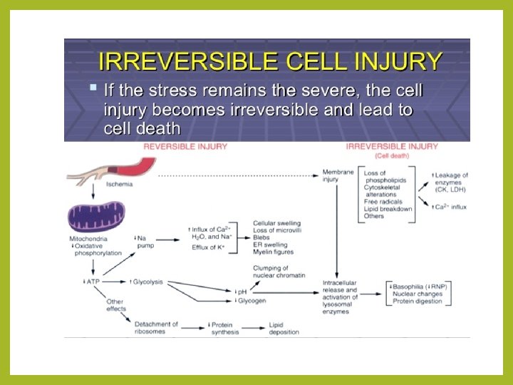

Mechanisms of Irreversible Injury • At what stage did the cell actually die? • And what is the critical biochemical event responsible for the 'point of no return'? • Two phenomena consistently characterize irreversibility. i. The first is the inability to reverse mitochondrial dysfunction (lack of oxidative phosphorylation and ATP generation) even after correction of the original injury (e. g. , restoration of blood flow), and ii. The second is the development of profound disturbances in membrane function.

Cell Membrane Damage • It is now clear that cell membrane damage is a central factor in the pathogenesis of irreversible cell injury. • There are several biochemical mechanisms that cause membrane damage: 1. Progressive loss of membrane phospholipids • Oxygen deprivation (hypoxia) releases sequestered calcium from mitochondria and endoplasmic reticulum, thus raising cytosolic calcium. • The increased cytosolic calcium concentration induced by ischaemia, following acute hypoxic injury, activates endogenous enzymes.

2. Mitochondrial dysfunction: • Whatever the mechanism of cell injury, loss of membrane integrity causes further influx of calcium from the extracellular space, where it is present in high concentration, into the cells. Calcium is taken up greedily by mitochondria. • Here it activates mitochondrial phospholipases and results in accumulation of free fatty acids. • Phospholipases and free fatty acids together cause changes in the permeability of the inner mitochondrial membrane.

3. Cytoskeletal abnormalities: Activation of proteases by increased intracellular calcium may cause damage to cytoskeleton. In the presence of cell swelling, this damage results in detachment of the cell membrane from the cytoskeleton, exposing it to stretching and rupture. 4. Toxic oxygen radicals: Partially reduced oxygen free radicals are highly toxic molecules that cause injury to cell membranes and other cellular constituents.

Thank you
- Slides: 32