Burns in Pregnancy Dr Muhammad El Hennawy Obgyn

Burns in Pregnancy • Dr Muhammad El Hennawy • Ob/gyn specialist • Rass el Barr Central Hospital and Dumyat Specialised Hospital • Dumyatt – EGYPT • www. geocities. com/mmhennawy

• Pregnant women rarely are burned seriously, but when they are, they have unique medical problems. • The mother and fetus are at great risk for fluid loss, hypoxemia, and sepsis.

• I have not been a large number of pregnant women with severe burns in my practice for twenty years But • It has been become apparent however that pregnant women suffer from smoke inhalation pneumonitis that evolves poorly as they do most all forms of pneumonitis • There is positive relationship between extent of burns , maternal mortality , infant mortality , and preterm labour

Burn Scar • Burn scar during pregnancy undergoes considerable softening and therfore can stretch • Skin contracture following scar abdominal burn may be painful during subsequent pregnancy and may be necessitate surgical decompression and split scar autograft • Loss or distortion of breast nipple may cause problem in breast feeding only if two nipples are involved but if one is affected the other breast give sufficient feeding without no problem

What are the different types of burns? • A burn injury usually results from an energy transfer to the body. There are many types of burns caused by thermal, radiation, chemical, or electrical contact. • thermal burns - burns due to external heat sources which raise the temperature of the skin and tissues and cause tissue cell death or charring. Hot metals, scalding liquids, steam, and flames, when coming in contact with the skin, can cause thermal burns. • radiation burns - burns due to prolonged exposure to ultraviolet rays of the sun, or to other sources of radiation such as x-ray. • chemical burns - burns due to strong acids or alkalis coming into contact with the skin and/or eyes. • electrical burns - burns from electrical current, either alternating current (AC) or direct current (DC).

Burn severity It is classified according to • depth of tissue injury, • total body surface area affected , • the presence or absence of an inhalation injury.

The Pregnancy Loss <20% BSA burn: no effect on fetal outcome • >30% BSA burn: increased risk of preterm labour • >40% BSA burn: high risk of fetal death • >50% BSA burn: consider elective cesarean section if fetus is still viable • The overall fetal and neonatal mortality rate is greater than 50% when the mother is burned over more than 60% of her body. • with the pregnancies spontaneously terminated within 10 days of sustaining the injury

Maternal Mortality • It was 47% • The most common cause of death is sepsis • Prophylactic systemic antibiotics should be given to minimise the development of sepsis

Site Of Managment • Patients are best managed in the obstetrics ward during the first 2 weeks of injury. • A multidisplinary approach is encouraged in managing cases of severe burns in pregnancy

Hospital addmission • • • It is recommended for smoke inhalation, electrical burns, burns of both hands or both feet, partial-thickness burns that cover more than 10% of the surface area, full-thickness burns on more than 2% of the surface area. The depth of the injury is estimated by appearance and sensation.

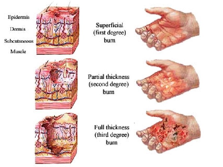
Degree Of Burns • first degree: only involves epithelial layer. Often very painful but resolves with no residual scarring. Skin is red and painful but blisters are not present • second degree: involves epithelium and part of dermis. Pain and scarring vary according to depth of burn. • In superficial second-degree burns damage is limited to epidermis and uppermost part of dermis. • Deep second-degree burns spare only the deepest portion of dermis • third degree: full thickness. Usually painless due to destruction of cutaneous innervation. Leads to scarring. Usually dry and have milky white or tanned leather appearance

A general estimate of the body surface area involved by a burn • • It is determined by the rule of nines: head and neck, 9%; upper extremities, 9% each; anterior torso, 18%; posterior trunk, 18%; lower extremities, 18% each; and genitalia, 1%. Another method is to equate the number of palmar surfaces the burn entails, each palmar surface being equal to 1. 25% of body surface

The Emergency Management • Fluid replacement, respiratory support, and initial wound care the emergency management goals in pregnant burn patients. • The loss of fluid through the denuded surface can be massive, and the amount often is underestimated in pregnant patients. • On arrival to the hospital and after the vital signs of the mother and fetus (monitor) are evaluated, a large-bore (ie, 18 -gauge) intravenous line is started. • In cases in which the burn covers more than 20% of the surface area, a central venous or Swan-Ganz catheter provides a better guide to fluid replacement. Lactated Ringer solution is started at 200 m. L/h until the fluid replacement volume is calculated. Insert a nasogastric tube for burns involving more than 20% of body surface area.

The Fluid Requirements • During late pregnancy, 5% is added if the anterior abdomen is involved. • The fluid requirements for the first 24 hours after a burn injury are calculated as follows: body surface area burned (%) multiplied by 2 -4 m. L/kg body weight. • For example, a 20% burn is calculated as 20 X 3 m. L X 70 kg = 4200 m. L. • Fluid requirements are met with lactated Ringer solution. • The free-water requirement (ie, 500 m. L) is supplied with 5% dextrose in water. • Fifty percent of the replacement fluid is administered in the first 8 hours and the remainder during the next 16 hours. • In the second 24 hours, colloids (albumin) are administered to maintain the serum albumin greater than 3 g/100 m. L.

• All metabolic changes of the burn disease are enhanced by the hypermetabolic state of gestation • . There is also an increase of the intravascular space in pregnant women of up to 40% in volume. • Extreme care should therefore be taken to initiate resuscitation therapy as soon as possible, since the mother's intravascular space is in equilibrium with the amniotic fluid. • Resuscitation should be vigorous to prevent intrauterine death of the child due to loss of amniotic fluid

Monitorig Fluid Replacement Fluid replacement is monitored by clinical and laboratory means. Systolic blood pressure should be greater than 110 mm Hg maternal heart rate less than 120 beats per minute, temperature less than 38°C, respiratory rate should be 12 -24 breaths per minute. Central venous pressure should be approximately 10 cm H 2 O, urine output should be greater than 0. 5 m. L/kg/h. The initial laboratory workup should include a complete blood count and determination of blood levels of electrolytes, glucose, albumin, urea nitrogen, and serum creatinine. Monitor these parameters on a serial basis (eg, q 4 -8 h).

Smoke Inhalation • Smoke inhalation is a major cause of morbidity and mortality in burn patients. • In pregnancy, the fetus is at special risk because of its relatively hypoxic state (ie, normal umbilical vein Pa. O 2 = 27 mm Hg). • The pathophysiology of inhalation injury relates to impaired maternal ventilation (eg, upper airway obstruction from edema), increased diffusion distance (eg, interstitial alveolar edema), and acute functional anemia from carbon monoxide poisoning. Carbon monoxide binds more efficiently to hemoglobin than does oxygen. • In addition to displacing oxygen, carbon monoxide impairs the release of oxygen from oxyhemoglobin. Very little carbon monoxide is needed to cause serious hypoxia. One part carbon monoxide per 1500 parts air can result in blood concentrations of carboxyhemoglobin of 5 -10%. Car exhaust is 5 -7% carbon monoxide. Carboxyhemoglobin values less than 15% usually are well tolerated, whereas values greater than 30% cause severe maternal syncope and fetal death.

• Inhalation injury should be suspected among patients who have a history of closed-space fire, facial injury, carbonaceous material in the oropharynx, or respiratory symptoms. Interstitial edema on chest x-ray film, a carboxyhemoglobin level greater than 10%, or abnormal arterial blood gas levels also aid in establishing the diagnosis of inhalation injury. Initial treatment of any burn patient should include an arterial blood sample for gases and carboxyhemoglobin, as well as a chest radiograph. Patients should be placed on 100% oxygen by mask for at least 3 hours or until the carboxyhemoglobin level is known. They should receive vigorous chest physiotherapy. Intubation and mechanical ventilation should be used early in the presence of upper airway obstruction or oxygenation failure

Sepsis • Sepsis is another major risk for the fetus and mother. • Initial wound care can be instrumental in the prevention of these complications. • On admission, the wound is cleaned with bland soap and water, and all dirt and loose devitalized tissue are removed. Blisters should be left intact if they are smaller than 5 cm in diameter. When burns involve the scalp, axilla, or pubic area, the hair should be clipped short until an adequate margin of unburned skin is obtained. • After cleaning and debridement, a topical agent is applied with a bulky dressing. Silver sulfadiazine cream is used most commonly, but the consideration in pregnant patients is that this drug can be absorbed. The sulfa derivative crosses the placenta and displaces bilirubin. • Should delivery ensue, hyperbilirubinemia is a risk for the neonate. Silver nitrate (0. 5%) also is used, but this agent requires continuous soaking (ie, q 2 h) and a bulky dressing. • Tetanus toxoid (0. 5 m. L) should be administered to all patients with burns

The general and topical treatment of bums • in the pregnant woman has to take into account the embryonal, foetal, and perinatal toxicity of the pharmacological therapy employed, • since what is beneficial for the mother may be harmful for the child. • Particularly difficult therapeutic courses have been found to cause serious malformations; and even the infusion of hypertonic glucose solutions can lead to secondary hyperinsulaemia with foetal macrosornia.

A Team Approach • After the initial management of a severely burned patient, her care requires a team approach with the obstetrician acting as a consultant. • Pregnant women with severe burns are best cared for in centers geared both to managing severe burns and to the possibilities of early delivery. The major long-term problems are healing, sepsis prevention, scar complications, nutritional support, and rehabilitation • surgeons and obstetricians should also be urged to work on an acceptable management protocol for burned pregnant patients. • individualization of its management is always necessary

Time Of Termination • Obstetrical management should be individualized • It is advocated that viable pregnancies should be terminated as soon as the mother is resuscitated following severe burn injury

The factors Determining Obstetric Procedures • the severity of the burn • foetal viability, which must be confirmed immediately. Such biophysical measurements to assess foetal health • The gestational period is in fact one of the factors determining obstetric procedures (no intervention, protection of pregnancy by tocoytic treatment, induction and/or acceleration of labour). • foetuses delivered before 24 weeks generally will not survive, • while those delivered after 32 weeks will do well with modem neonatal intensive care if born without hypoxia or birth trauma. • The most difficult to manage are foetuses of between 24 and 32 weeks' gestational age, where ex utero survival is difficult to predict. In such cases, therefore, when pre-term labour occurs, tocolysis procedures are initiated

Events determining spontaneous uterine activity Synthesis and release of prostaglandins both by the burned skin and as a result of dehydration if not appropiately corrected
 in women")
• a more advanced state of pregnancy (2 nd-3 rd quarter) in women with over 50% TBSA bums had an unfavourable effect on mother unless delivery was immediate, as the burn created an unfavourable environment for the foetus; • in burn in less than 40% TBSA pregnancy and its continuation had no effect on prognosis in the mother and every attempt had to be made to interrupt inception of labour if the foetus was too immature to survive

Total % burn < 30 Age of gestation First trimester No obstetric interference Second trimester No obstetric interference Third trimester 30 -50 Induce labour / caesarian section Less than 36 wks Conservative approach and monitoring of heart rate Foetal monitoring by ultrasound 3 -4 wks Second trimester Foetal monitoring every 3 -4 wks. Tocolytic therapy More than 32 wks Deliver foetus within 48 h Less than 32 wks Careful foetal monitoring First trimester Terminate pregnancy Second trimester Terminate pregnancy Third trimester > 70 More than 36 wks First trimester Third trimester 50 -70 Management If baby is viable Induce labour / caesarian section within 24 h Intrauterine death No active intervention up to 4 wks / monitoring of foetus of haemocoagulation factors First trimester No treatment Second trimester No treatment Third trimester Caesarian section as an emergency procedure at the

• In order to reduce pharmacological therapy to the minimum possible • and to accelerate the burn healing process (and thus improve prognosis), • the majority of authors are favourable to early surgical intervention

the patient was subjected to a caesarian section and free skin graft in burned area

• On the basis of my experience and from other literatures and studies • I recommend that women in second and third trimester of pregnancy with burn more than fifty persent of her body should be delivered immediately as maternal death is otherwise almost certain and fetal survival is not improved by waiting • Maternal prognosis is markdly worse than for non pregnant woman suffering otherwise comperable burn

Prevention • by teaching pregnant women in the antenatal care clinic, for example, • to avoid contact with kerosene for cooking and heating • to apply proper first-aid measures, such as prompt cooling of the burn with cold water, to reduce the depth of the injury should a burn occur.
- Slides: 32