
BURN LECTURE M Catherine Hough RN Ph D
BURN LECTURE M. Catherine Hough RN, Ph. D University of North Florida College of Health Department of Nursing
REVIEW OF SKIN FUNCTIONS • Functions of the Skin – Protection – Heat Regulation – Sensory perception – Excretion – Vitamin D Production – Expression
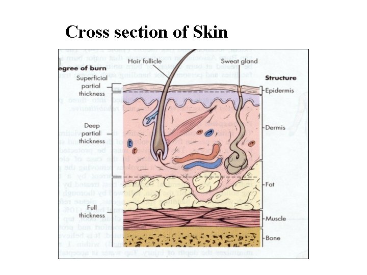
Cross section of Skin
CLASSIFIATION OF BURNS Rx of burn is R/T the severity of the burn severity is determined by: • depth of the burn • extent o the burn (% of total body surface area (TBSA) • location of the burn • patients risk factors
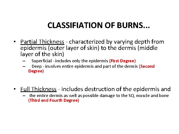
CLASSIFIATION OF BURNS. . . • Partial Thickness - characterized by varying depth from epidermis (outer layer of skin) to the dermis (middle layer of the skin) – – Superficial - includes only the epidermis (First Degree) Deep - involves entire epidermis and part of the dermis (Second Degree) • Full Thickness - includes destruction of the epidermis and – the entire dermis as well as possible damage to the SQ, muscle and bone (Third and Fourth Degree)
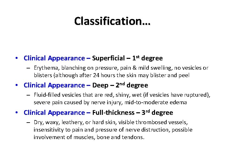
Classification… • Clinical Appearance – Superficial – 1 st degree – Erythema, blanching on pressure, pain & mild swelling, no vesicles or blisters (although after 24 hours the skin may blister and peel • Clinical Appearance – Deep – 2 nd degree – Fluid-filled vesicles that are red, shiny, wet (if vesicles have ruptured), severe pain caused by nerve injury, mid-to-moderate edema • Clinical Appearance – Full-thickness – 3 rd degree – Dry, waxy, leathery, or hard skin, visible thrombosed vessels, insensitivity to pain and pressure of nerve distruction, possible involvement of muscles, bone and tendons.
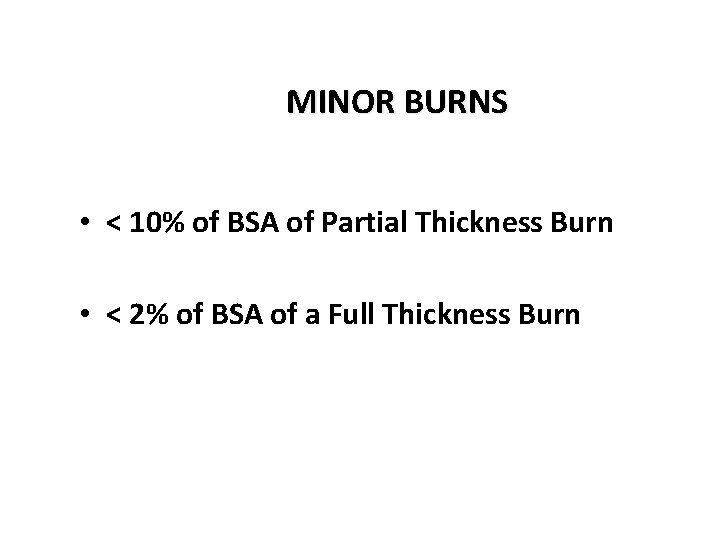
MINOR BURNS • < 10% of BSA of Partial Thickness Burn • < 2% of BSA of a Full Thickness Burn
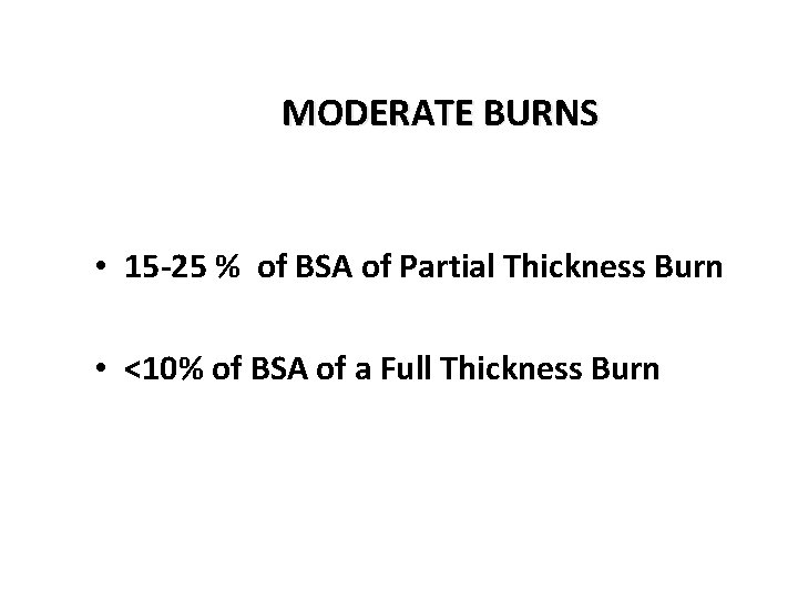
MODERATE BURNS • 15 -25 % of BSA of Partial Thickness Burn • <10% of BSA of a Full Thickness Burn
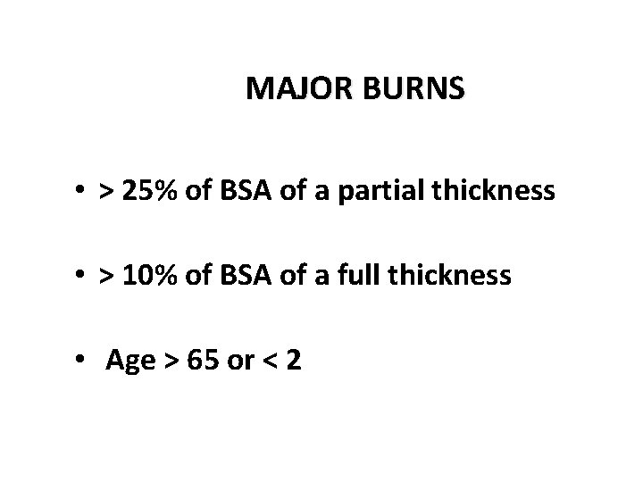
MAJOR BURNS • > 25% of BSA of a partial thickness • > 10% of BSA of a full thickness • Age > 65 or < 2
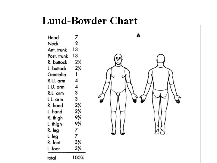
Lund-Bowder Chart
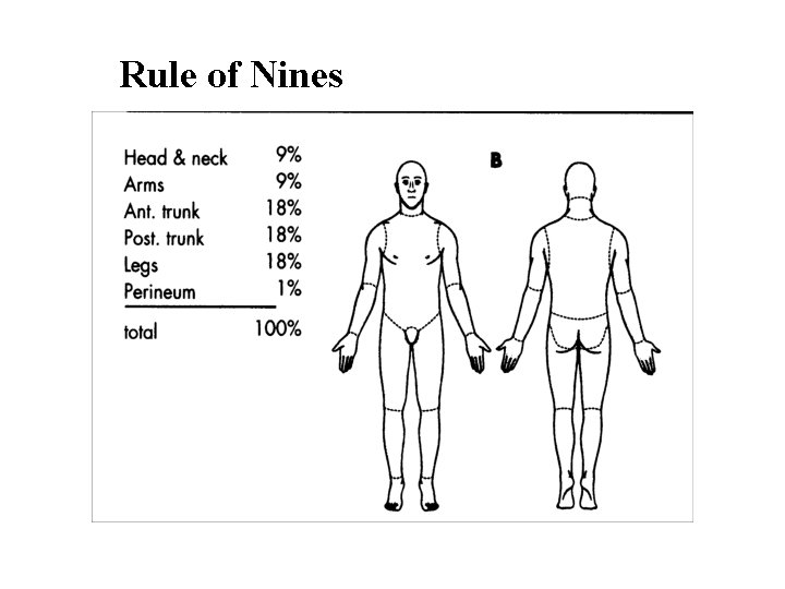
Rule of Nines
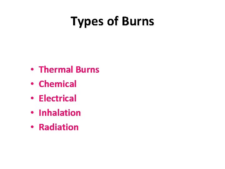
Types of Burns • • • Thermal Burns Chemical Electrical Inhalation Radiation
PERIODS OF TREATMENT • Emergent • Acute • Rehabilitation
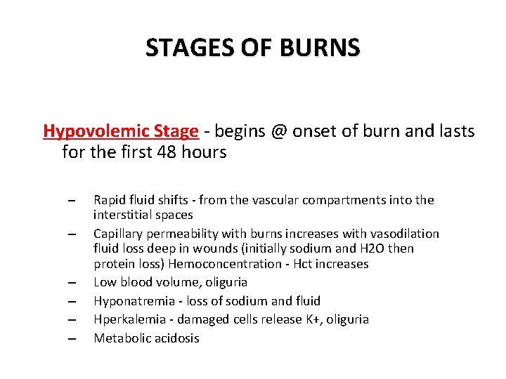
STAGES OF BURNS Hypovolemic Stage - begins @ onset of burn and lasts for the first 48 hours – – – Rapid fluid shifts - from the vascular compartments into the interstitial spaces Capillary permeability with burns increases with vasodilation fluid loss deep in wounds (initially sodium and H 2 O then protein loss) Hemoconcentration - Hct increases Low blood volume, oliguria Hyponatremia - loss of sodium and fluid Hperkalemia - damaged cells release K+, oliguria Metabolic acidosis
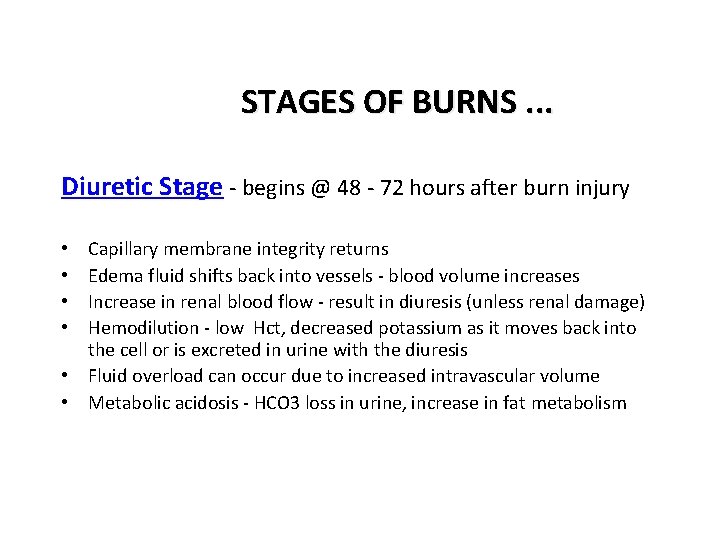
STAGES OF BURNS. . . Diuretic Stage - begins @ 48 - 72 hours after burn injury Capillary membrane integrity returns Edema fluid shifts back into vessels - blood volume increases Increase in renal blood flow - result in diuresis (unless renal damage) Hemodilution - low Hct, decreased potassium as it moves back into the cell or is excreted in urine with the diuresis • Fluid overload can occur due to increased intravascular volume • Metabolic acidosis - HCO 3 loss in urine, increase in fat metabolism • •
I. EMERGENT PERIOD • First 24 - 48 hours • Maintain airway, fluids, analgesia, temperature, wound • Assessment: – Objective: how burn occurred, when, duration, type of agent – Subjective: previous medical problems, size and depth of burn, age, body part involved, mechanism of injury
EMERGENT PERIOD. . . Factors determining severity of burns: • size of burn • depth of burn • age • body part effected • mechanism of injury • history of cardiac, pulmonary, renal, or hepatic diseases • injuries sustained @ time of burns • duration of contact with burning agent • size & depth of burn • “Rule of Nines”
NURSING DIAGNOSIS • • Airway clearance Ineffective fluid volume (deficit or excess) Hypothermia High risk for pain (with partial thickness burns) Skin integrity, impaired Anxiety Knowledge deficit
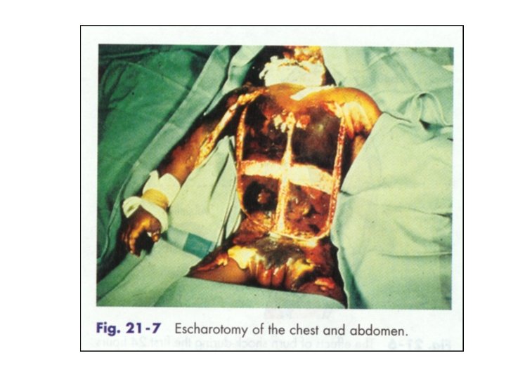
INTERVENTIONS • Maintain patent airway - watch for laryngeal edema – Escharotomy may be needed – 100% Fi. O 2 mask – intubation for inhalation is often required – may inquire emergent tracheostomy – may require ventilatory assistance
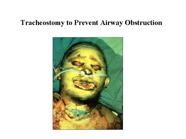
Tracheostomy to Prevent Airway Obstruction
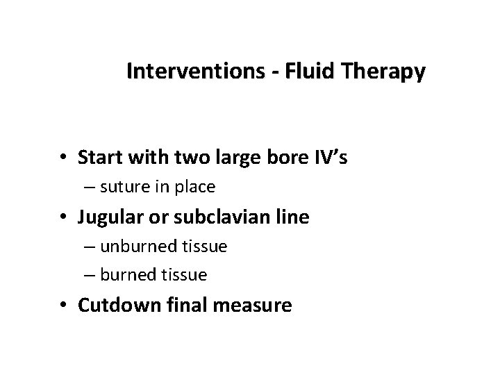
Interventions - Fluid Therapy • Start with two large bore IV’s – suture in place • Jugular or subclavian line – unburned tissue – burned tissue • Cutdown final measure
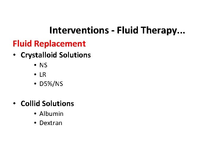
Interventions - Fluid Therapy. . . Fluid Replacement • Crystalloid Solutions • NS • LR • D 5%/NS • Collid Solutions • Albumin • Dextran
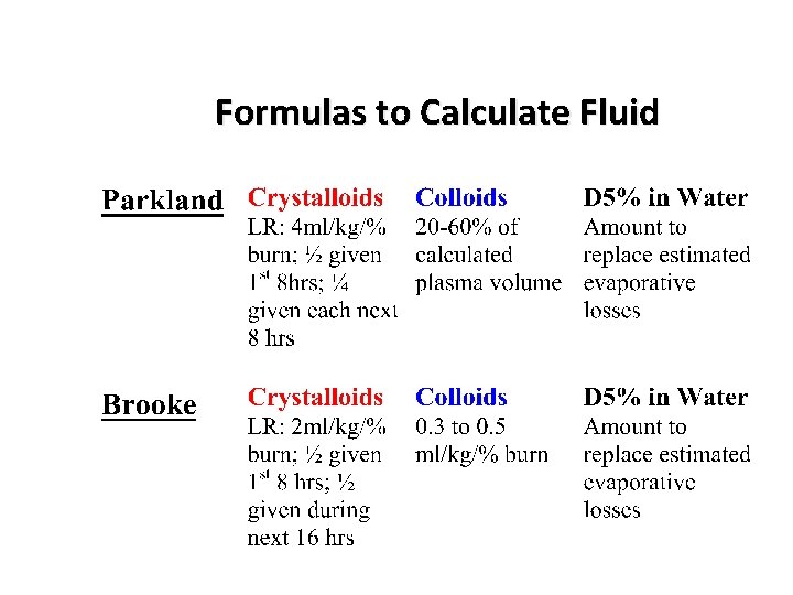
Formulas to Calculate Fluid
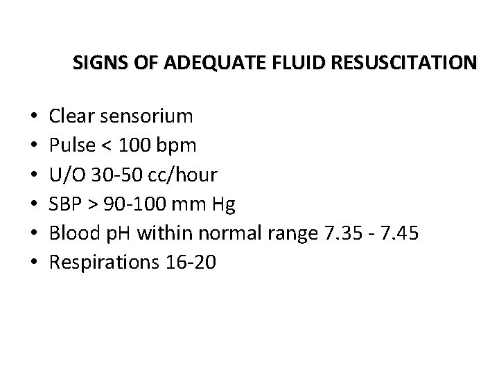
SIGNS OF ADEQUATE FLUID RESUSCITATION • • • Clear sensorium Pulse < 100 bpm U/O 30 -50 cc/hour SBP > 90 -100 mm Hg Blood p. H within normal range 7. 35 - 7. 45 Respirations 16 -20
II. ACUTE PERIOD • End of emergent period until burns heal • Focus shifts to care of wounds and prevention of complications • Actual range of phase depends on degree and extent of burn • Assessment: Subjective - pain and anxiety Objective - complete assessment every 8 hours, dietary intake, motor ability, I&O, weight
NURSING DIAGNOSIS • • Skin integrity, impaired Infection, high risk for altered nutrition Pain, acute (with partial thickness burns) Fluid Volume Deficit Anxiety Hypothermia
Pain Control • Morphine Sulfate 5 -10 mg IV every 1 -3 hours • Combination therapy for painful procedures: – Diprivan – Valium – Haldol – Versed –…
NURSING DIAGNOSIS. . . Impaired skin integrity R/T thermal injury Coping, ineffective individual/family Body Image Disturbance Altered nutrition: less than body requirements R/T increased catabolism and metabolism • Mobility, Impaired R/T pain, impaired joint movement, scar formation • Self-care Deficit • High risk for infection R/T denuded skin, presence of pathogenic organism, & altered immune response • •
INTERVENTIONS • Releiving anxiety, denial, regression, anger, depression • Wounds - refer to wound care • Nutrition (Nutritional assessment, pre albumin levels, large protein requirement, carbohydrates and fats for energy, mega vitamins, TPN, enteral tube feedings any follow (~5, 000 kcal/day) • Pain - around the clock management • Prevention of infection - refer to wound care
ORGANISMS: • Staphylococcus aureus • Pseudomonas Infection is usually the cause of any deterioration
SIGNS OF SEPSIS: • Change in sensorium • • • Fever Tachyapnea Paralytic ileus Abdominal distention Oliguria
WAYS TO PREVENT INFECTIONS: • • • Gowns, masks, gloves Sterile linen Person with URI should not come in contact with patient
WOUND CARE Goals: • • clean & debride the area of necrotic tissue minimize further destruction of viable skin promote wound re-epithelialization promote patient comfort
WOUND CARE: • Burn wound is unique • Burn wound sepsis – gram + – gram (pseudomonas) – fungal (candida albicans)
WOUND CARE. . . • Nutrition – collagen primary structure in healing by secondary intention – need increased protein – may need up to double the normal calorie requirements • Inadequate blood supply • Burn wound disorders – scarring, contractures, keloids, failure to heal
WOUND CARE. . . • GOALS: • close wound ASAP • prevent infection • reduce scarring and contractures • provide for comfort
WOUND CARE. . . • Wound cleaning: • at bed side hyrotherapy tanks, tubbing, tables • Debridement: • mechanical, surgical, enzymatic • Topical antibacterial therapy • sulfonamide spray
WOUND CARE. . . Open Technique or Exposed - more often used with burns effecting the: – – face neck perineum broad areas of the trunk • Partial thickness - exudate dries in 48 to 72 hours forming a hard crust that protects the wound. • Full thickness - dead skin is dehydrated and converted to black leathery escar in 48 to 72 hours. Loose escare is gradually removed with hydrotherapy &/or debridement
WOUND CARE. . . • Closed Technique • Wound is washed and sterile dressings changed (may be q shift, daily) • Dressing consists of gauze &/or ace wraps impregnated with topical ointments
WOUND CARE. . . • Semi-Open consists of covering the wound with topical antimicrobial agents and gauze ADVANTAGE: – speeds debridement – develops granulation tissues faster – makes skin grafting possible sooner
WOUND CARE. . . Biological Dressings: • Homeografts - same species (cadaver skin) • temporary (3 days to 2 weeks) then body rejects • Heterografts - another species (pig skin) • temporary coverage (3 days to 2 weeks) • Autografts - patients own skin • can be temporary or permanent coverage • Cultured Epithelial Autographs • permanent
Wound Care - GRAFTING • Indications for Grafting: – – full thickness burns priority areas (face) wound bed pink firm, free of exudate bacterial count < 100, 000/gram of tissue • Care of Grafts - assess, assess
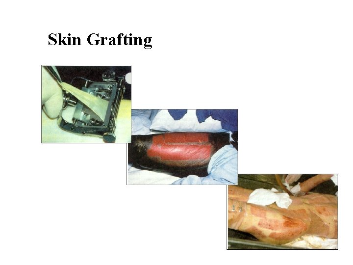
Skin Grafting
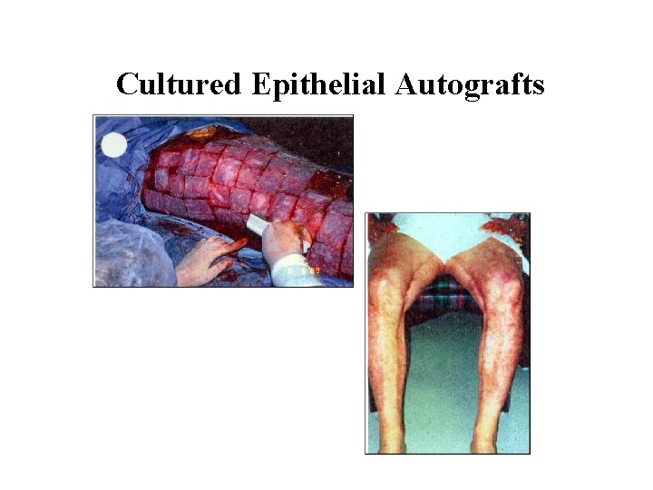
Cultured Epithelial Autografts
III. REHABILITATION PERIOD • Care of healing skin - wash daily, cover with cocoa butter or other barrier • Pressure garments, ace wraps - helps prevent scaring and contractures • Promote mobility - positioning, exercise, splinting, ADL • Rehab period can last for months to even years
Primary Prevention Strategies Safety Education: • Wear sun-screen • Fireproof your home – Install smoke alarms – check routinely – Plan emergency exits – Have regular fire drills • Check wiring in home; safety caps on unused outlets if you have children • Teach children safety rules for matches, fires, electrical outlets, cords, etc.
- Slides: 47