
Building of statistical models Guido Gerig Department of

Building of statistical models Guido Gerig Department of Computer Science, UNC, Chapel Hill 1
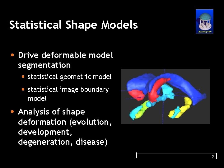
Statistical Shape Models • Drive deformable model segmentation • statistical geometric model • statistical image boundary model • Analysis of shape deformation (evolution, development, degeneration, disease) 2
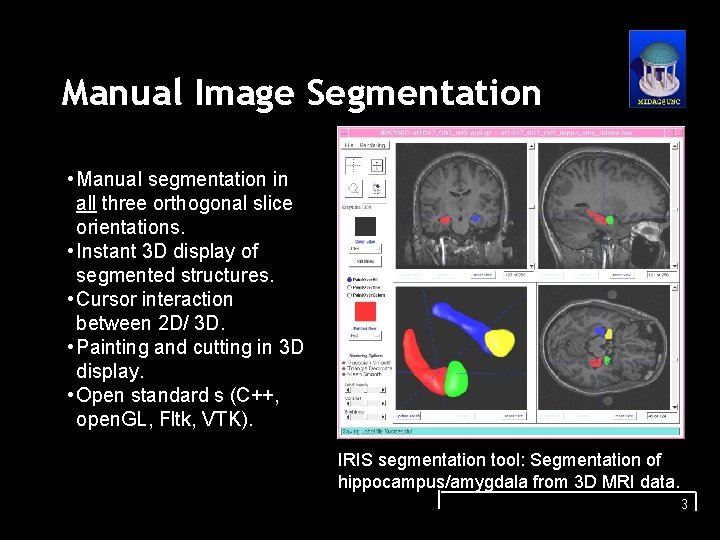
Manual Image Segmentation • Manual segmentation in all three orthogonal slice orientations. • Instant 3 D display of segmented structures. • Cursor interaction between 2 D/ 3 D. • Painting and cutting in 3 D display. • Open standard s (C++, open. GL, Fltk, VTK). IRIS segmentation tool: Segmentation of hippocampus/amygdala from 3 D MRI data. 3
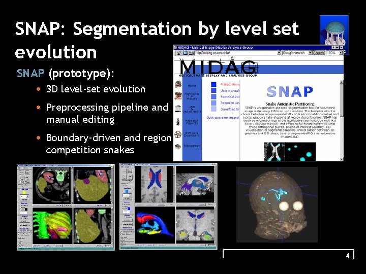
SNAP: Segmentation by level set evolution SNAP (prototype): • 3 D level-set evolution • Preprocessing pipeline and manual editing • Boundary-driven and regioncompetition snakes 4
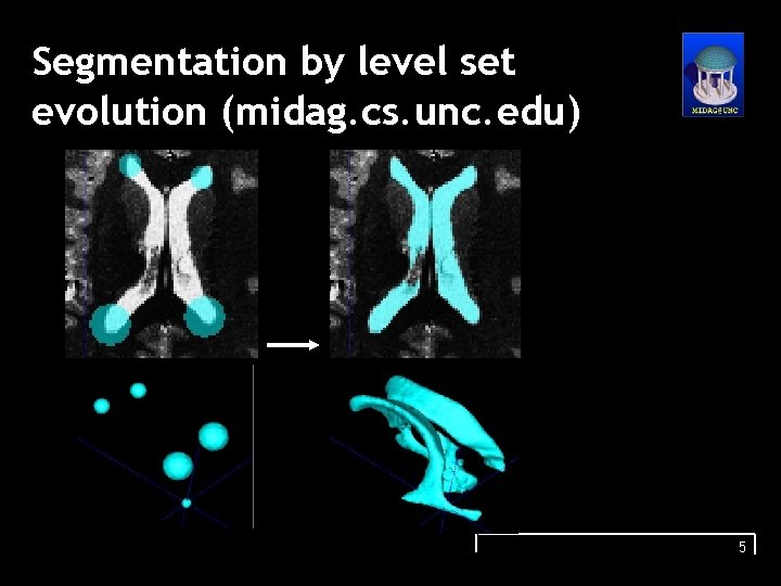
Segmentation by level set evolution (midag. cs. unc. edu) 5
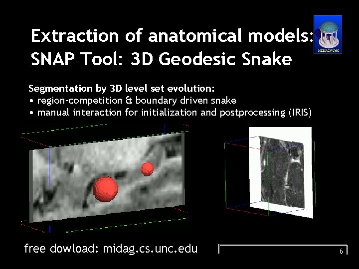
Extraction of anatomical models: SNAP Tool: 3 D Geodesic Snake Segmentation by 3 D level set evolution: • region-competition & boundary driven snake • manual interaction for initialization and postprocessing (IRIS) free dowload: midag. cs. unc. edu 6
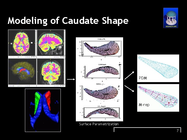
Modeling of Caudate Shape PDM M-rep Surface Parametrization 7
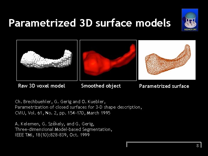
Parametrized 3 D surface models Raw 3 D voxel model Smoothed object Parametrized surface Ch. Brechbuehler, G. Gerig and O. Kuebler, Parametrization of closed surfaces for 3 -D shape description, CVIU, Vol. 61, No. 2, pp. 154 -170, March 1995 A. Kelemen, G. Székely, and G. Gerig, Three-dimensional Model-based Segmentation, IEEE TMI, 18(10): 828 -839, Oct. 1999 8
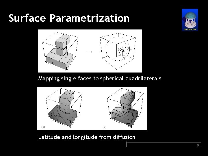
Surface Parametrization Mapping single faces to spherical quadrilaterals Latitude and longitude from diffusion 9
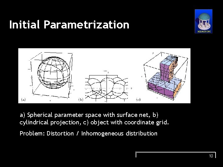
Initial Parametrization a) Spherical parameter space with surface net, b) cylindrical projection, c) object with coordinate grid. Problem: Distortion / Inhomogeneous distribution 10
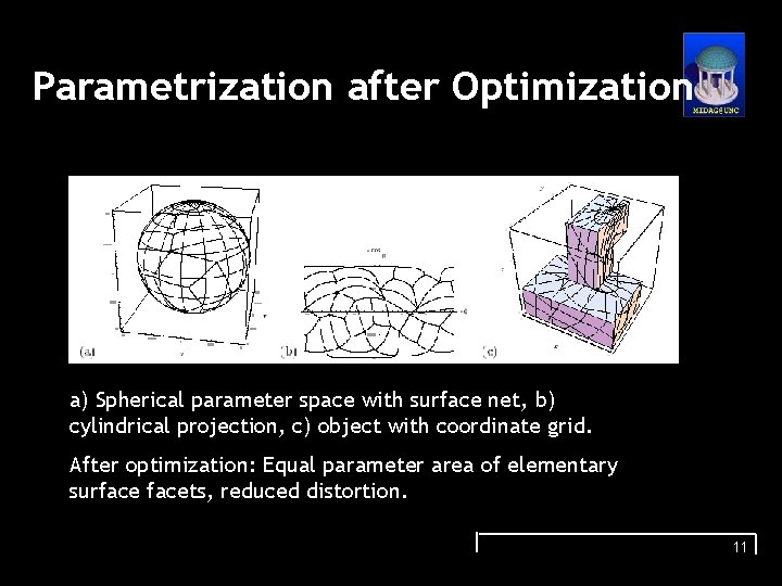
Parametrization after Optimization a) Spherical parameter space with surface net, b) cylindrical projection, c) object with coordinate grid. After optimization: Equal parameter area of elementary surfacets, reduced distortion. 11
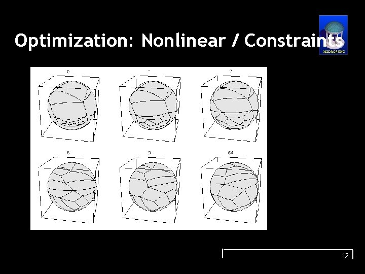
Optimization: Nonlinear / Constraints 12
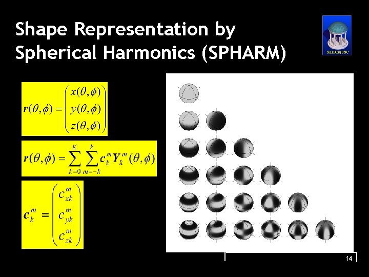
Shape Representation by Spherical Harmonics (SPHARM) 14
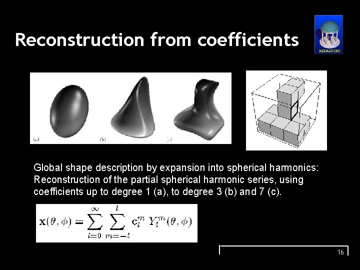
Reconstruction from coefficients Global shape description by expansion into spherical harmonics: Reconstruction of the partial spherical harmonic series, using coefficients up to degree 1 (a), to degree 3 (b) and 7 (c). 16
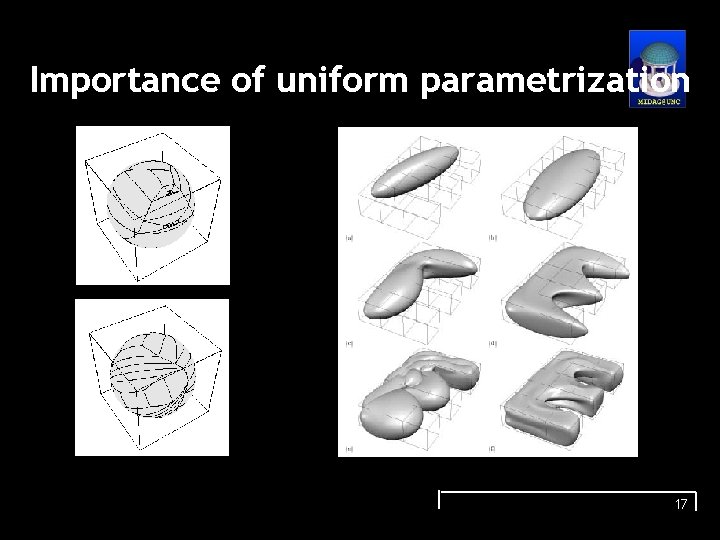
Importance of uniform parametrization 17
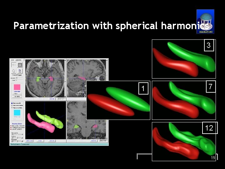
Parametrization with spherical harmonics 3 1 7 12 19
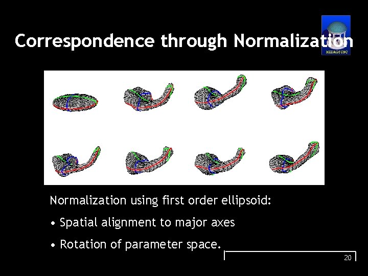
Correspondence through Normalization using first order ellipsoid: • Spatial alignment to major axes • Rotation of parameter space. 20
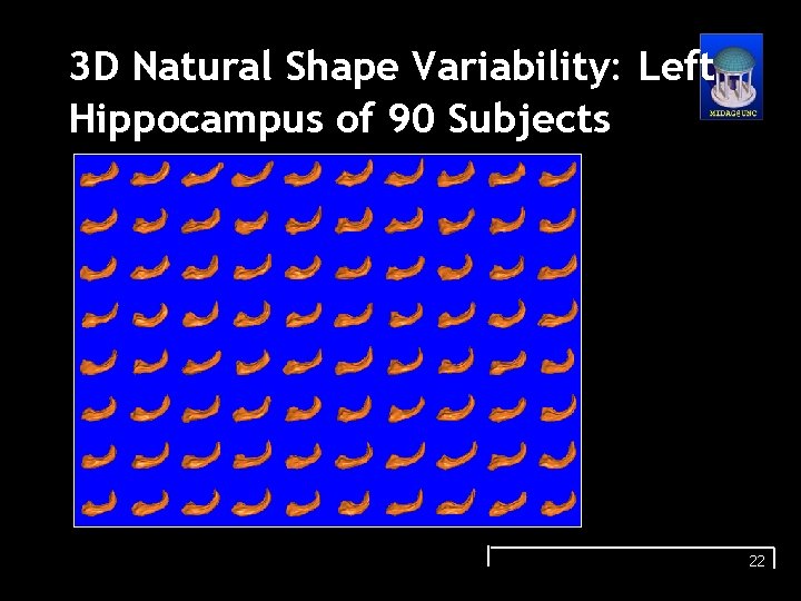
3 D Natural Shape Variability: Left Hippocampus of 90 Subjects 22
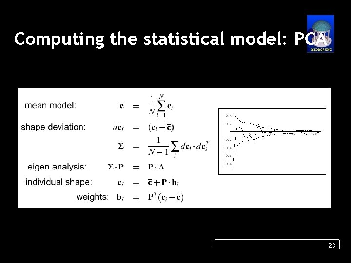
Computing the statistical model: PCA 23
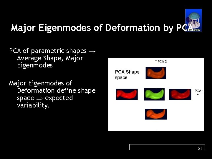
Major Eigenmodes of Deformation by PCA of parametric shapes Average Shape, Major Eigenmodes of Deformation define shape space expected variability. 26
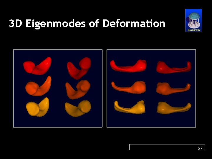
3 D Eigenmodes of Deformation 27
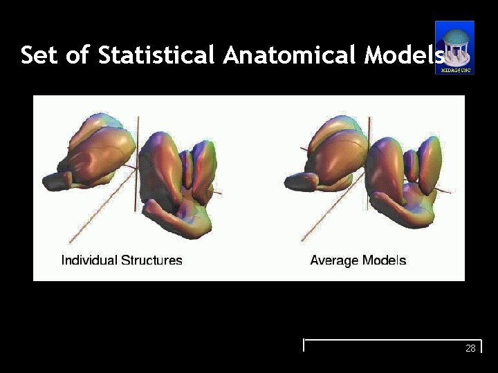
Set of Statistical Anatomical Models 28
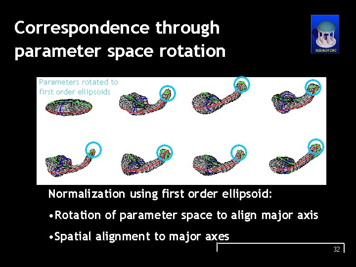
Correspondence through parameter space rotation Parameters rotated to first order ellipsoids Normalization using first order ellipsoid: • Rotation of parameter space to align major axis • Spatial alignment to major axes 32
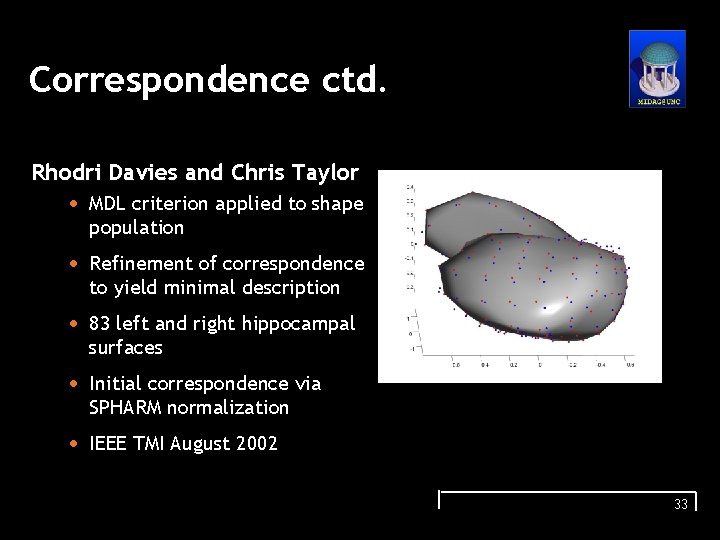
Correspondence ctd. Rhodri Davies and Chris Taylor • MDL criterion applied to shape population • Refinement of correspondence to yield minimal description • 83 left and right hippocampal surfaces • Initial correspondence via SPHARM normalization • IEEE TMI August 2002 33
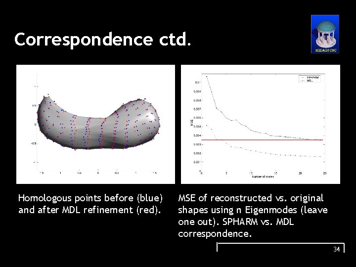
Correspondence ctd. Homologous points before (blue) and after MDL refinement (red). MSE of reconstructed vs. original shapes using n Eigenmodes (leave one out). SPHARM vs. MDL correspondence. 34
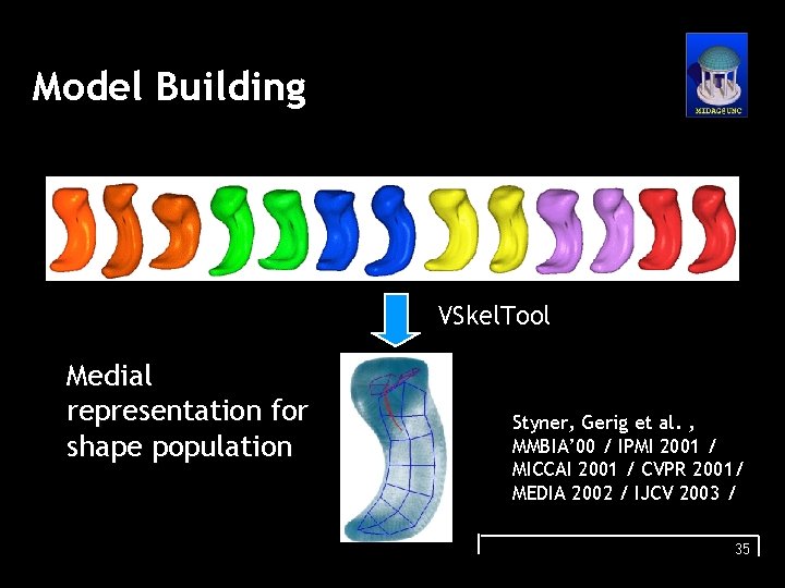
Model Building VSkel. Tool Medial representation for shape population Styner, Gerig et al. , MMBIA’ 00 / IPMI 2001 / MICCAI 2001 / CVPR 2001/ MEDIA 2002 / IJCV 2003 / 35
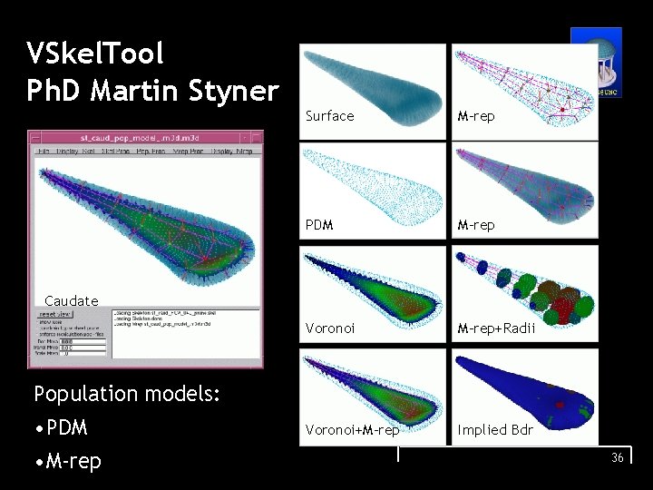
VSkel. Tool Ph. D Martin Styner Surface M-rep PDM M-rep Voronoi M-rep+Radii Voronoi+M-rep Implied Bdr Caudate Population models: • PDM • M-rep 36
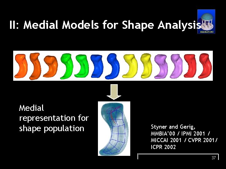
II: Medial Models for Shape Analysis Medial representation for shape population Styner and Gerig, MMBIA’ 00 / IPMI 2001 / MICCAI 2001 / CVPR 2001/ ICPR 2002 37
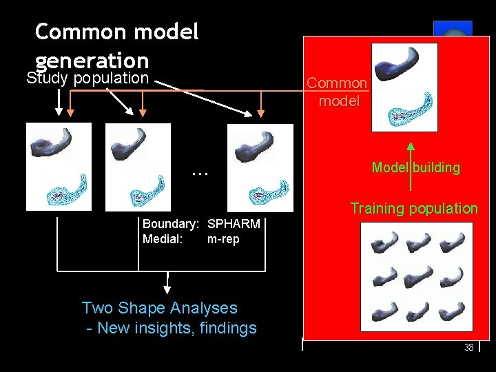
Common model generation Study population Common model . . . Boundary: SPHARM Medial: m-rep Model building Training population Two Shape Analyses - New insights, findings 38
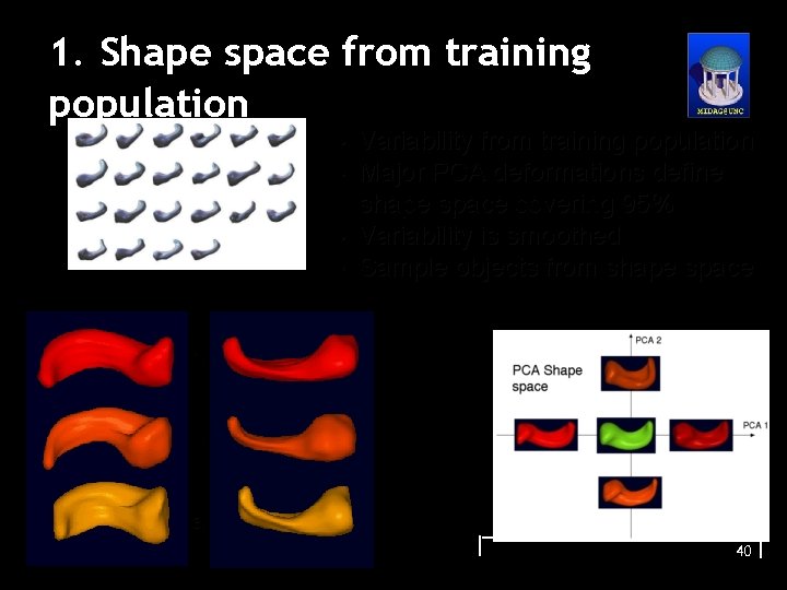
1. Shape space from training population • • Variability from training population Major PCA deformations define shape space covering 95% Variability is smoothed Sample objects from shape space 1. 2. 3. 40
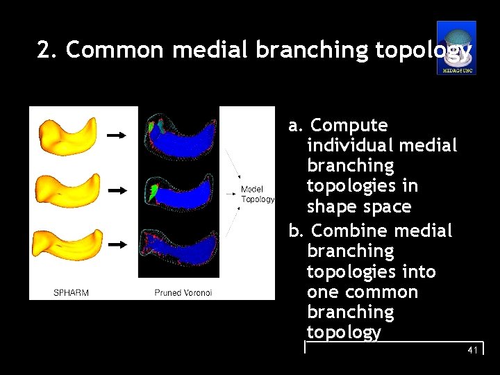
2. Common medial branching topology a. Compute individual medial branching topologies in shape space b. Combine medial branching topologies into one common branching topology 41
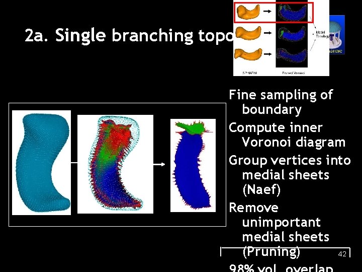
2 a. Single branching topology Fine sampling of boundary Compute inner Voronoi diagram Group vertices into medial sheets (Naef) Remove unimportant medial sheets (Pruning) 42
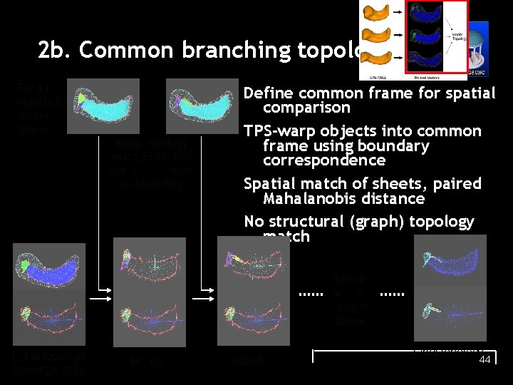
2 b. Common branching topology For all objects in shape space Warp topology using SPHARM correspondence on boundary Define common frame for spatial comparison TPS-warp objects into common frame using boundary correspondence Spatial match of sheets, paired Mahalanobis distance No structural (graph) topology match Match whole shape space Initial topology (average case) Match Final topology 44
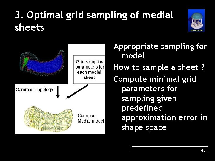
3. Optimal grid sampling of medial sheets Appropriate sampling for model How to sample a sheet ? Compute minimal grid parameters for sampling given predefined approximation error in shape space 45
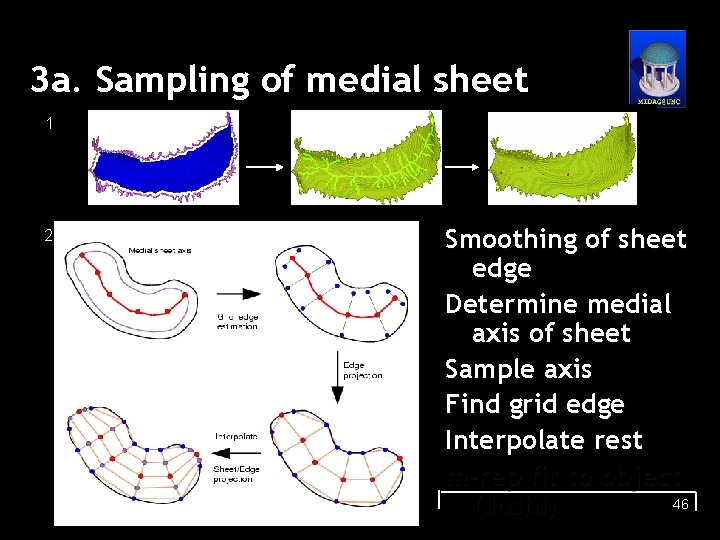
3 a. Sampling of medial sheet 1 2 Smoothing of sheet edge Determine medial axis of sheet Sample axis Find grid edge Interpolate rest m-rep fit to object 46 (Joshi)
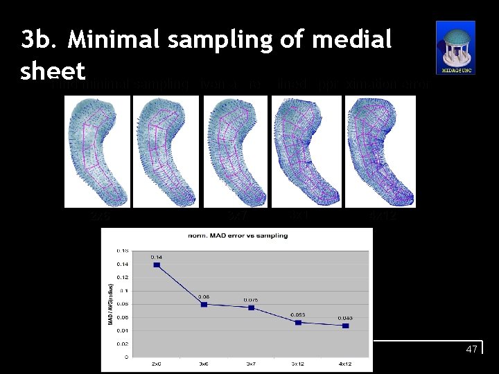
3 b. Minimal sampling of medial sheet • Find minimal sampling given a predefined approximation error 2 x 6 3 x 7 3 x 12 47
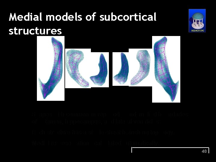
Medial models of subcortical structures Shapes with common m-rep model and implied boundaries of putamen, hippocampus, and lateral ventricles. Each structure has a single-sheet branching topology. Medial representations calculated automatically. 48
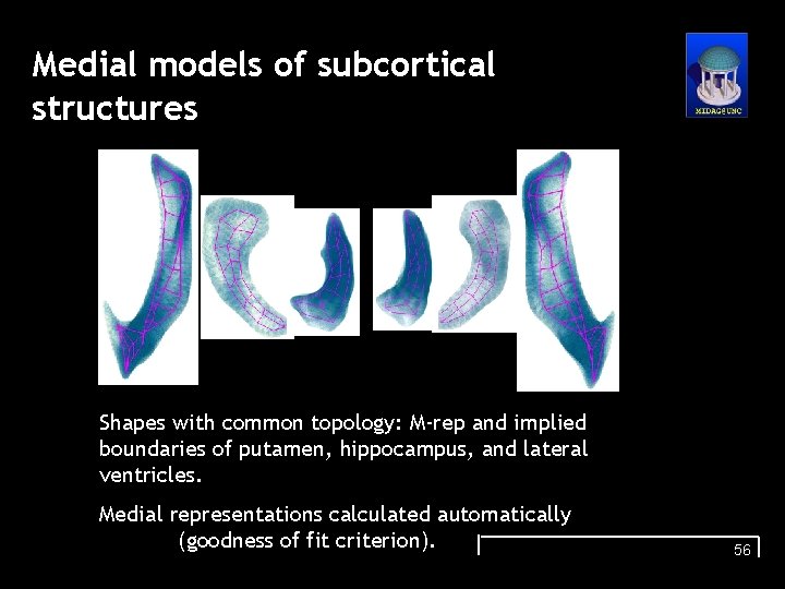
Medial models of subcortical structures Shapes with common topology: M-rep and implied boundaries of putamen, hippocampus, and lateral ventricles. Medial representations calculated automatically (goodness of fit criterion). 56
- Slides: 38