Bronchi Right principal bronchus Shorter wider and more

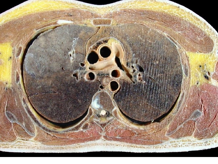
支气管Bronchi Right principal bronchus 右主支气管 • Shorter, wider, and more vertical than the left , is about 2. 5 cm long, Leaves the extend line of the middle line of trachea at 22~ 25 o angle • Foreign bodies are therefore more likely to lodge in this bronchus or one of its branches Left principal bronchus 左主支气管 • Narrower, longer, and more horizontal than the right is about 5 cm long, leaves the extend line of the middle line of trachea at about 35~ 36 o angle
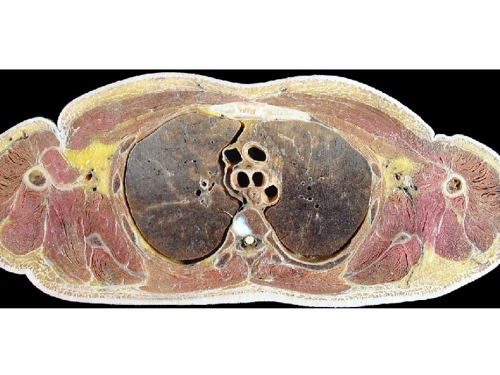

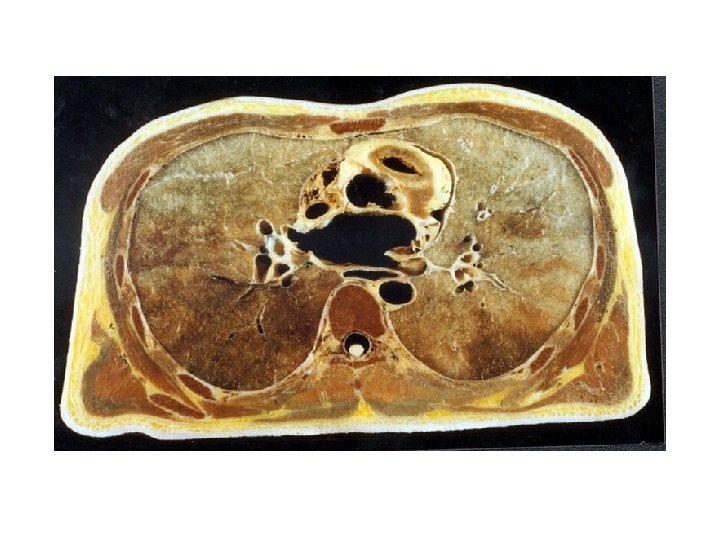
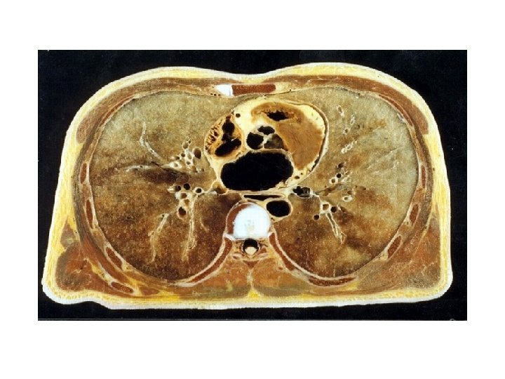
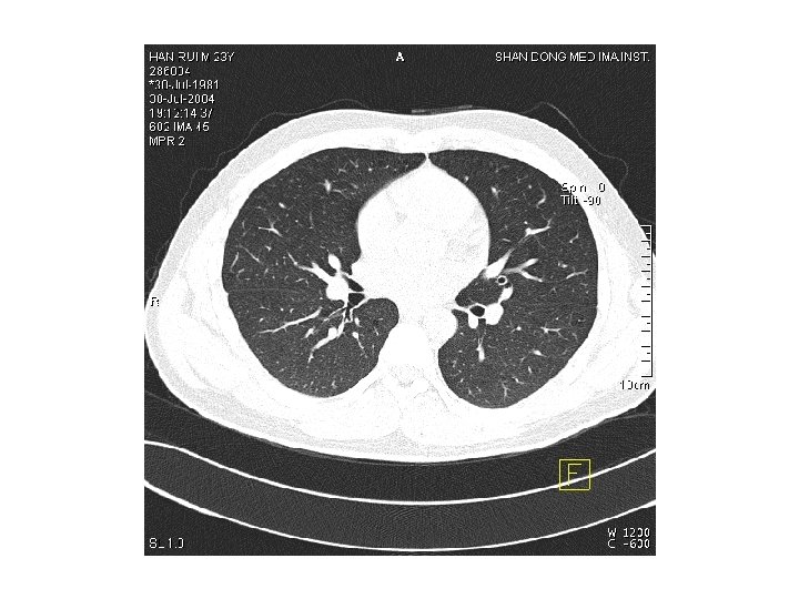
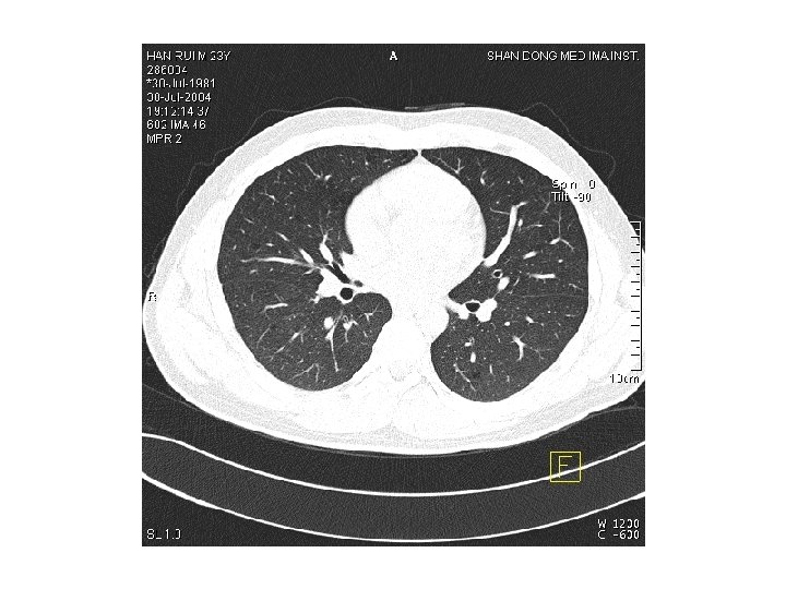
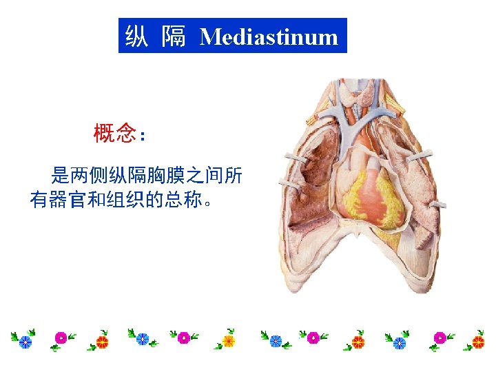
肺The Lungs Position: located in the thoracic cavity by both sides of mediastinum General features • Cone-shaped, the right lung is shorter and broader, the left one is longer and narrower • Apex of lung-rises 2 ~ 3 cm above the medial third of clavicle into neck • Base-concave, related to diaphragm, also called diaphragmatic surface • Costal surface-large, convex, related to thoracic wall

• Medial surface-concave, related to mediastinum and vertebrae – Hilum of lung 肺门:area on medial surface where structures in root enter or leave lung – Root of lung 肺根 • Contents – Principal bronchus – Pulmonary artery and vein – Nerves and lymphatics • Surrounded by connective tissue • Order of structures in the root of lung – From before backward: V. A. B. – From above downward: » R. -B. A. V. » L. -A. B. V.


Borders – Posterior-blunt – Inferior- sharp – Anterior-sharp • • cardiac notch心切迹 lingual in left lung 左肺小舌 Lobes and Fissure • Right lung – Two fissures : horizontal and oblique – Three lobes : superior, middle, inferior • Left lung – One fissure : oblique – Two lobes : superior and inferior

支气管树Bronchial tree Each principal bronchus divides into lobar bronchi (two on the left, three on the right), each of which supplies a lobe of lung. Each lobar bronchus then divided into segmental bronchi, which supply specific segments of the lung.

Bronchopulmonary segments支气管肺段 • Wedge shaped, with the base lying peripherally and the apex lying towards the root of lungs, ten in each lung • Each with a segmental bronchus and branches of pulmonary artery • The veins lie both in and between segments


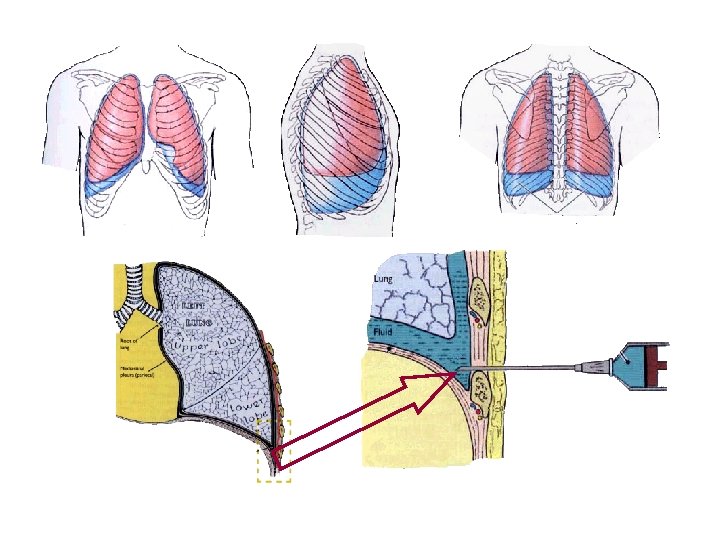
胸膜The Pleura General features • Serous membranes forming closed sacs • Two layers – Visceral pleura-adheres to lung, continuous with parietal pleura at root of lung – Parietal pleura-lines the thoracic cavity

Two pleural layers continue with each other at root of lung forming closed potential space-pleural cavity 胸膜腔 – Contains a small amount pleural fluid – Subatmospheric pressure in it

Named parts of parietal pleura • Cupula of pleura 胸膜顶 -extends up into the neck, over the apex of lung, 2~ 3 cm above the medial third of clavicle • Costal pleura 肋胸膜 - lines the inner surface of the wall of the chest • Mediastinal pleura 纵隔胸膜 – Lines mediastinum – Pulmonary ligament 肺韧带 -redundant pleura at root of lung, which extends downward, allows movement of structures forming root of lung • Diaphragmatic pleura 膈胸膜- Lines diaphragm

Pleura recesses 胸膜隐窝- potential spaces of pleural cavity which lungs are not occupied in quiet respiration • Costodiaphragmatic recesse肋 膈隐窝-are the slit-like intervals between costal and diaphragmatic pleurae on each side, the lowest point of pleural cavity • Costomediastinal recess 肋 纵隔隐窝-on the left side between the mediastinal pleural and costal pleura

The surface projection of lower border of lung and pleurae Lower border Midclavicular lines Midaxillary lines Sides of the vertebral column Lungs 6 th rib 8 th rib 10 th rib Pleura 8 th rib 10 th rib 12 th rib
- Slides: 24