Brief ear anatomy Otitis Externa Acute Otitis media

Brief ear anatomy Otitis Externa Acute Otitis media

Objectives • Gross applied anatomy of the ear • Nerve supply of the external and middle ears and the principles of referred earache • Central connection of the vestibulocochlear nerve • Physiology of the external, middle and inner ears

ANATOMY OF THE EAR

ANATOMY OF THE EAR • External Ear • Middle Ear Cleft • Inner Ear

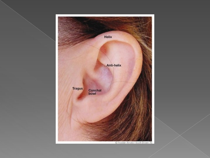
EXTERNAL EAR Auricle External Canal

The Auricle

Perichondritis Erysipelas

The External Auditory Canal

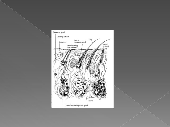
The External Auditory Canal Lateral Third: Medial Two Thirds: • Cartilagenous • Bony • Hair follicles • Develops after birth • Ceruminous glands • Sebacous glands

Relations of EAM

ANATOMY OF THE EAR • External Ear • Middle Ear Cleft • Inner Ear
 Tube • Tympanum (Middle Ear Cavity) • Mastoid")
MIDDLE EAR CLEFT • Eustachian (Pharyngo-tympanic) Tube • Tympanum (Middle Ear Cavity) • Mastoid Antrum and Air Cells
 Tube")
MIDDLE EAR CLEFT • Eustachian (Pharyngo-Tympanic) Tube
 tube • Tympanic cavity (Middle ear cavity)")
MIDDLE EAR CLEFT • Eustachian (Pharyngo. Tympanic) tube • Tympanic cavity (Middle ear cavity)

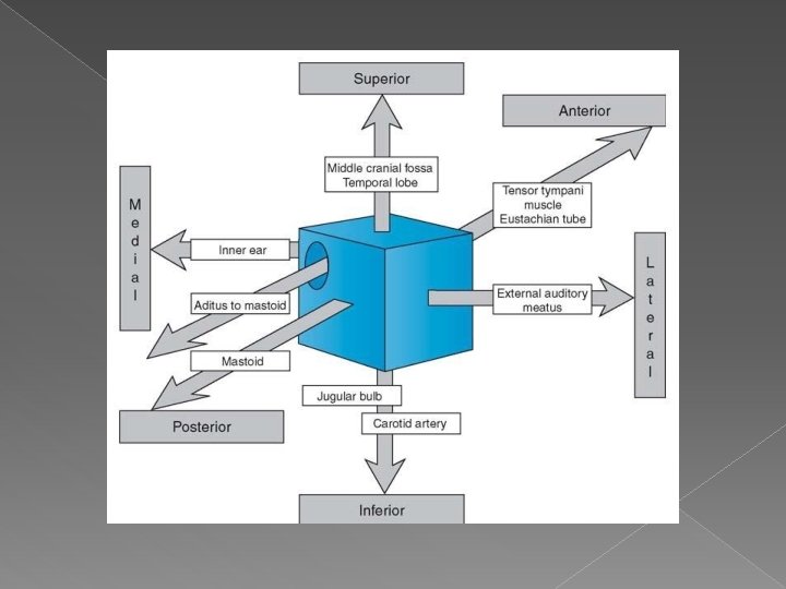
WALLS OF TYMPANIC CAVITY • Roof • Floor • Anterior wall • Posterior wall • Lateral wall • Medial wall

WALLS OF TYMPANIC CAVITY Tegmen • Roof • Floor

WALLS OF TYMPANIC CAVITY • Roof • Floor • Anterior wall • Posterior wall

WALLS OF TYMPANIC CAVITY • Roof • Floor Membrana Flaccida • Anterior wall • Posterior wall • Lateral wall Membrana Tensa
 • Anterior wall • Posterior")
WALLS OF TYMPANIC CAVITY • Roof • Floor Epitympanum(attic) • Anterior wall • Posterior wall • Lateral wall Mesotympanum Hypotympanum

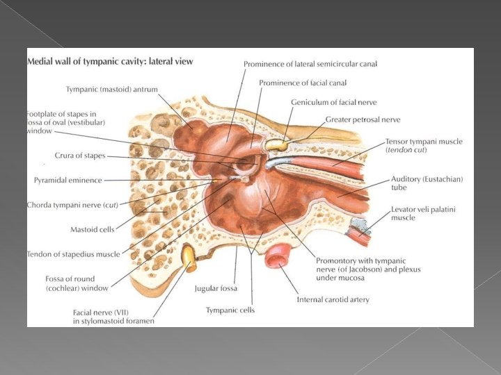
WALLS OF TYMPANIC CAVITY • Roof • Floor • Anterior wall • Posterior wall • Lateral wall • Medial wall
 Tube • Tympanic (Middle Ear) Cavity • Mastoid")
MIDDLE EAR CLEFT • Eustachian (Pharyngo-tympanic) Tube • Tympanic (Middle Ear) Cavity • Mastoid antrum and air cells

Mastoid Antrum & Air cells

Relationships of the mastoid antrum.

Mastoid Antrum & Air cells

CONTENTS OF MIDDLE EAR CAVITY • Air • Ossicles Malleus, Incus, & Stapes • Muscles Tensor Tympani & Stapedius • Nerves Chorda Tympani & Tympanic Plexus

LINING OF MIDDLE EAR Mucous membrane : ciliated columnar anteriorly and cuboidal or flat elsewhere

SENSARY SUPPLY OF MIDDLE AND EXTERNAL EAR • Cervical II & III ( great auricular and lessor occipital) • V cranial nerve ( auriculotemporal) • IX cranial nerve (tympanic or Jacobson’s) • X cranial nerve ( auricular or Arnold’s) • ? VII cranial nerve

Referred Earache • Pain in the ear due to a disease in an area supplied by a nerve that also supply the ear.

Referred Earache • Cervical II & III – Cervical spondylosis, neck injury etc. • V cranial nerve – Dental infections, sinonasal diseases etc. • IX cranial nerve – Tonsillitis, post-tonsillectomy, carcinoma etc. • X cranial nerve – Tumors of hpopharynx, larynx & esophagus

ANATOMY OF INNER EAR

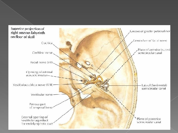
Osseous Labyrinth • Bony Cochlea • Vestibule • Bony semicircular canals

CONTENTS OF THE BONY LABYRINTH • Perilymph • Membranous labyrinth – Cochlear duct – Saccule and utricle – Membranous semicircular ducts

CONTENTS OF MEMBRANOUS LABYRINTH • Endolymph • Sensory epithelium – Cochlea: organ of Corti – Utricle & saccule: maculae – Semicircular canals: cristae

INNER EAR SENSORY EPITHELIUM • Cochlea: organ of Corti • Utricle & saccule: maculae • Semicircular canals: cristae

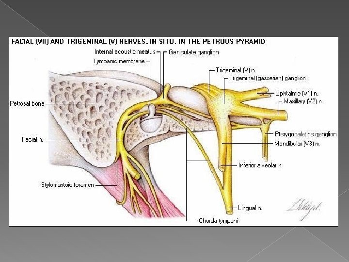
THE VESTIBULO-COCHLEAR NERVE

CENTRAL CONNECTIONS OF COCHLEAR NERVE

CENTRAL CONNECTIONS OF VESTIBULAR NERVE

PHYSIOLOGY OF THE EAR

FUNCIONS OF THE EXTERNAL EAR • Protection of the middle ear – Curvature – Cerumen • Auditory functions: – Sound conduction – Increase sound pressure by the resonance function

FUNCTIONS OF THE EUSTACHIAN TUBE Protection Ventilation Drainage

FUNCIONS OF THE MIDDLE EAR • Conduction of sound • Transformer mechanism – Hydraulic action – Ossicular leverage

FUNCIONS OF THE MIDDLE EAR • Conduction of sound • Transformer mechanism – Hydraulic action – Ossicular leverage • Protection to the inner ear – stapedial reflex

FUNCIONS OF THE INNER EAR • Hearing Function: – Transduction of sound to action potentials
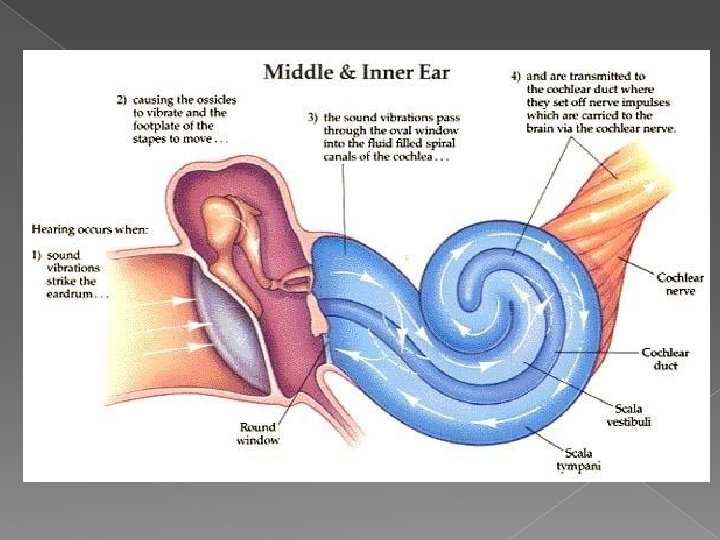

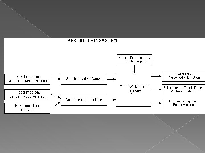
FUNCIONS OF THE INNER EAR • Hearing Function: – Transduction of sound to action potentials • Vestibular Function: – Participate in maintaining body balance

THE BALANCE SYSTEM C. Cortex BRAIN-STEM Cerebellum

THE BALANCE SYSTEM Vestibular Proprioceptive Visual INPUT C. Cortex BRAIN-STEM Cerebellum

THE BALANCE SYSTEM Vestibular Proprioceptive Visual INPUT C. Cortex BRAIN-STEM Cerebellum OUTPUT Ocular muscles Postural muscles


Objectives • Recognize the congenital anomalies of the external ear • Diagnose and treat wax accumulation • Diagnose and treat the common external ear inflammatory conditions • Discuss the pathology, clinical features and management of AOM

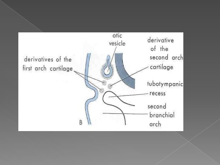
DEVELOPMENT OF THE EAR • External ear: 1 st pharyngeal cleft & arch • Middle ear: 1 st pharyngeal pouch & 1 st and 2 nd arches
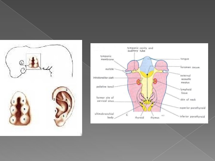

DEVELOPMENT OF THE EAR • External ear: 1 st pharyngeal cleft & arch • Middle ear: 1 st pharyngeal pouch & 1 st and 2 nd arches • Inner ear: Ectoderm of hindbrain

DISEASES OF THE EXTERNAL EAR

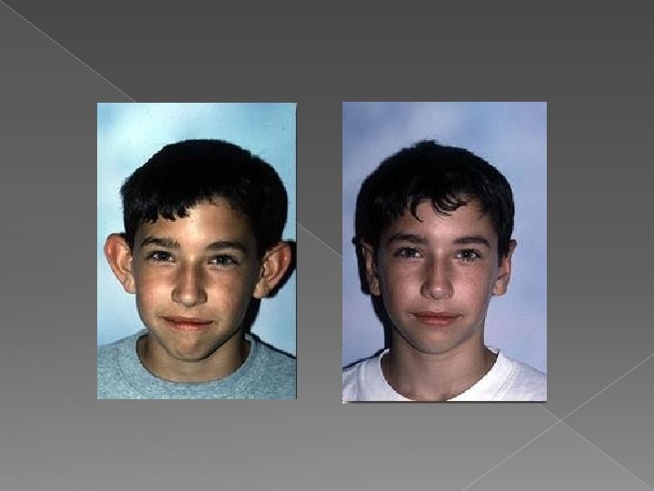
CONGENITAL MALFORMATIONS • Anotia & microtia • Accessory auricle • Preauricular sinus • Protruding ear

TRAUMA TO THE AURICLE • Lacerations • Hematoma auris

Complication Cauliflower ear
 or")
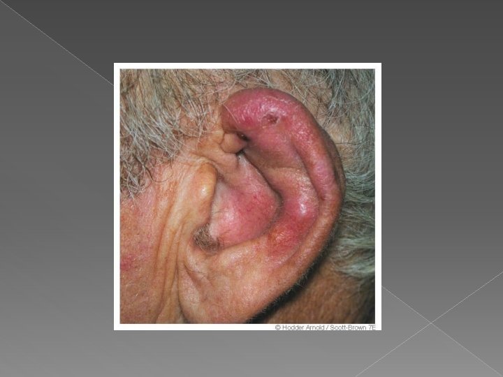
PERICHONDRITIS OF THE PINNA • Usually follow trauma (hematoma auris, surgical, frostbite, burn) or otitis externa • Commonly caused by Pseudomonas • Fever, pain, redness and swelling • Treatment is by antibiotics.

OTITIS EXTERNA

DEFINITION � An acute or chronic infection of the whole or a part of ear canal the skin of the external

CAUSES OF OTITIS EXTERNA

CAUSES OF OTITIS EXTERNA INFECTIVE REACTIVE

CAUSES OF OTITIS EXTERNA INFECTIVE Bacterial Fungal REACTIVE Viral

CAUSES OF OTITIS EXTERNA INFECTIVE Bacterial Fungal REACTIVE Viral Eczematous Seborrheic

CAUSES OF OTITIS EXTERNA INFECTIVE Bacterial Fungal Staph. arues Pseudomonos Others REACTIVE Viral Eczematous Seborrheic

CAUSES OF OTITIS EXTERNA INFECTIVE Bacterial REACTIVE Fungal Viral Staph. arues Aspirigillus Niger Pseudomonos Candida Albicans Others Eczematous Seborrheic

CAUSES OF OTITIS EXTERNA INFECTIVE Bacterial REACTIVE Fungal Viral Eczematous Staph. arues Aspirigillus Niger Herpes zoster Pseudomonos Candida Albicans Others Seborrheic

CLINICAL FEATURES OF OTITIS EXTERNA • • • Itching Pain Tenderness and swelling Otorrhea Deafness Changes in the lumen and skin of EAM
")
CLINICAL TYPES OF OTITIS EXTERNA • Localize O. E ( furuncle)
 • Diffuse infective")
CLINICAL TYPES OF OTITIS EXTERNA • Localize O. E ( furuncle) • Diffuse infective O. E.
 • Diffuse infective")
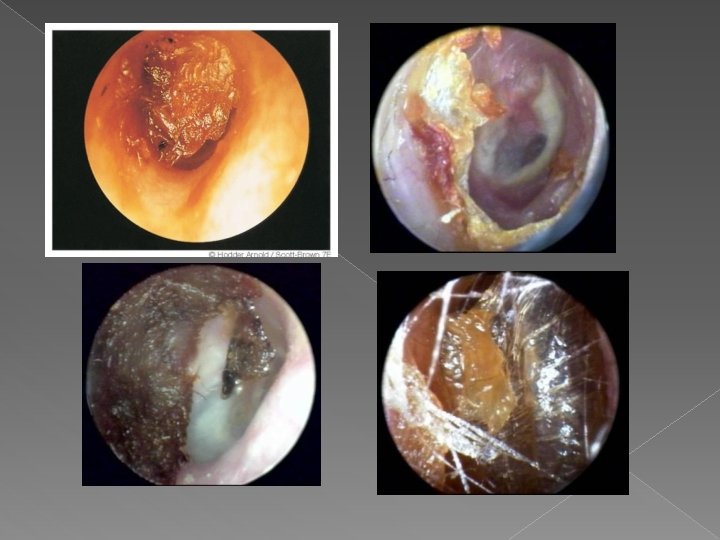
CLINICAL TYPES OF OTITIS EXTERNA • Localize O. E ( furuncle) • Diffuse infective O. E. • Otomycosis
 Diffuse infective")
CLINICAL TYPES OF OTITIS EXTERNA • • Localize O. E ( furuncle) Diffuse infective O. E. Otomycosis Bullous myringitis
 • Diffuse infective")
CLINICAL TYPES OF OTITIS EXTERNA • Localize O. E ( furuncle) • Diffuse infective O. E. • Otomycosis • Bullous myringitis • Herpetic O. E.
 • Diffuse infective")
CLINICAL TYPES OF OTITIS EXTERNA • Localize O. E ( furuncle) • Diffuse infective O. E. • Otomycosis • Bullous myringitis • Herpetic O. E • Eczematous and seborrheic O. E.
 • Diffuse infective")
CLINICAL TYPES OF OTITIS EXTERNA • Localize O. E ( furuncle) • Diffuse infective O. E. • Otomycosis • Bullous myringitis • Herpetic O. E • Eczematous and seborrheic O. E.

MANAGEMENT OF OTITIS EXTERNA • Swab for culture and sensitivity • Ear toilet • Keep the ear dry • Local medications • Systemic medications • Surgery may be required in chronic cases
 OTITIS EXTERNA • An acute Pseudomonas infection of the skin of the")
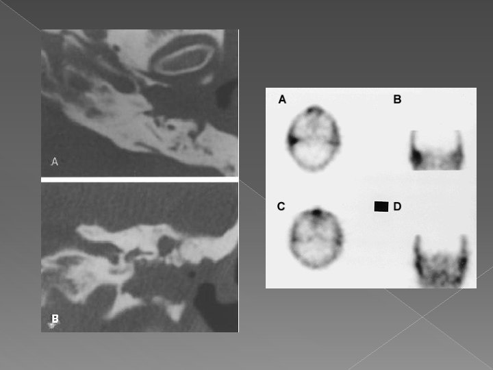
NECROTIZING (MALIGNANT) OTITIS EXTERNA • An acute Pseudomonas infection of the skin of the external ear canal which has spread to the adjacent bone. It occurs mostly in elderly diabetic patients.

Complications

DIAGNOSIS • Diabetes • Advanced age • Severe otalgia • Granulation tissue • Cranial nerve involvement • Radiology

TREATMENT • Control of diabetes • Anti Pseudomonas antibiotics • Local treatment and debridement • The role of surgery remains controversial

MISCELLANEOUS CONDITIONS OF THE EXTERNAL EAR

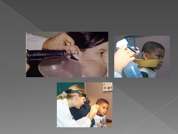
WAX • Mixture of ceruminous and sebaceous glands secretion • Normally is expelled from the canal aided by movements of the jaw • When accumulated it may cause deafness, earache or tinnitus

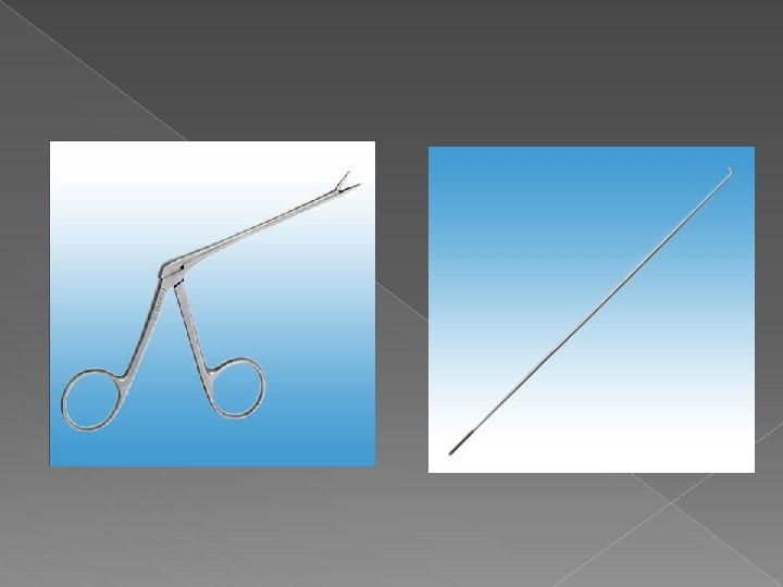
WAX � Treatment is by removal using syringing, suction or instrumentation

KERATOSIS OBTURANS • Accumulation of desquamated epithelium in the bony canal • It may be associated with sinusitis, bronchiectasis or primary ciliary dyskinesia • Usually cause deafness and pain • Treatment is periodic removal

ACUTE OTITIS MEDIA

DEFINITION Acute infection of the mucous membrane lining of the middle ear cleft

PREDISPOSING FACTORS • • Age Maleness Bottle feeding Climate Crowded living conditions Heredity Associated conditions: cleft palate, immuno-deficiency, down syndrome, cystic fibrosis , and ciliary dyskinesia ,

ROUTE OF INFECTION • Eustachian tube • External auditory canal • Blood borne

BACTERIOLOGY • Streptococcus pneumonia • Haemophilus influenzae • Branhamella catarrhalis • Streptococcus pyogens • Staphylococcus aureus

PATHOLOGY

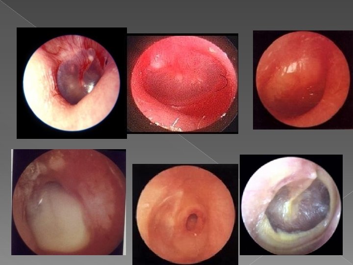
CLINICAL PICTURE • Tubal occlusion • Suppurative inflammation • Tympanic membrane rupture • Resolution

CLINICAL PICTURE • Tubal occlusion Discomfort, autophony, retracted drum • Suppurative inflammation • Tympanic membrane rupture • Resolution

CLINICAL PICTURE • Tubal occlusion Discomfort, autophony, retracted drum • Suppurative inflammation Fever, sever earache, deafness, bulging drum • Tympanic membrane rupture • Resolution

CLINICAL PICTURE • Tubal occlusion Discomfort, autophony, retracted drum • Suppurative inflammation Fever, sever earache, deafness, bulging drum • Tympanic membrane rupture – Otorrhea. . . temp. & earache subside • Resolution

CLINICAL PICTURE • Tubal occlusion Discomfort, autophony, retracted drum • Suppurative inflammation Fever, sever earache, deafness, bulging drum • Tympanic membrane rupture – Otorrhea. . . temp. & earache subside • Resolution

TREATMENT • Symptomatic • Antimicrobials – Amoxycillin/clavulanic acid – Tri-methoprimsulphamethoxazole – Cefaclor, cefixime – Erythromycin-sulfisoxazole • Decongestant • Myringotomy • Eartoilet and local antibiotics

RECURRENT ACUTE OTITIS MEDIA

DEFINITION • Three or more attacks over a 6 -months period

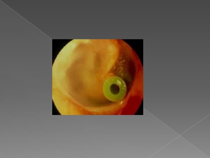
TREATMENT • Long-term low dose antimicrobials • Ventilation tube insertion

THANK YOU
- Slides: 116