Breast Surgery Introduction Prof Mohamed Gaber Professor of
 Prof. Mohamed Gaber Professor of Surgery & Surgical Oncology Faculty of")
Breast Surgery (Introduction) Prof. Mohamed Gaber Professor of Surgery & Surgical Oncology Faculty of Medicine, Alexandria University

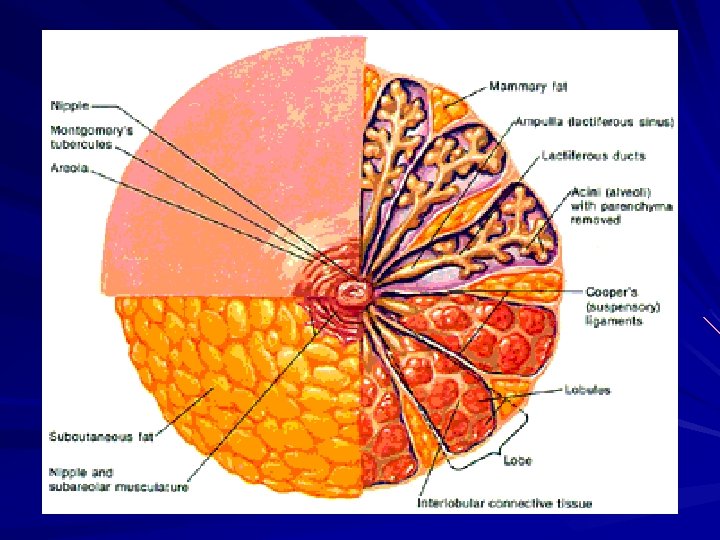
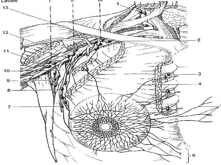
Anatomy Modified Sweat Gland between the skin on the pectoral Fascia and chest wall 2 nd rib Ant. Lateral axillary Border 6 th rib line of sternum It consists of 12 -20 lactiferous ducts which open into the nipple by separate openings. Fibrous tissue septa around the breast lobules pass between the skin and pectoral fascia (Cooper’s Ligament)

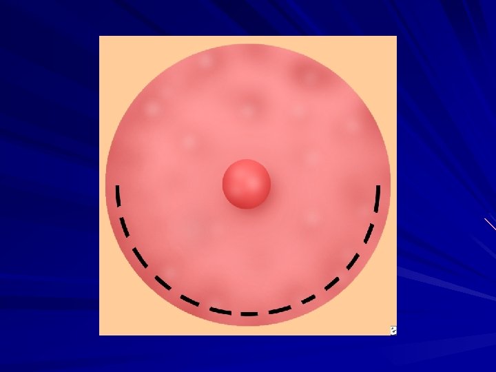
The nipple is a protuberant part covered by thick wrinkled skin, and contains muscle fibers which facilitates its contraction. The nipple is surrounded by the areola, which is circular pigmented skin. It contains sweat glands & sebaceous glands ( Montgomery glands). It contains contractile smooth muscle fibers also.
 Ø Lateral Thoracic")
Blood supply Arteries: Ø Internal Mammary A. ( subclavian A. ) Ø Lateral Thoracic A. ( Axillary A. ) Ø Pectoral Branches of the thoraco-acromial A. ( Axillary A. )
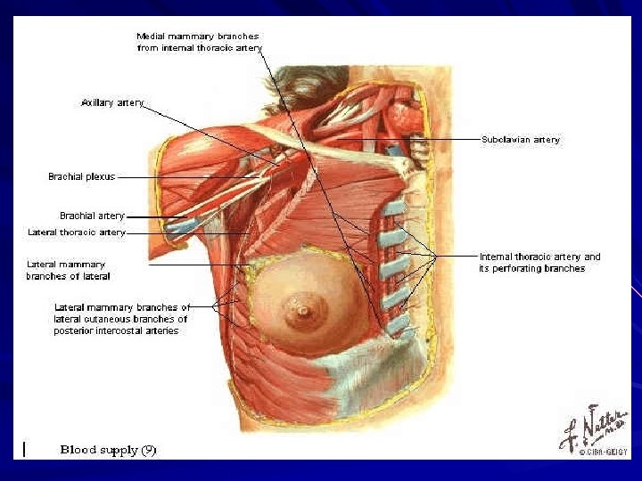
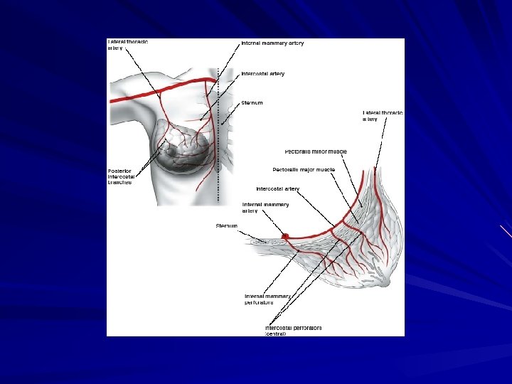

Veins: ØInternal Mammary vein. Ø Deep mammary veins intercostal vein Posterior vertebral vein.

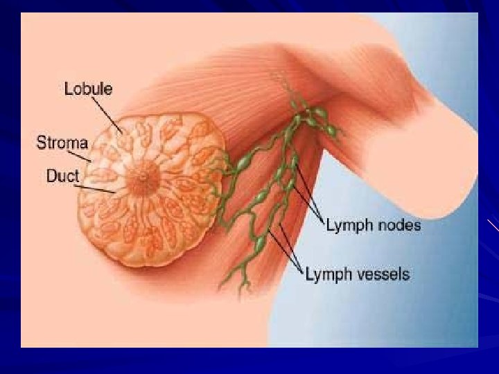
Lymphatics: The breast is rich in lymphatics. The lymphatics pass from the subareolar region and the rest of the breast tissue along the lactiferous ducts in the breast tissue.

Afterwards 3 main pathways could be identified: 1. Most of the lymphatics pass along the lateral thoracic vessels to the pectoral group, then to the central, followed by the apical group. 2. Lymphatics draining the posterior part pass along the thoraco-acromial vessels to the inter pectoral nodes, then to the apical group. 3. Medial half lymphatics pass to the internal mammary nodes (first 3 spaces)

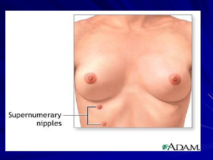
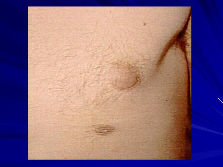
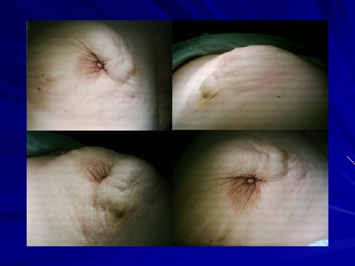
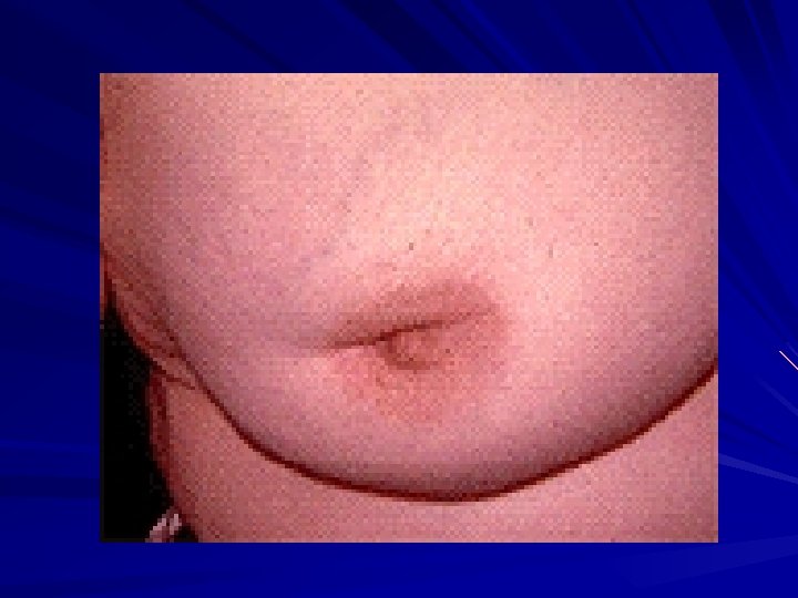
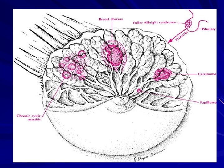
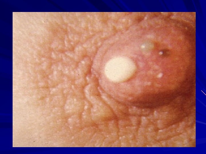
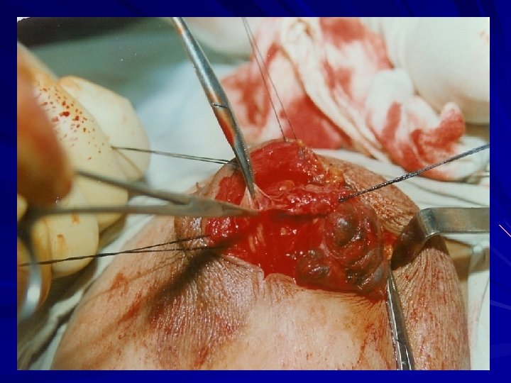
Diseases of the nipple Athelia : Congenital absence. Supernumerary nipples : along the nipple line. Retraction : Congenital : Short lactiferous ducts or acquired : Cancer or MDE 4) Fissure : during lactation acute mastitis. 5) Paget’s disease of the nipple. 1) 2) 3)
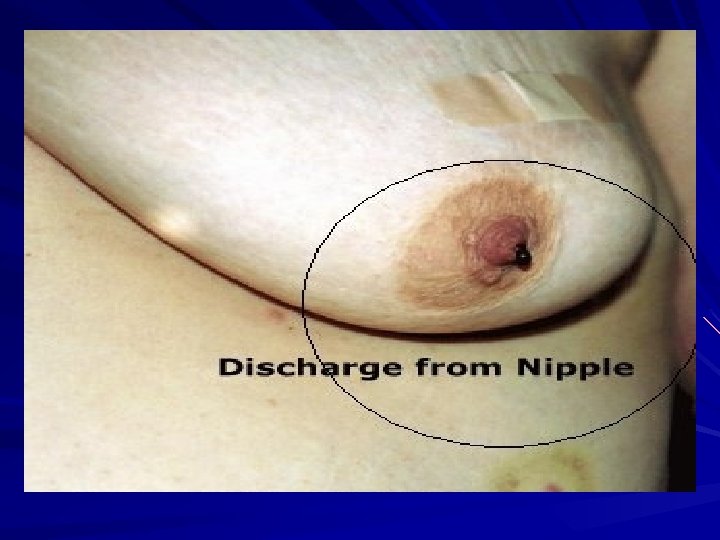
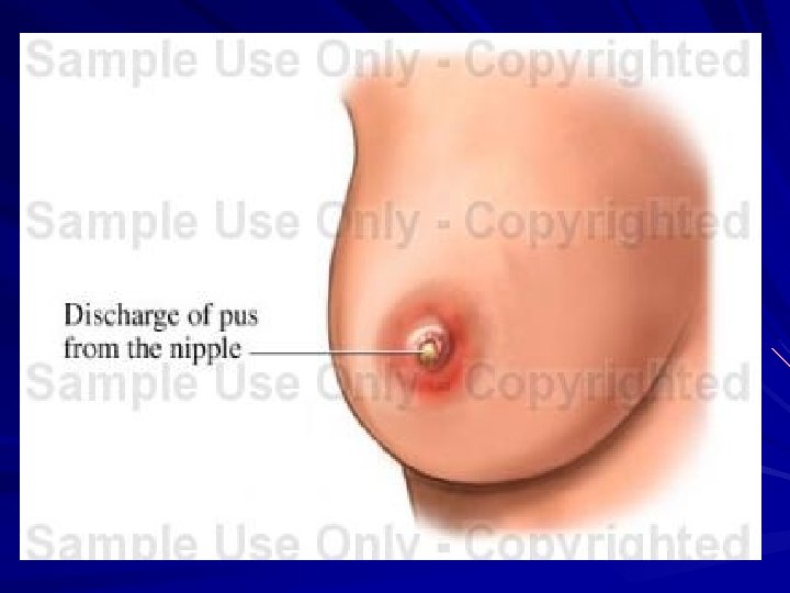
 Nipple discharge: Types & causes: § Clear : Fibroadenosis. § Blood : duct")
6) Nipple discharge: Types & causes: § Clear : Fibroadenosis. § Blood : duct papilloma or carcinoma. § Green- Black: fibroadenosis or MDE § Creamy ( pus ) : abscess. § White : before delivary and after lactation and in hyperprolactinemia.
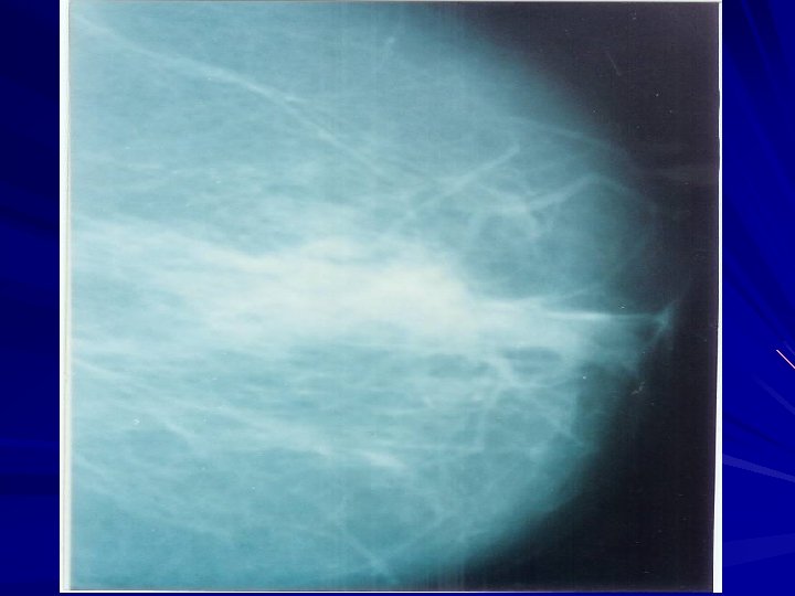

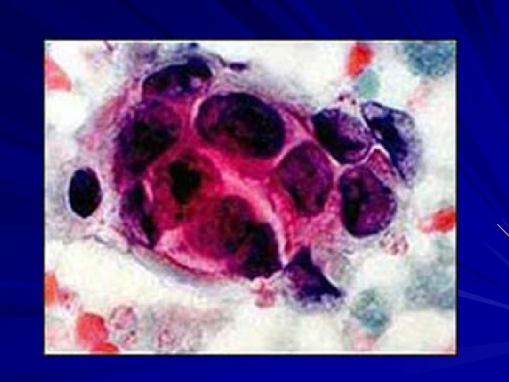
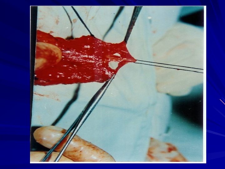
Management Policy : § Associated with a lump : remove the lump Biopsy manage accordingly. § No lump: Localized to one duct Microdochectomy. From many ducts Smear cytology Malignant cells ( mastectomy ) Non- Malignant cells ( observe ) § Many duct discharge over 45 years Major duct excision in patients

- Slides: 29