
BREAST 34 Fibrocystic Diseasechange Pathology Harmone induced acinar

BREAST. 3&4.



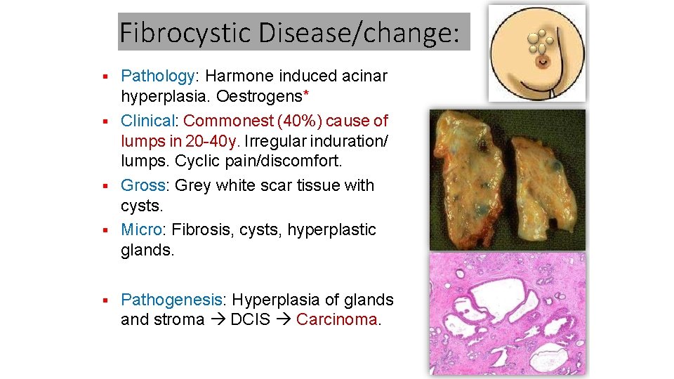
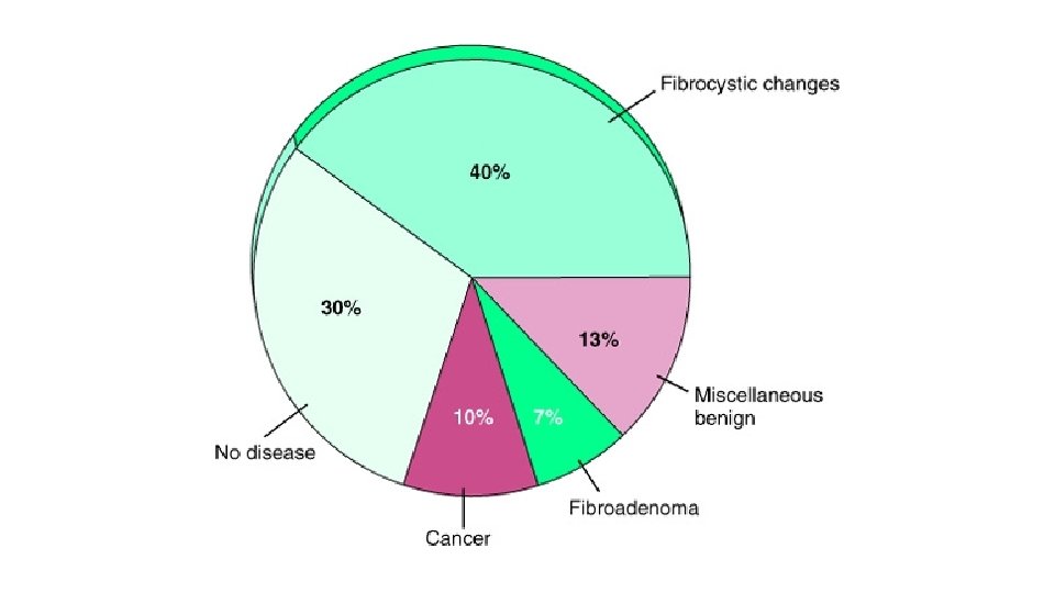
Fibrocystic Disease/change: Pathology: Harmone induced acinar hyperplasia. Oestrogens* Clinical: Commonest (40%) cause of lumps in 20 -40 y. Irregular induration/ lumps. Cyclic pain/discomfort. Gross: Grey white scar tissue with cysts. Micro: Fibrosis, cysts, hyperplastic glands. Pathogenesis: Hyperplasia of glands and stroma DCIS Carcinoma.
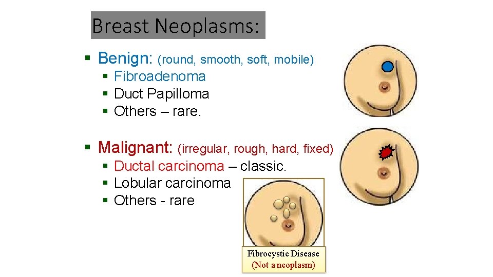
Breast Neoplasms: Benign: (round, smooth, soft, mobile) Fibroadenoma Duct Papilloma Others – rare. Malignant: (irregular, rough, hard, fixed) Ductal carcinoma – classic. Lobular carcinoma Others - rare Fibrocystic Disease (Not a neoplasm)


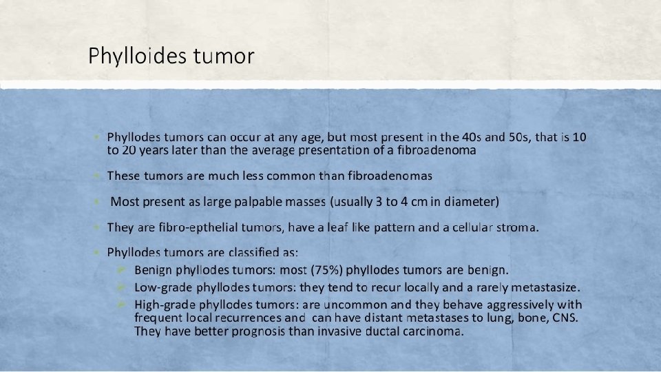
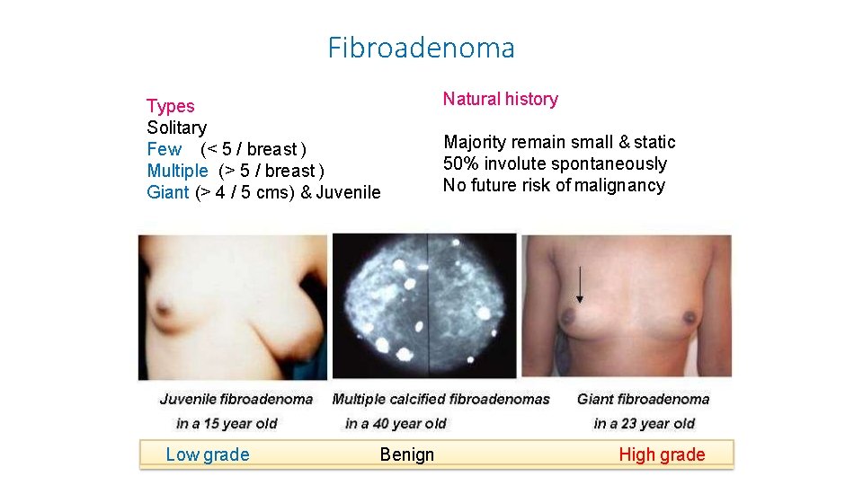
Fibroadenoma Types Solitary Few (< 5 / breast ) Multiple (> 5 / breast ) Giant (> 4 / 5 cms) & Juvenile Low grade Benign Natural history Majority remain small & static 50% involute spontaneously No future risk of malignancy High grade
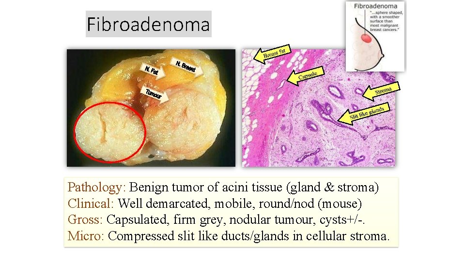
Fibroadenoma Pathology: Benign tumor of acini tissue (gland & stroma) Clinical: Well demarcated, mobile, round/nod (mouse) Gross: Capsulated, firm grey, nodular tumour, cysts+/-. Micro: Compressed slit like ducts/glands in cellular stroma.
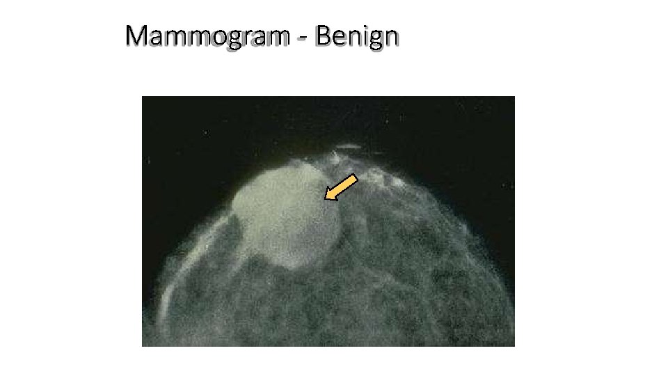
Mammogram - Benign
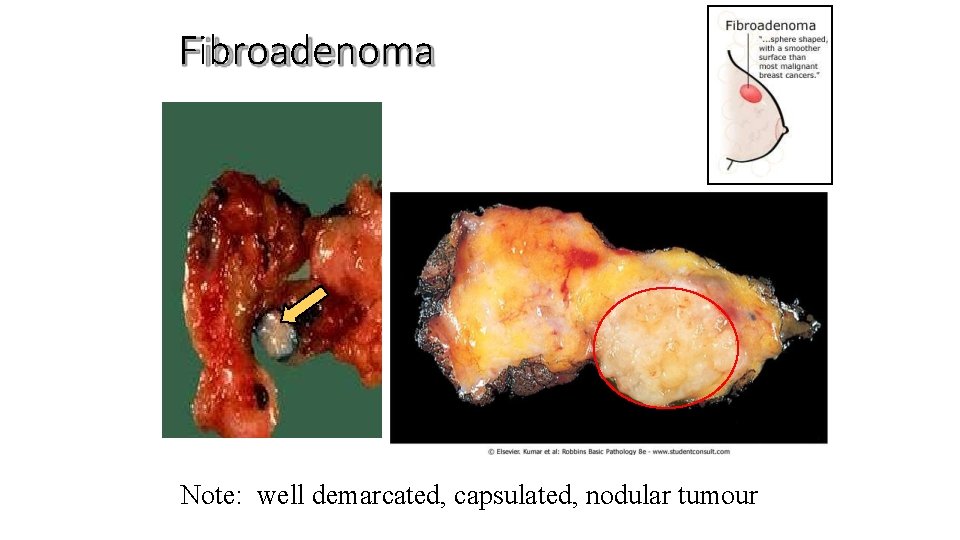
Fibroadenoma Note: well demarcated, capsulated, nodular tumour
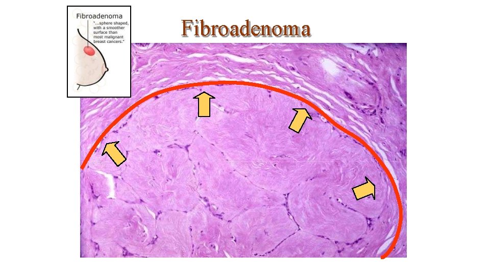
Fibroadenoma
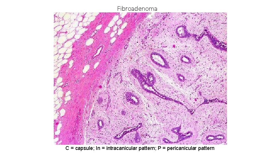
Fibroadenoma In P P In P C = capsule; In = intracanicular pattern; P = pericanicular pattern
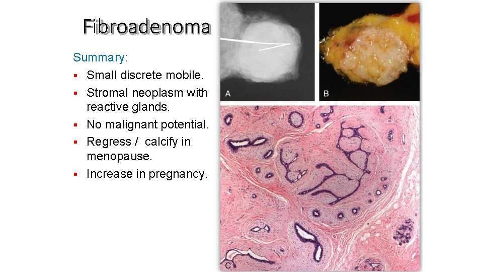
Fibroadenoma Summary: Small discrete mobile. Stromal neoplasm with reactive glands. No malignant potential. Regress / calcify in menopause. Increase in pregnancy.
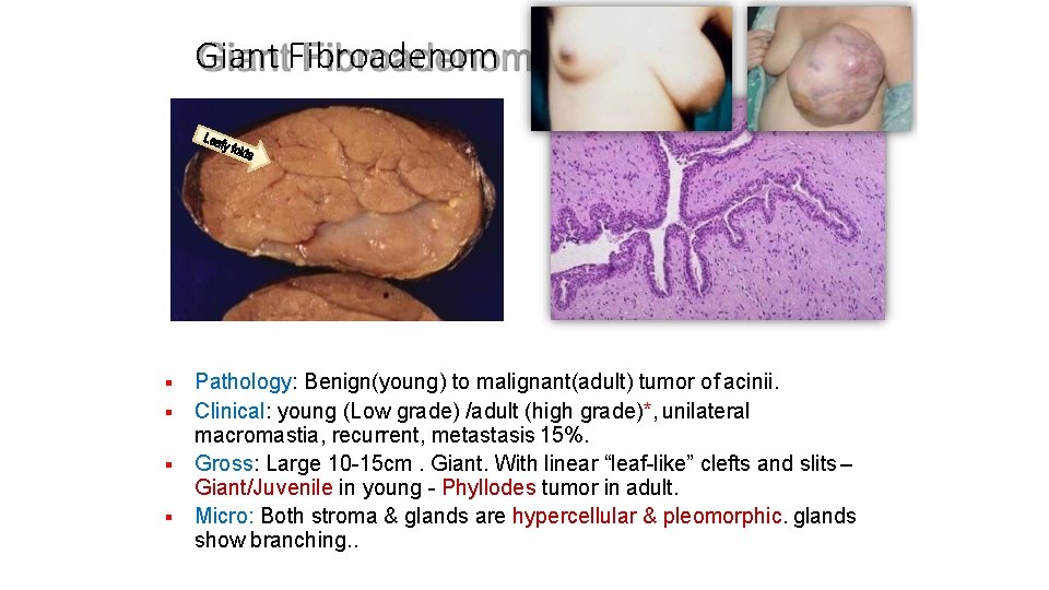
Giant Fibroadenom a Pathology: Benign(young) to malignant(adult) tumor of acinii. Clinical: young (Low grade) /adult (high grade)*, unilateral macromastia, recurrent, metastasis 15%. Gross: Large 10 -15 cm. Giant. With linear “leaf-like” clefts and slits – Giant/Juvenile in young - Phyllodes tumor in adult. Micro: Both stroma & glands are hypercellular & pleomorphic. glands show branching. .
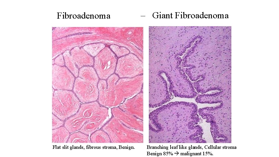
Fibroadenoma Flat slit glands, fibrous stroma, Benign. – Giant Fibroadenoma Branching leaf like glands, Cellular stroma Benign 85% malignant 15%.
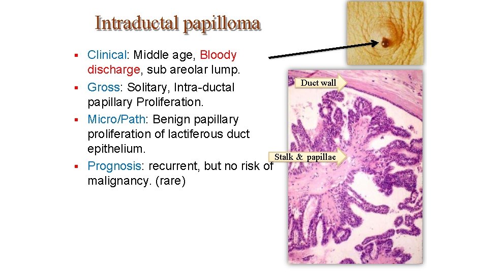
Intraductal papilloma Clinical: Middle age, Bloody discharge, sub areolar lump. Duct wall Gross: Solitary, Intra-ductal papillary Proliferation. Micro/Path: Benign papillary proliferation of lactiferous duct epithelium. Stalk & papillae Prognosis: recurrent, but no risk of malignancy. (rare)
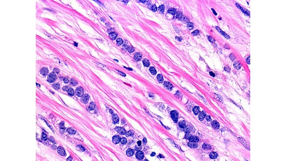
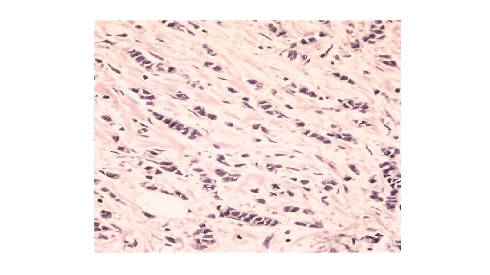

MALE BREAST

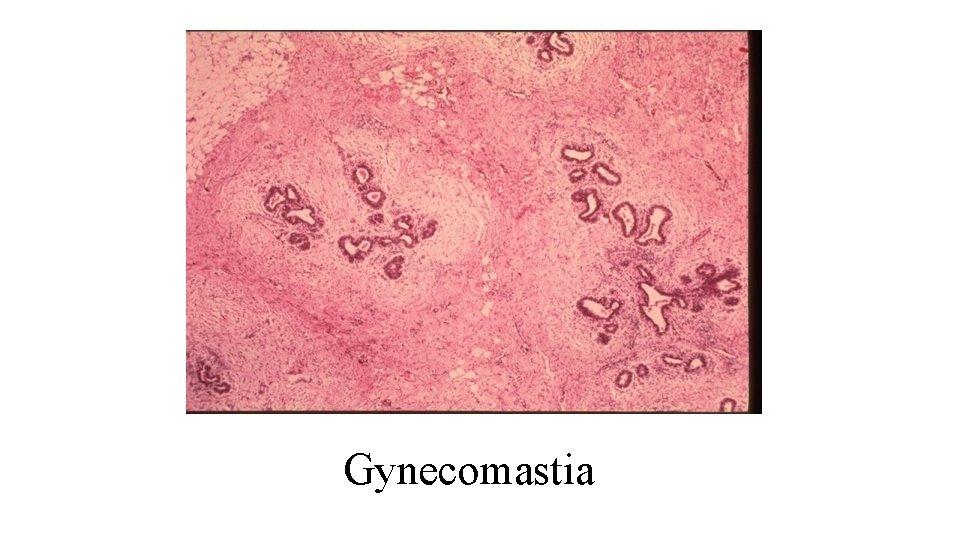
Gynecomastia
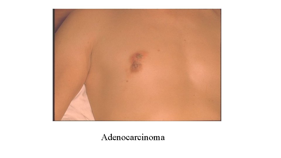
Adenocarcinoma

Knowledge is a burden, If it robs you of innocence, If it makes you feel you are special, If it gives you an idea you are wise, If it is not integrated into life, If it does not bring you joy, If it does not set you free. Sri Ravi Shankar, Humanitarian and founder of the Art of Living Foundation, India.
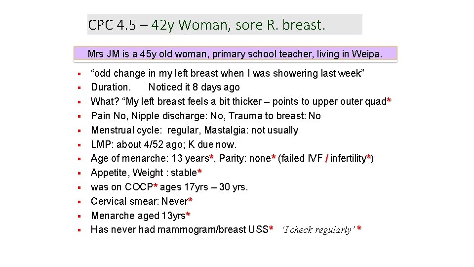
CPC 4. 5 – 42 y Woman, sore R. breast. Mrs JM is a 45 y old woman, primary school teacher, living in Weipa. “odd change in my left breast when I was showering last week” Duration. Noticed it 8 days ago What? “My left breast feels a bit thicker – points to upper outer quad* Pain No, Nipple discharge: No, Trauma to breast: No Menstrual cycle: regular, Mastalgia: not usually LMP: about 4/52 ago; K due now. Age of menarche: 13 years*, Parity: none* (failed IVF / infertility*) Appetite, Weight : stable* was on COCP* ages 17 yrs – 30 yrs. Cervical smear: Never* Menarche aged 13 yrs* Has never had mammogram/breast USS* ‘I check regularly’ *
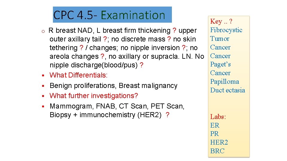
CPC 4. 5 - Examination o Key. . ? Fibrocystic R breast NAD, L breast firm thickening ? upper Tumor outer axillary tail ? ; no discrete mass ? no skin Cancer tethering ? / changes; no nipple inversion ? ; no areola changes ? , no axillary or supracla. LN. No Cancer Paget’s nipple discharge(blood/pus) ? Cancer What Differentials: Papilloma Benign proliferations, Breast malignancy Duct ectasia What further investigations? Mammogram, FNAB, CT Scan, PET Scan, Biopsy + immunochemistry (HER 2) ? Labs: ER PR HER 2 BRC
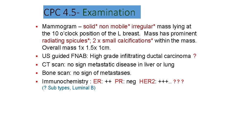
CPC 4. 5 - Examination Mammogram – solid* non mobile* irregular* mass lying at the 10 o’clock position of the L breast. Mass has prominent radiating spicules*; 2 x small calcifications* within the mass. Overall mass 1 x 1. 5 x 1 cm. US guided FNAB: High grade infiltrating ductal carcinoma ? CT scan: no sign metastatic disease in liver or lung Bone scan: no sign of metastases. Immunochemistry : ER: ++ PR: neg HER 2: +++. . ? ? ? (? Sub types, Luminal B)
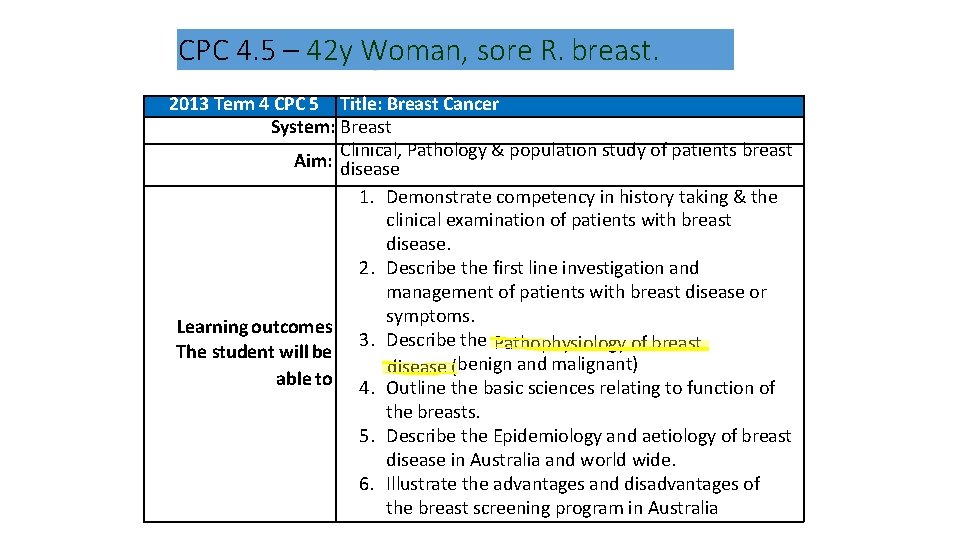
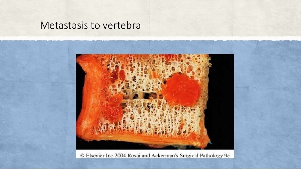
CPC 4. 5 – 42 y Woman, sore R. breast. 2013 Term 4 CPC 5 Title: Breast Cancer System: Breast Clinical, Pathology & population study of patients breast Aim: disease 1. Demonstrate competency in history taking & the clinical examination of patients with breast disease. 2. Describe the first line investigation and management of patients with breast disease or symptoms. Learning outcomes 3. Describe the Pathophysiology of breast The student will be disease (benign and malignant) able to 4. Outline the basic sciences relating to function of the breasts. 5. Describe the Epidemiology and aetiology of breast disease in Australia and world wide. 6. Illustrate the advantages and disadvantages of the breast screening program in Australia
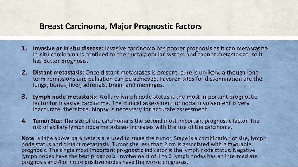
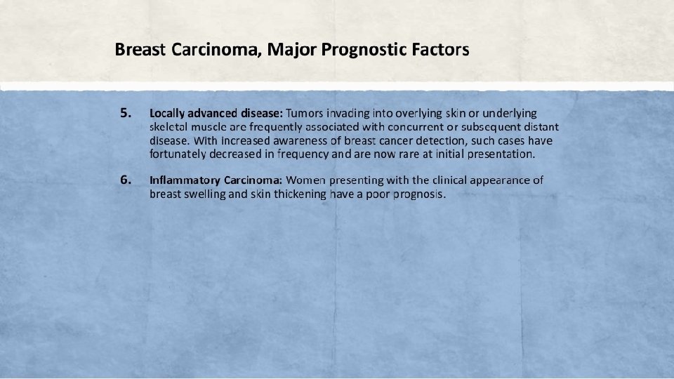
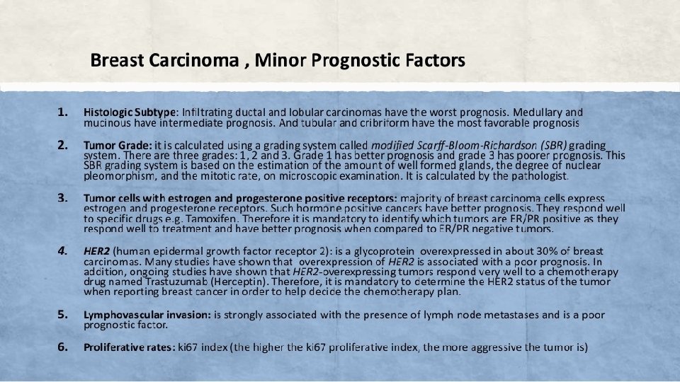
CPC 4. 5 - Core Learning Issues Pathology Major CLI: • • • Pathology of Breast – overview, classification & common dis. . Breast Lumps - Differential diagnosis. Trauma, infections & Inflam. – Mastitis, fat necrosis, abscess. Hyperplasia – Fibrocystic disease Tumours – Benign – Fibroadenoma, giant fibroadenoma. Breast cancer – etiology, pathogenesis, morphology & complications, Laboratory diagnosis, including markers. Pathology Minor CLI: • • Duct ectasia, Breast Cysts. Paget’s disease. Gynecomastia & male breast disorders.
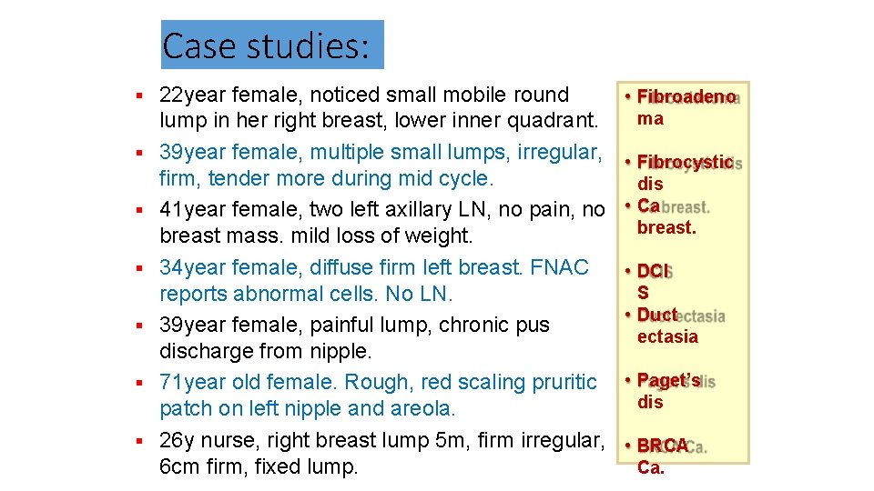
Case studies: 22 year female, noticed small mobile round lump in her right breast, lower inner quadrant. 39 year female, multiple small lumps, irregular, firm, tender more during mid cycle. 41 year female, two left axillary LN, no pain, no breast mass. mild loss of weight. 34 year female, diffuse firm left breast. FNAC reports abnormal cells. No LN. 39 year female, painful lump, chronic pus discharge from nipple. 71 year old female. Rough, red scaling pruritic patch on left nipple and areola. 26 y nurse, right breast lump 5 m, firm irregular, 6 cm firm, fixed lump. • Fibroadeno ma • Fibrocystic dis • Ca breast. • DCI S • Duct ectasia • Paget’s dis • BRCA Ca.
Self assessment: Clinical features of benign, malignant & reactive… Breast cancer screening guidelines. Hyperplasia / tumour features. Familial vs Non familial breast Ca features. Screening Mammogram – policy, procedure & interpretation. Fibrocystic disease, fibroadenoma & cancer. Breast cancer common types & features (gross, microscopy, complications etc. ) Duct carcinoma, lobular carcinoma, other types. BRCA testing in familial breast ca.
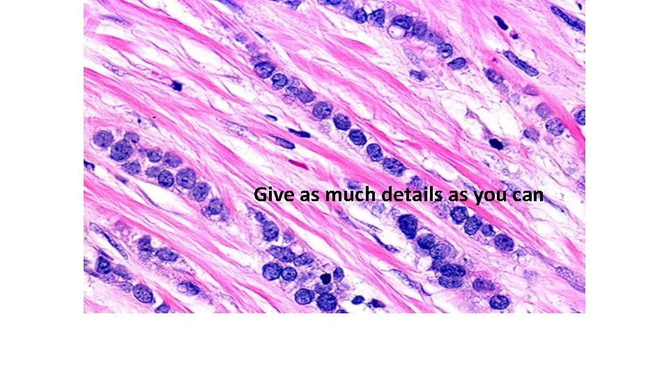
Give as much details as you can
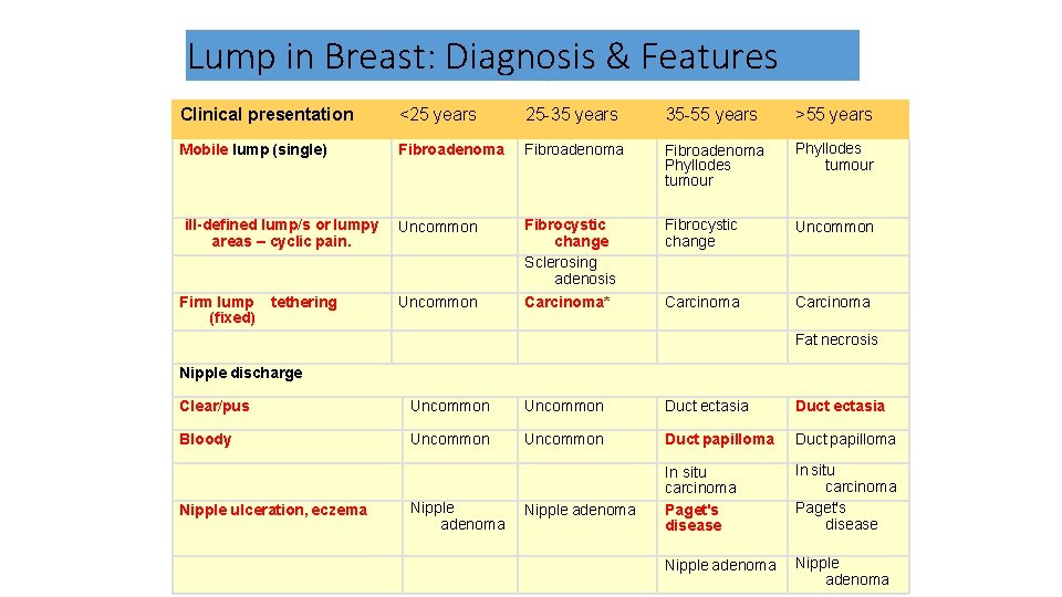
Lump in Breast: Diagnosis & Features Clinical presentation <25 years 25 -35 years 35 -55 years >55 years Mobile lump (single) Fibroadenoma Phyllodes tumour Uncommon Fibrocystic change Sclerosing adenosis Carcinoma* Fibrocystic change Uncommon Carcinoma ill-defined lump/s or lumpy areas – cyclic pain. Firm lump (fixed) tethering Uncommon Fat necrosis Nipple discharge Clear/pus Uncommon Duct ectasia Bloody Uncommon Duct papilloma In situ carcinoma Paget's disease Nipple adenoma Nipple ulceration, eczema Nipple adenoma
- Slides: 50