Branches of Coeliac trunk 1 Left gastric artery

 Left gastric artery, 2) Splenic artery and 3) Hepatic")
Branches of Coeliac trunk 1) Left gastric artery, 2) Splenic artery and 3) Hepatic artery

Coeliac Trunk - The artery of the foregut (abdominal part of the esophagus, stomach, 1 st part and upper 1/2 of the 2 nd part of the duodenum, pancreas (upper 1/2 of the head, body, and tail), liver, gall bladder and spleen. ** Origin, from the front of the abdominal aorta at the level of the T 12. ** Branches 1 - Left gastric artery. 2 - Hepatic artery. 3 - Splenic artery • Left gastric artery supplies 1) abdominal part of the esophagus. 2) Cardiac end of the stomach and Upper part of the body of the stomach along lesser curvature. • Splenic Artery ** Origin, it is the largest branch of the coeliac trunk. ** Course, - It runs as a tortuous course along the upper border of pancreas behind the stomach. • Why tortuous: Protects the artery during gastric distention and splenic enlargement. 2 - Slows the rate of blood flow to the spleen. ** Branches 1 - Pancreatic branches to the pancreas. 2 - Left gastroepiploic artery runs along the greater curvature of the stomach. 3 - Short gastric arteries to the fundus of the stomach. 4 - Terminal 5 to 6 splenic branches which pass into the hilum of the spleen

Hepatic artery

** Branches of Hepatic Artery 1 - Right gastric artery runs on the lesser curvature of the stomach. - It ends by anastomosing with left gastric artery. 2 - Gastroduodenal artery descends behind the first part of the duodenum. divides into two: a- Right gastroepiploic artery runs along the greater curvature of the stomach. - It ends by anastomosing with the left gastro-epiploic artery. b- Superior pancreaticoduodenal artery runs between the duodenum and head of pancreas. It ends by anastomosing with the inferior pancreaticoduodenal artery. 3 - Supraduodenal artery to the first part of the duodenum. 4 - Left terminal hepatic branch to the left lobe of the liver, 5 - Right terminal hepatic branch to the right lobe of the liver. 6 - Cystic artery to the gall bladder from the right hepatic branch

Clinical notes 1 - If the hepatic artery is ligated proximal to the right gastric branch, - A collateral circulation to the liver through Left gastric, splenic and superior mesenteric arteries. 2 - If the hepatic artery is ligated distal to the gastroduodenal branch, hepatic necrosis commonly occurs. - A collateral circulation to the liver is only carried by the inferior phrenic arteries.

** Origin, from the front of the abdominal aorta at L 1. ** Course - Its origin behind by the body of the pancreas. - It crosses in front of, 1 - Uncinate process of pancreas. 2 - Third part of duodenum. 3 - Abdominal aorta. 4 - Inferior vena cava. 5 - Right psoas major separated from it by a) Right ureter and b) Right gonadal vessels infront the ureter. Superior mesenteric artery
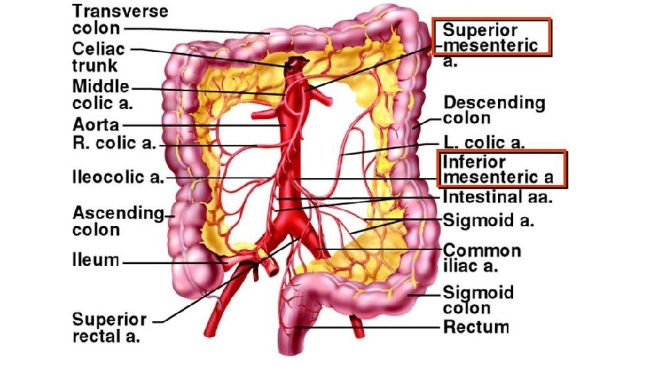

Superior Mesenteric Artery - This is the artery of the midgut (lower 1/2 of the 2 nd part, 3 rd, 4 th of the duodenum, pancreas (lower 1/2 of the head, uncinate processes), jejunum, ileum, caecum, ascending colon and right 2/3 of the transverse colon) ** Branches 1 - Inferior pancreatico-duodenal artery: - It ends by anastomosing with the superior pancreatico-duodenal artery. - It supplies the duodenum below the major papilla and lower half of the head of pancreas. 2 - Jejunal to the jejunum. 3 - Ileal ranches to the ileum. 4 - Ileocolic artery 5 - Right colic artery to the ascending colon and right colic flexure. - It divides into: a- Ascending branch anastomoses with the right branch of the middle colic artery. b- Descending branch anastomoses with the ascending branch of the ileocolic artery. 6 - Middle colic artery, to the right 2/3 of the transverse colon. It divides into a- Right branch anastomoses with the ascending branch of right colic artery. b- Left branch anastomoses with the ascending branch of the left colic artery.

- Ileocolic artery gives 1 - Ileal branches to the terminal part of the ileum. 2 - Anterior caecal artery to the caecum. 3 - Posterior caecal artery to the caecum. 4 - Appendicular artery to the appendix. 5 - Ascending (colic) branch to the lower part of the ascending colon.

Inferior Mesenteric Artery This is the artery of the hindgut (left 1/3 of the transverse colon, descending colon, pelvic colon, rectum and anal canal). ** Origin, from the abdominal aorta at the level of the 3 rd lumbar vertebra. ** Termination: It continues as the superior rectal artery. ** Branches 1 - Left colic (upper) artery It divides into, a- Ascending branch anastomoses with the left branch of the middle colic artery b- Descending branch anastomoses with the highest sigmoid artery. 2 - Sigmoid arteries (2 -3), anastomoses with the left colic and superior rectal arteries. 3 - Superior rectal (Hemorrhoidal) artery to the rectum and anal canal. - It anastomoses with the middle and inferior rectal arteries

** PORTAL VEIN is formed by the union of the Superior mesenteric and Splenic veins behind the neck of the pancreas (S+S=P). - It is about 2 inches long. - The portal vein and its tributaries have NO valves

** Tributaries of the portal vein 1 - Superior mesenteric vein. 2 - Splenic vein. 3 - Right gastric vein. 4 - Left gastric vein. 5 - Paraumbilical vein to left terminal branch. 6 - Cystic vein to the right terminal branch. - Inferior mesenteric vein ends in the splenic vein.

Portosystemic anastomosis

Portosystemic Anastomoses ** The anastomosis between the portal venous systems and systemic veins. 1 - At the lower end of the oesophagus, between a- Tributaries of the left gastric vein (portal). b- Tributaries of the hemi-azygos vein (systemic), - In case of portal hypertension leading to enlargement of these anastomoses oesophageal varices. 2 - In the wall of the anal canal, between a- Tributaries of the superior rectal vein (portal). b- Tributaries of the middle and inferior rectal veins (systemic). - In case of portal hypertension leading to enlargement of these anastomoses piles. 3 - Around the umbilicus, between a- Para-umbilical vein (portal). b- Superficial veins of the anterior abdominal wall (systemic). - In case of portal hypertension leading to enlargement of these anastomoses caput Medusae. 4 - At the bare area of the liver, between a- Portal capillaries (portal). b- Inferior phrenic veins of the diaphragm (systemic). 5 - At the posterior abdominal wall, between a- Veins of the colon (portal). b- Veins of the posterior abdominal wall and renal veins (systemic)

Ascites Caput Medusae

Th ank Qu you est ion s I/Azzam - 2004
- Slides: 17