BRAINSTEM DR GAUTHAM KAMBLE BRAIN STEM Midbrain Pons

BRAINSTEM DR GAUTHAM KAMBLE

BRAIN STEM Midbrain Pons Medulla

• Stalk like part of brain which connects spinal cord with forebrain • Located in posterior cranial fossa • Consists of midbrain, pons and medulla oblongata • Midbrain is continuous above with the cerebral hemispheres • Medulla is continuous below with the spinal cord

MIDBRAIN

PONS

MEDULLA

• Posteriorly pons and medulla are separated from the cerebellum by fourth ventricle • Anteriorly lies on clivus

• Cranial nerves are attached to brainstem

Midbrain, pons and medulla oblongata are connected to cerebellum by superior, middle and inferior cerebellar peduncles respectively Superior cerebellar peduncles Middle cerebellar peduncle Inferior cerebellar peduncles

Functions : 1. 2. 3. 4. Passage to various ascending and descending tracts Contains important autonomic reflex centres Contains reticular activating system which controls consciousness Contains important nuclei of last 10 cranial nerves

MEDULLA OBLONGATA • Direct upward continuation of spinal cord • Extends from foramen magnum to lower border of pons • Almost vertical and lies between the clivus and vallecula of cerebellum behind • Shaped like a truncated cone (bulb like) • 3 x 2 x 1. 25 cms • Contains cardiac centre, vasomotor centre and respiratory centre • Provides attachment to last four cranial nerves

EXTERNAL FEATURES • Divided into 2 halves by anterior median fissure and posterior median sulcus • Anterior median fissure is continuous below with corresponding fissure of spinal cord • Above it ends in a triangular depression – foramen caecum

• In lower part – decussation of pyramids • Posterior median sulcus continues below with the corresponding sulcus and present in lower half only • Above its lips diverge to form boundaries of triangular area, lower part of fourth ventricle

• Each half made up of two sulci – anterolateral and posterolateral sulci • Anterolateral sulcus lies in line with ventral roots of spinal nerves • Along the anterolateral sulcus emerge rootlets of XII cranial nerve • Posterolateral sulcus lies in line with dorsal roots of spinal nerves • Along the posterolateral sulcus emerge rootlets of IX, X and XI cranial nerve

Features on ventral aspect : 1. Pyramids : - Longitudinal elevations between anterior median fissure and anterolateral sulcus - Produced by bundles of corticospinal fibres - Most of these fibres (75%) cross to opposite side at pyramidal decussation 2. Olives: - Oval elevation between anterolateral and posterolateral sulcus - Produced due to underlying inferior olivary nucleus

3. Inferior cerebellar peduncle: - Thick bundle of fibres which lie posterolateral to olive - Connects medulla with cerebellum

Features on dorsal aspect : Can be divided into 2 parts i. e lower closed part and upper open part CLOSED PART: - Between posterior median sulcus and posterolateral sulcus - Presents 2 longitudinal elevations fasciculus gracilis fasciculus cuneatus - Upper ends expand to form 2 tubercles i. e gracile and cuneate tubercle - Tuber cinereum- elevation lateral to cuneate tubercle produced by spinal nucleus of trigeminal nerve

OPEN PART: ØForms lower part of floor of fourth ventricle ØShows a number of features ØMedian sulcus , hypoglossal and vagal triangle , vestibular areas, area postrema, stria medullaris

INTERNAL STRUCTURE • Contains white matter which surrounds the central grey matter • But grey matter is broken up into nuclei that are separated by nerve fibres • Internal structure is well studied by examining transverse sections • Studied at 3 levels: a. At level of pyramidal decussation b. At level of sensory decussation c. At the level of olives
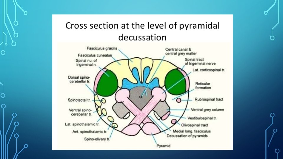

TRANSVERSE SECTION AT LEVEL OF PYRAMIDAL DECUSSATION • Section passes through inferior half of medulla • Central canal is surrounded by central grey matter • Behind central grey matter occupied by fasciculus gracilis and fasciculus cuneatus • Central grey matter shows extensions- nucleus gracilis and nucleus cuneatus • Apex of posterior horn gets swollen up and forms spinal nucleus of trigeminal nerve • Spinal tract of trigeminal nerve caps the nucleus

• Decussation of pyramid forms most important feature • Due to this ventral horns are separated from central grey matter • Area between ventral grey column and spinal nucleus of trigeminal nerve occupied by reticular formation
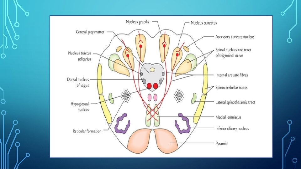

TRANSVERSE SECTION AT LEVEL OF SENSORY DECUSSATION • Section passes through middle of medulla • Nucleus gracilis and nucleus cuneatus become more pronounced and separated from central grey matter • Internal arcuate fibres arising from cells of gracile and cuneate nuclei pass forwards and medially and decussate with opposite side – sensory decussation • They ascend as medial lemniscus • Medial fibres come to lie anteriorly in medial lemniscus

• Accessory cuneate nucleus – dorsolateral to cuneate nuclei • Spinal nucleus and tract of trigeminal nerve- lie ventrolateral to cuneate nucleus • Lower part of inferior olivary nucleus • Pyramids lie on either side of anterior median fissure • Central grey matter shows 3 nuclei – hypoglossal nuceus, dorsal nucleus of vagus, nucleus of tractus solitaries • Medial longitudinal bundle- posterior to medial lemniscus
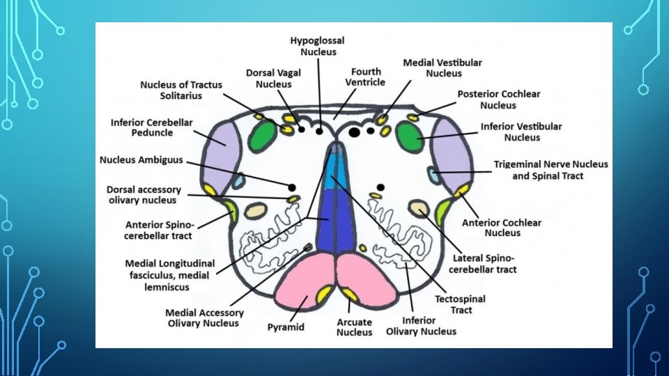

TRANSVERSE SECTION AT LEVEL OF OLIVES • Section passes across floor of fourth ventricle and middle of olives • Central grey matter is spread over floor of fourth ventricle • Contains nuclei of cranial nerves- hypoglossal nucleus, nucleus intercalatus, dorsal nucleus of vagus, vestibular nuclei • Either side of midline- medial longitudinal fasciculus, tectospinal tract, medial lemniscus, pyramidal tract • Inferior olivary nucleus- crumpled bag like appearance

BLOOD SUPPLY • 2 vertebral arteries • Anterior and posterior spinal arteries • Posterior inferior cerebellar arteries
: - Dorsolateral part is supplied by")
APPLIED ANATOMY 1. LATERAL MEDULLARY SYNDROME(SYNDROME OF WALLENBERG): - Dorsolateral part is supplied by posterior inferior cerebellar artery Thrombosis of posterior inferior cerebellar artery affects a wedge shaped area on dorsolateral aspect of medulla Signs and symptoms: - Contralateral loss of pain and temperature in trunk and limbs- spinothalamic tract - Ipsilateral loss of pain and temperature over face- spinal nucleus and tract of trigeminal nerve - Ipsilateral paralysis of muscles of palate, pharynx and larynx- nucleus

• Ipsilateral ataxia- inferior cerebellar peducle and cerebellum • Giddiness- vestibular nuclei • Horner’s syndrome- descending sympathetic pathway
: - Paramedian region is supplied by")
2. MEDIAL MEDULLARY SYNDROME (DEJERINE’S ANTERIOR BULBAR SYNDROME): - Paramedian region is supplied by branches of vertebral artery - Vascular lesion in this region causes the syndrome Signs and symptoms: - Contralateral hemiplegia(arm and leg)- pyramid - Ipsilateral paralysis and atrophy of half of tongue- hypoglossal nerve - Contralateral loss of position and vibration sense- medial lemniscus
- Slides: 31