
Brain Tumors Done by Aseel Sadaqa Brain Tumors

Brain Tumors Done by: Aseel Sadaqa
Brain Tumors • A brain tumor is a mass or growth of abnormal cells in your brain. • Categorized by primary site location ( supratentorial or infratentorial ) • Named by cell type involved , ( astrocytoma formed by mutated astrocyte ) • Severity is calssified by WHO scale ( grade from 1 -4 based on morphologic and functional features of the tumor cell , grade 4 being most abnormal looking cell and most agressive
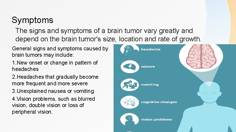
Symptoms The signs and symptoms of a brain tumor vary greatly and depend on the brain tumor's size, location and rate of growth. General signs and symptoms caused by brain tumors may include: 1. New onset or change in pattern of headaches 2. Headaches that gradually become more frequent and more severe 3. Unexplained nausea or vomiting 4. Vision problems, such as blurred vision, double vision or loss of peripheral vision.
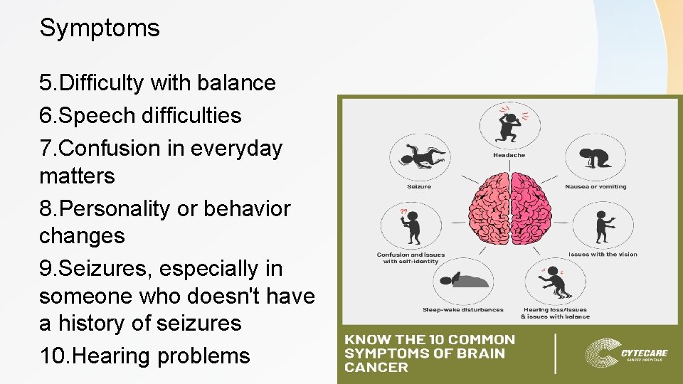
Symptoms 5. Difficulty with balance 6. Speech difficulties 7. Confusion in everyday matters 8. Personality or behavior changes 9. Seizures, especially in someone who doesn't have a history of seizures 10. Hearing problems
Causes 1)Brain tumors that begin in the brain Primary brain tumors originate in the brain itself or in tissues close to it, such as in the brain-covering membranes (meninges), cranial nerves, pituitary gland or pineal gland. Primary brain tumors begin when normal cells acquire errors (mutations) in their DNA. These mutations allow cells to grow and divide at increased rates and to continue living when healthy cells would die. The result is a mass of abnormal cells, which forms a tumor.
Causes • 2)Secondary (metastatic) brain tumors that result from cancer that starts elsewhere in your body and then spreads (metastasizes) to your brain. Secondary brain tumors most often occur in people who have a history of cancer. But in rare cases, a metastatic brain tumor may be the first sign of cancer that began elsewhere in your body. In adults, secondary brain tumors are far more common than are primary brain tumors. Any cancer can spread to the brain, but common types include: Breast cancer, Kidney cancer, Lung cancer
Risk Factors • Risk factors • In most people with primary brain tumors, the cause of the tumor is not clear. But doctors have identified some factors that may increase your risk of a brain tumor. • Risk factors include: 1) Exposure to radiation. People who have been exposed to a type of radiation called ionizing radiation have an increased risk of brain tumor. Examples of ionizing radiation include radiation therapy used to treat cancer and radiation exposure caused by atomic bombs. 2) Family history of brain tumors. A small portion of brain tumors occurs in people with a family history of brain tumors or a family history of genetic syndromes that increase the risk of brain tumors.
Gliobastoma Multiform • • most commen brain primary tumor. Astrocyte origin. Express a GFAP ( Glial fibrillary acidic protein). Grade 4 , highly malignant , rapidlly progressive , usually fatal >1 year median survival, the older the patient the worse the prognosis. • It can cause worsening headaches, nausea, vomiting and seizures. • Found in cerebral hemisphers. Because of their quick growth and invasion to nearby tissue , it tends to rapidlly cross the corpus callosum ( midline strcture seprating 2 hemosphers ) “ butterfly glioma”.
Gliobastoma Multiform • “ Pseudopalisading” cancer cells typically recruite BV to provide nerishment in a process called angiogensis , but gliobalstoma proliferates so fast that even with angiogensis the neuitrint demand outpaces blood supply , as a result becuse the blood supply serves priphral tumor cells first , the tumor cells in the center die first because the are the furthest from blood supply , the remaining viable tumor collect along edges of necrotic region ( neucli are lining up on the edges because they are bing pushed out from something in the middle which is the necotic cells )

Pseudopalisading butterfly glioma

• Diagnosis : Neurologic exam , imaging test , biobsy • Treatment : can be very difficult to treat and a cure is often not possible. Treatments may slow progression of the cancer and reduce signs and symptoms. ( Surgery , chemotherapy , radiation therapy , Supportive (palliative) care)
Meningioma • A meningioma is a tumor that arises from the meninges (the membranes that surround your brain and spinal cord). Although not technically a brain tumor, it is included in this category because it may compress or squeeze the adjacent brain, nerves and vessels. • Second most common , female <male. • Arachnoid cell origin. • Typically benign , often asymptomatic, may present with seizures if enlarges enough to case symptoms. • Risk factor : prior rdiation to the head ( in childhood malignancies , latency period ~ 20 y) • Most often occurs near surface of brain and in parasagital region. • Grade 1 -3 , relatively slow growing.
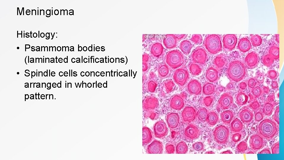
Meningioma Histology: • Psammoma bodies (laminated calcifications) • Spindle cells concentrically arranged in whorled pattern.
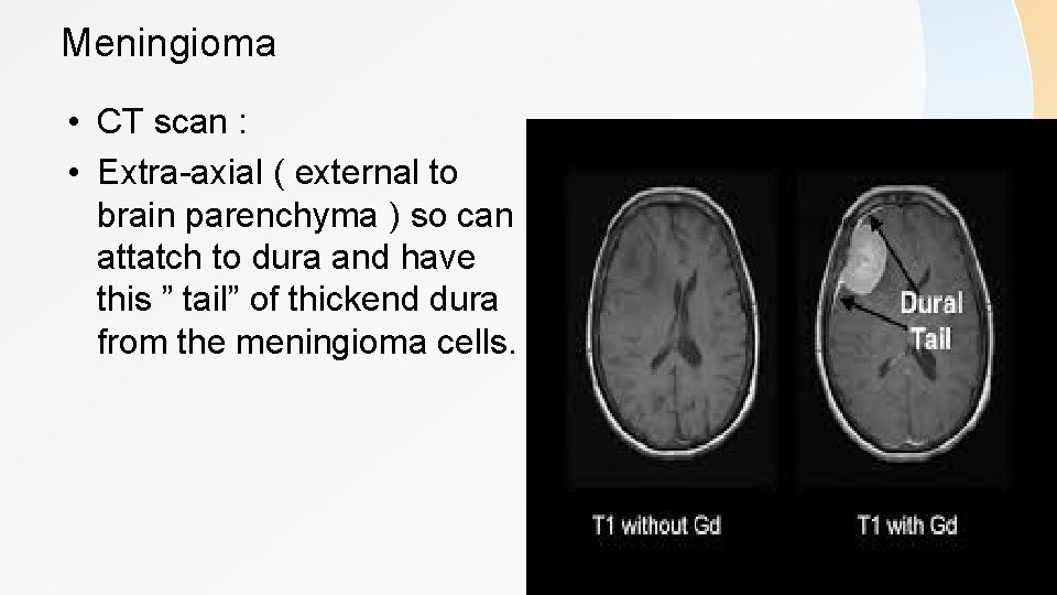
Meningioma • CT scan : • Extra-axial ( external to brain parenchyma ) so can attatch to dura and have this ” tail” of thickend dura from the meningioma cells.
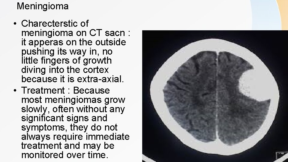
Meningioma • Charecterstic of meningioma on CT sacn : it apperas on the outside pushing its way in, no little fingers of growth diving into the cortex because it is extra-axial. • Treatment : Because most meningiomas grow slowly, often without any significant signs and symptoms, they do not always require immediate treatment and may be monitored over time.
Schwannoma • Third most common adult primary brain tumor. • Schwan cells origin. • Clasically at cerebellopontine angle involving both CN 7 and 8 , but can be along any periphral nerve. Often localized to CN 8 in internal acoustic meatus , leading to vestibular schwannoma that presents as hearing loss and tinnitus. • Bilateral schwannomas found in neurofibromatosis type 2. • Treatment : Surgery for resection and radiation.
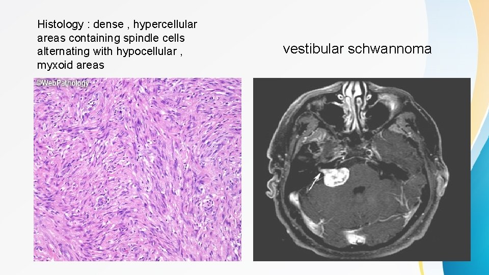
Histology : dense , hypercellular areas containing spindle cells alternating with hypocellular , myxoid areas vestibular schwannoma
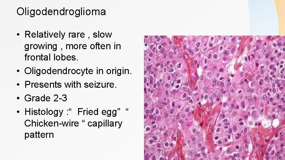
Oligodendroglioma • Relatively rare , slow growing , more often in frontal lobes. • Oligodendrocyte in origin. • Presents with seizure. • Grade 2 -3 • Histology : “ Fried egg” “ Chicken-wire “ capillary pattern
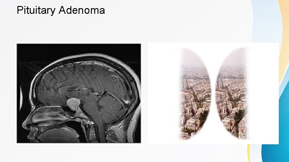
Pituitary Adenoma • Formed by hormone secreting cells of anterior pituitary , may be nonfunctioning or hyperfunctioning. • classified by the hormone that is released as tumor forms, and by size of tumor. • The non-functioning type 1. presents with mass effect (like : bitemporal hemianopia , due to pressure on optic chiasm you can't see in the outer portion of both your eyes. ) • 2. Pituitary apoplexy is characterized by a sudden onset of headache, visual symptoms, altered mental status, and hormonal dysfunction due to acute hemorrhage or infarction of a pituitary gland. • 3. Hypopituitrism
Pituitary Adenoma • Hyperplasia of only one type of endocrine cells found in pituitary , most commenly form lactotrophes , causes hyperprolactinemia present at galactorrhea, amenorehea , decrease in bone density , decreased libido and infertility in men. • Somatotrophes : acromegaly , gigantisim. • Corticotrophes : cushing disease • Rarely from thyrotrophes and gonadotrophes
Pituitary Adenoma

CHILDHOOD CNS TUMOR S
Childhood cns tumors • • Pilocytic astrocytoma Medulloblastoma Ependymoma Craniopharyngioma Cerebellar
Pilocytic astrocytoma • • Most common brain tumor in children Low grade astrocytoma Usually in posterior fossa (cerebellum) Usually benign without mets Well-circumscribed , cystic or solid Often successfully treated with surgery Contain rosenthal fibers GFAP positive
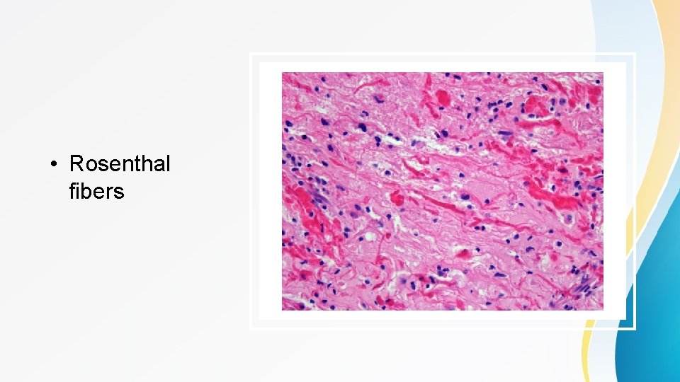
• Rosenthal fibers
Medulloblastoma • Highly malignant primary brain tumor • Usually occurs in children • Usually occurs in cerebellum often in midline (truncal ataxia) • Type of primitive neuroectodermal tumor (PNET)
Medulloblastoma • Treatment : surgery , radiation , chemo • 75% children survive to adulthood Many with complications of treatment • Can compress 4 th ventricle hydrocephalus • Can spread to CSF ü Nodules in dura of spinal cord : "Drops metastasis " ü Tends to occur in lower spinal cord, cauda equina ü Back pain , focal neuro lesions can occur
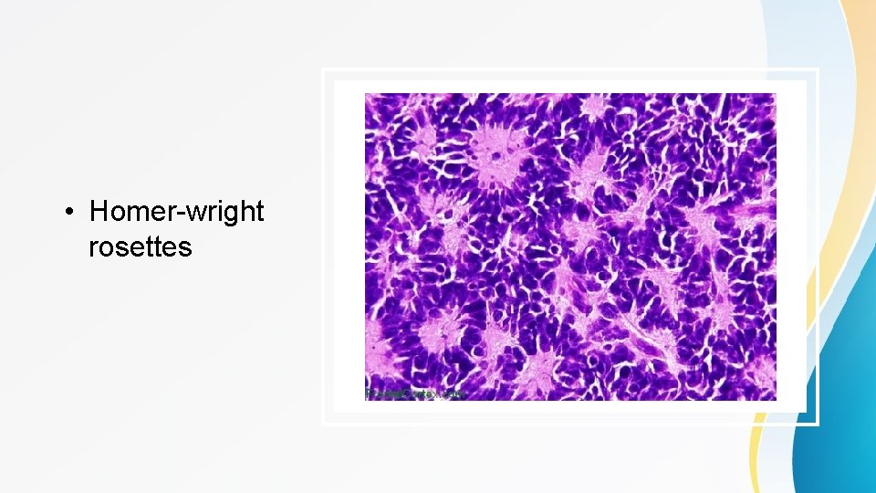
• Homer-wright rosettes
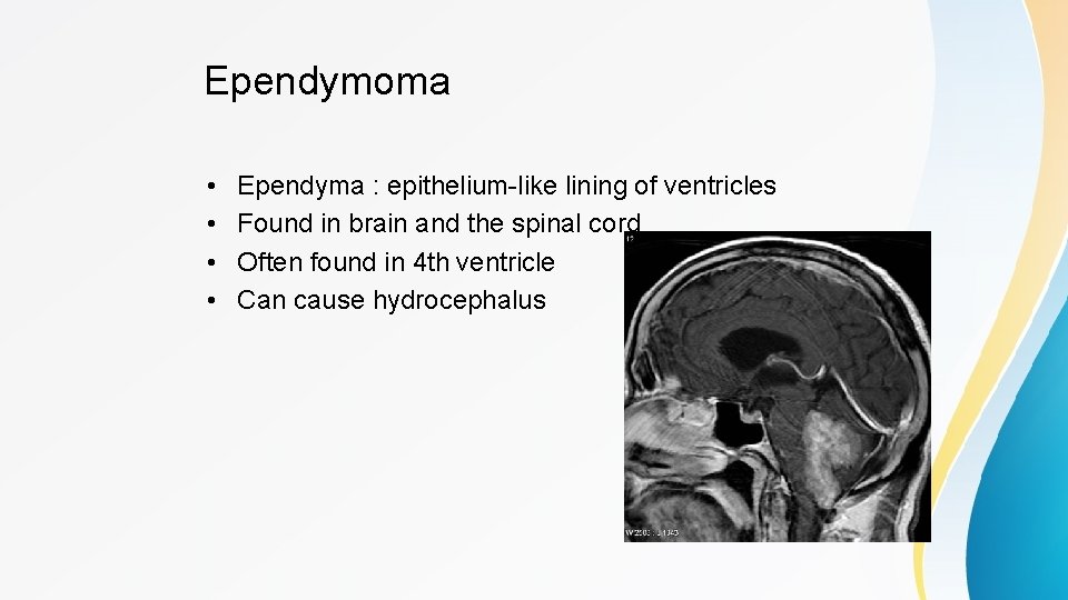
Ependymoma • • Ependyma : epithelium-like lining of ventricles Found in brain and the spinal cord Often found in 4 th ventricle Can cause hydrocephalus
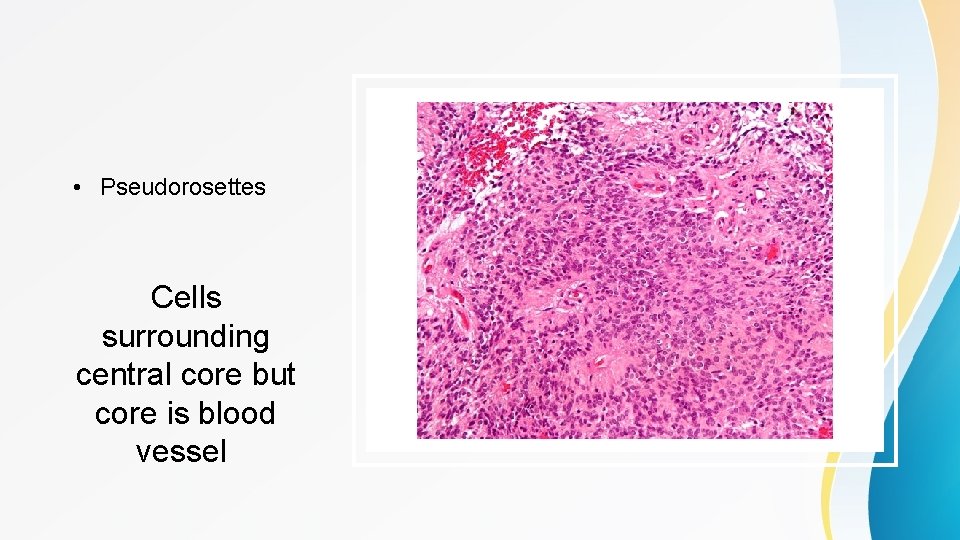
• Pseudorosettes Cells surrounding central core but core is blood vessel
Hemangioblastoma • Very rare , slow growing CNS tumor • Often cerebellar , also brainstem and spinal cord • Weel-circumscribed , highly vascular
Hemangioblastoma • Two key facts to know : 1. Can produce EPO >> polycythemia (increased Hct ) 2. Occur in von hippel lindau syndrome : o Autosomal dominant disease o Tumor suppressor gene mutation o LOTS of tumors : (hemangioblastomas of brain and spine , retinal angioma , renal cell carcinoma , pheochromocytoma )
Craniopharyngioma • Mostly children 10 -14 years old ( rarely younger adults ) • Suprasellar Anywhere pituitary gland >> base 3 rd ventricle • Benign • Symptoms from compression : o Visual field defects o Hormonal imbalance o Behavioral change (frontal lobe dysfunction )
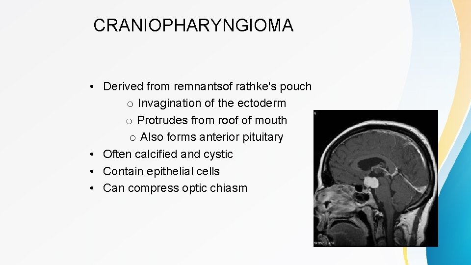
CRANIOPHARYNGIOMA • Derived from remnantsof rathke's pouch o Invagination of the ectoderm o Protrudes from roof of mouth o Also forms anterior pituitary • Often calcified and cystic • Contain epithelial cells • Can compress optic chiasm
Pineal tumors • Rare germ cell tumors or parenchymal tumors • Compression pretectal area of midbrain • Parinaud syndrome : o Paralysis of upward gaze o Pseudo argyll robertson pupil o React to accommodation but not light • Can compress cerebral aqueduct Hydrocephalus , papilledema
Stereotactic biopsy • • Stereotactic biopsy involves localization of the tumour with a stereotactic frame applied to the head of the patient using the CT scan or MRI. • • The three-dimensional coordinates of the tumour are ascertained. • • The surgeon chooses the point of entry and the desired path through the brain and a computer program determines the necessary angles for the biopsy probe and the depth to the
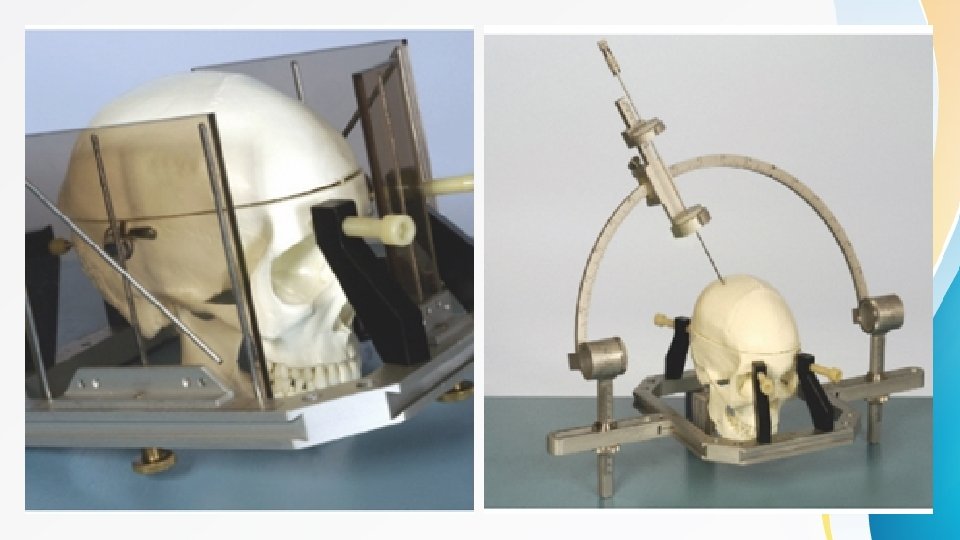
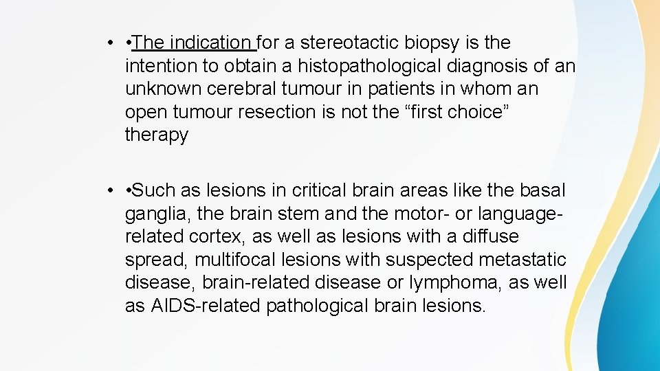
• • The indication for a stereotactic biopsy is the intention to obtain a histopathological diagnosis of an unknown cerebral tumour in patients in whom an open tumour resection is not the “first choice” therapy • • Such as lesions in critical brain areas like the basal ganglia, the brain stem and the motor- or languagerelated cortex, as well as lesions with a diffuse spread, multifocal lesions with suspected metastatic disease, brain-related disease or lymphoma, as well as AIDS-related pathological brain lesions.

Thank you Raghad al haj hasan
- Slides: 39