brain spine Coverings of the Brain Meninges skin

brain spine

Coverings of the Brain. Meninges skin skull dura mater arachnoid layer pia mater cerebral cortex

Menenges: 1. Covers and protects CNS 2. Protects blood vessels and encloses venus sinuses 3. Contains CSF 4. Forms partition within the skull

Cerebruspinal Fluid Brain Ventricles CSF Spinal Cord Rt. Ventricle Lf. Ventricle Anterior View Saggital View

Ventricles

CSF • 150 ml in adult • contains: glucose, proteins, lactic acid, urea, cations, anions, WBC Functions: 1. Reduces wt. of brain by 97% 2. Prevents head injury 3. Supplies brain with nutrition 4. Transports hormones along ventricular channels

Occipital Lobe Frontal Lobe Parietal Lobe The Cerebrum Temporal Lobe Brainstem The Brain Cerebellum

cerebrum corpus callosum thalamus hypothalamus Pineal gland cerebellum pituitary pons spinal cord medulla oblongata

Cerebrum • • Involved with higher brain functions. Processes sensory information. Initiates motor functions. Integrates information.

Cerebrum Cross-Section cerebral cortex white matter corpus callosum basal ganglia ventricles

Motor, Sensory & Association Cortex

Primary Sensory Cortex

Primary Motor Cortex

Right-Left Specialization of the Cerebrum left side – language development – mathematical & learning capabilities – sequential thought processes right side – visual spatial skills – musical and artistic activities – intuitive abilities

Diencephalon hypothalamus pituitary

Diencephalon

Thalamus • Relay center for sensory tracts from the spinal cord to the cerebrum. • Contains centers for sensation of pain, temperature, and touch. • Involved with emotions and alerting or arousal mechanisms.

The Reticular Formation

Hypothalamus Regulates: • autonomic control center- blood pressure, rate and force of heart contraction, center for emotional response and behavior • body temperature • water balance and thirst • sleep/wake cycles • appetite • sexual arousal • control of endocrine functioning: Acts on the pituitary gland through the release of neurosecretions.

Hypothalamus

The Limbic System

Midbrain • • • Contains ascending and descending tracts to the cerebrum and thalamus. Reflex center for eye muscles. Also involved with processing visual and auditory information (connects head movements with visual and auditory stimuli).

Pons • Connects the two halves of the cerebellum. • Regulates breathing.

Medulla Oblongata • Composed of nerve tracts to and from the brain (these tracts cross over left to right and right to left) • May be regarded as an extension of the spinal cord • Almost all of the cranial nerves arise from this region

Medulla Oblongata Contains control centers for many subconscious activities • Respiratory rate • Heart rate • Arteriole constriction • Swallowing • Hiccupping • Coughing • Sneezing

Cerebellum • Controls and coordinates muscular activity. • Important in equilibrium, posture and movement.

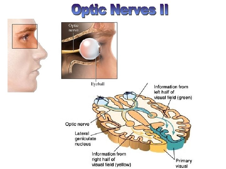
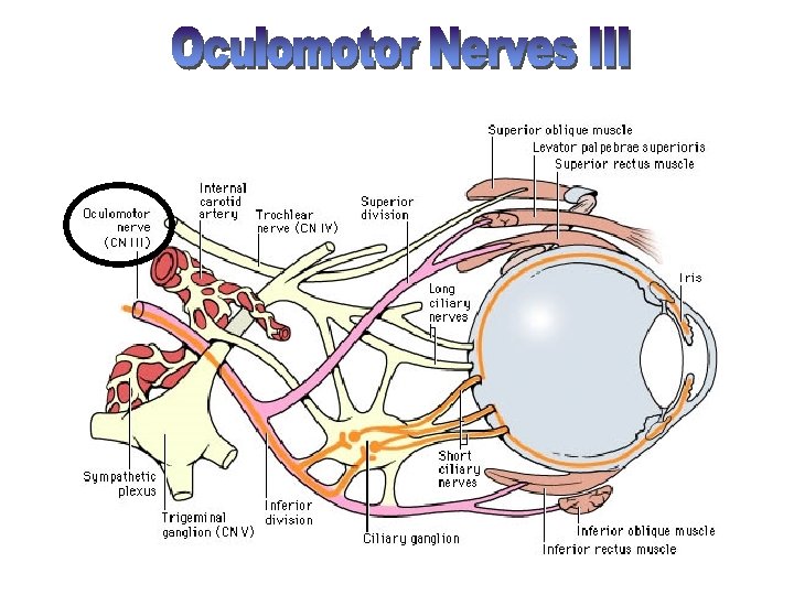
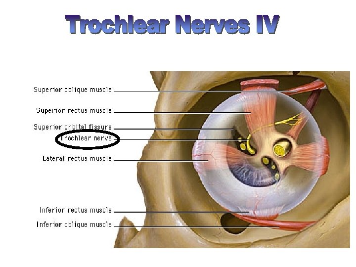
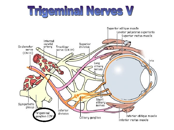
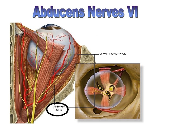
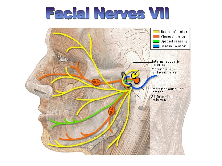
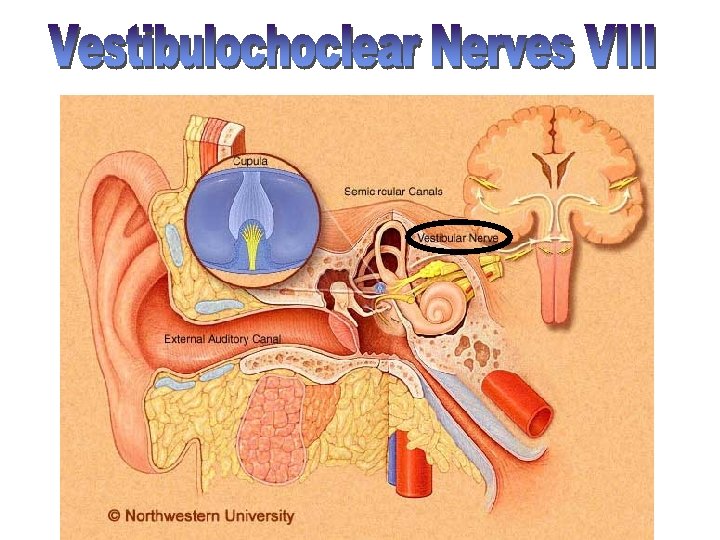
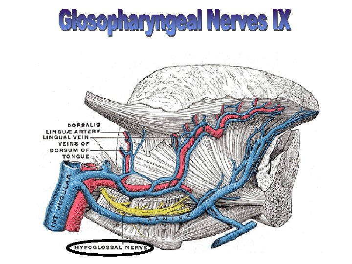
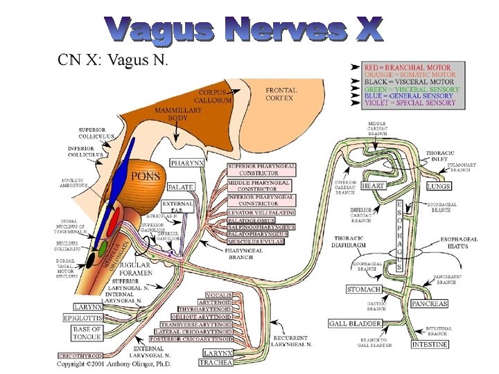
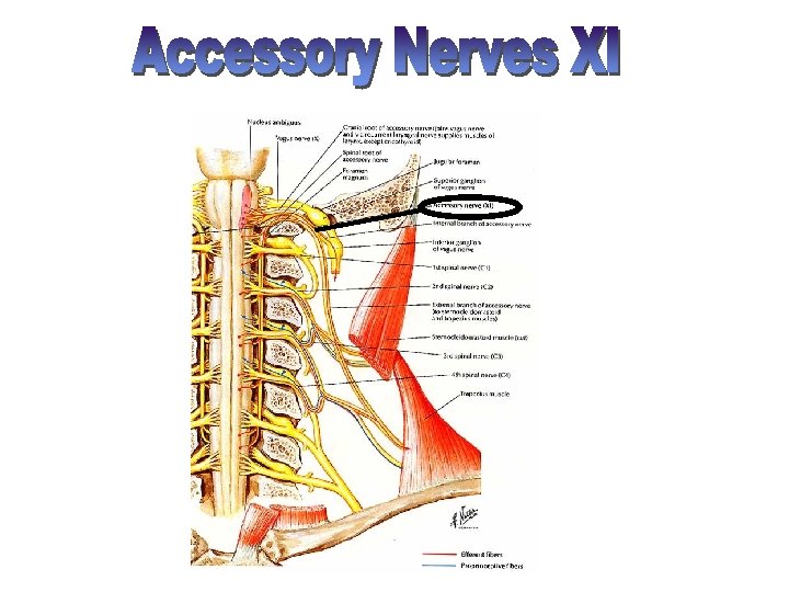
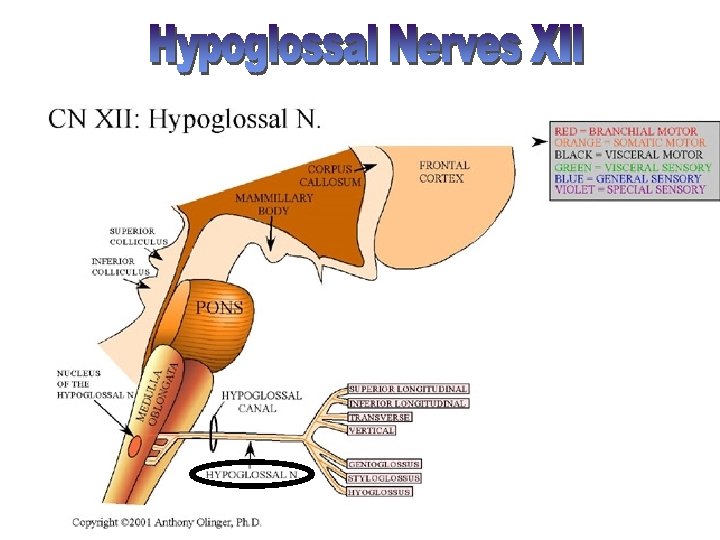
On Old Olympus Towering Tops A Fat Voracious German Viewed A Hop 1. 2. 3. 4. 5. Olfactory- smell Optic- vision Oculomotor- 4 of the 6 extrinsic eye muscles Trochlear- extrinsic eye muscles Trigeminal- sensory fibers to the face and motor fibers to the chewing muscles 6. Abducens- controls eye muscles that turn the eye laterally 7. Facial- facial expression 8. Vestibulocochlear- hearing and balance 9. Glosopharyngeal- tongue and pharynx 10. Vagus- from medulla- acetylcholine slows heart & breathing 11. Accessory- accessory part of vagus nerve 12. Hypoglossal- moves muscles under tongue

Olfactory Optic Oculomotor Trochlear Trigeminal Abducens Facial Vestibulocochlear Glossopharyngeal Vagus Accessory Hypoglossal

Olfactory tract Olfactory bulb Filaments of olfactory nerve Olfactory receptor cell

Traumatic Brain Injuries • Concussion • Contusion • Subdural or subarachnoid hemorrhage • Contrecoup injury
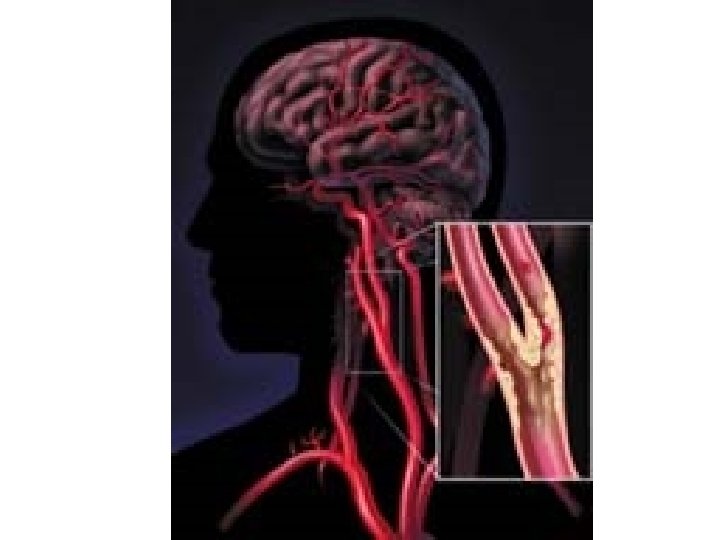
 • • • Ischemia Thrombus Embolism Arteriosclerosis Stroke")
Cerebrovascular Accidents (CVAs) • • • Ischemia Thrombus Embolism Arteriosclerosis Stroke

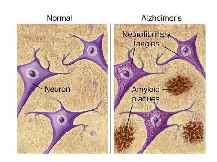
Degenerative brain diseases • • Alzheimer’s Down’s Parkinson’s Huntington’s Chorea MS Epilepsy Schizophrenia

PET Scans F-Dopa deficiency
- Slides: 50