Brain Neurotransmitters Dr Salah Elmalik objectives By the

Brain Neurotransmitters Dr. Salah Elmalik

objectives By the end of this lecture you are expected to: to Describe the functions of glutamergic system Describe the functions of NTs of the brain (the noradrenergic & serotonergic cholinergic, dopaminergic, GABAergic systems) Appreciate that many drugs and CNS disorders affect function of brain neurotransmitters

Brain Neurotransmitters Chemical substances released by electrical impulses into the synaptic cleft from synaptic vesicles of presynaptic membrane Diffuses to the postsynaptic membrane Binds to and activates the receptors Leading to initiation of new electrical signals or inhibition of the post-synaptic neuron
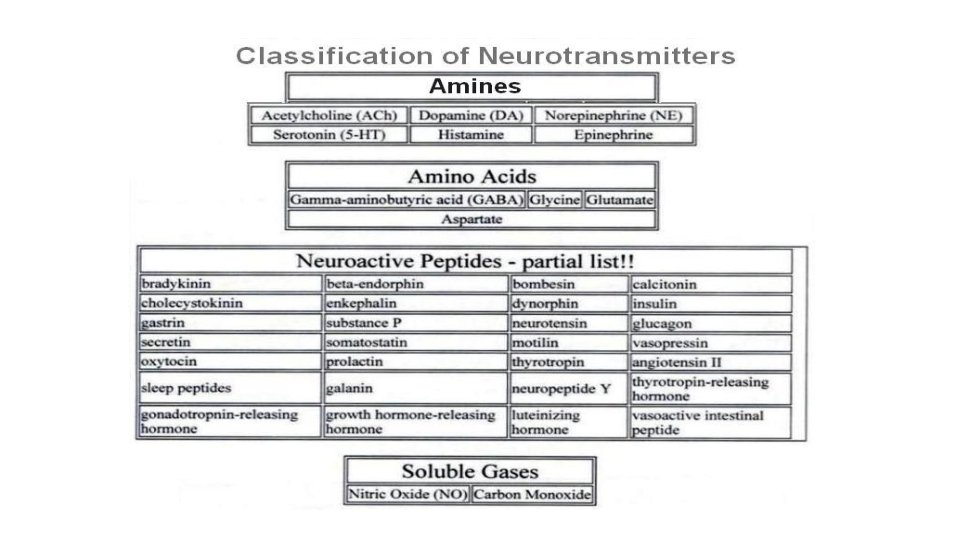

Some of the Brain Neurotransmitters 1. 2. 3. 4. 5. 6. Ach Glutamate GABA Norepinephrine (NE)/Epinephrine Serotonin Dopamine

Classes of Receptors • Metabotropic = trans membrane receptor acts through a second messenger • Ionotropic = Ligand gated ion channel
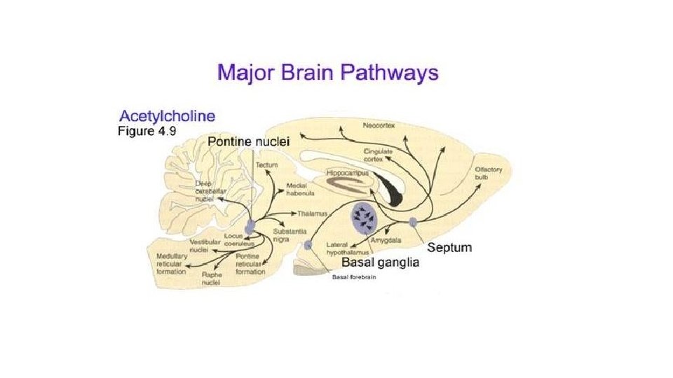

Cholinergic System Acetylcholine is the major neurotransmitter in the peripheral nervous system In the brain , cholinergic ( ACh producing ) neurons are present mainly in 2 areas : 1) Basal Forebrain ( namely Nucleus Basalis of Myenert and septal nuclei) (2) Mesopontine tegmental area which is alas called ponto-mesencephalic cholinergic complex
 • (The muscle-type can")
Acetylcholine Receptors Acts on 2 cholinergic receptors: • ❶Nicotinic (ionotropic) • (The muscle-type can be selectively blocked by curare, the neuronal-type by hexamethonium) • Excitatory • ❷Muscarinic (metabotropic) (antagonist- Atropine): ●Excitatory or inhibitory ● Five subtypes (M 1 -M 5): all are found in the brain but M 1 is abundant.

Muscarinic Receptors M 1 receptors most involved in cognitive functioning (evidence from Knockout mice and pharmacologic human studies with M 1 blocking drugs) M 2 blocking agents may facilitate cognition in animals (but these drugs are not being used in humans at this point). M 3 receptors do not seem to play much of a role in cognition (animal studies). M 4 and M 5 functions in the brain are unknown

Ach Functions & Disorders ACh influences mental processes such as Learning Memory Alertness sleep. Alzheimer’s Disease- the most common form of dementia that is associated with acetylcholine loss Damage to Ach producing cells in the basal forebrain Bipolar disorder Mood swings Depression Inhibitors of acetylcholinesterase in the brain are the main drugs used to treat Alzheimer’s disease.

Glutaminergic System Glutamate is the most commonly found NT in the brain (king of NTs, ~50% neurons). Glutamate is the major excitatory neurotransmitter of the brain and spinal cord, responsible for 75% of the excitatory transmission in the brain Glutamate (can cause excitotoxicity) is converted in astrocytes into glutamine (not toxic) and passed onto glutaminergic neurons Wide spread, but high levels in hippocampus; hypo function of NMDA receptors in this area and prefrontal cortex is associated with schizophrenia

Glutamate Receptors Are widely distributed in the brain; they are of two types: 1. Metabotropic receptors (G protein- coupled receptors): m. Glu. R Found in hippocampus, cerebellum and the cerebral cortex • act through second messengers which activate biochemical cascades, leading to modification of other proteins such as ion channels. 2. Ionotropic receptors (ligand-gated ion channels). Three types: • AMPA receptors (α-amino-3 -hydroxy-5 -methylisoxazole - 4 -propionate) Kainate receptors (kainite is an acid isolated from seaweed), NMDA receptors (for N-methyl D-aspartate); play a role in long term potentiation so they are involved in learning and memory

NMDA Receptors • Permits passage of Na+ and large amounts of Ca 2+. They are unique: Glycine is essential for their normal response to glutamate. The channel is blocked by Mg 2+ ion at normal membrane potentials This blockade is removed by depolarization (caused by e. g. AMPA) Excitatory post synaptic potential induced by activation of NMDA receptor is slower than that elicited by activation of AMP and kainate receptors
 : are major")
Functions & Disorders Of Glutamate • Glutamic acid (and aspartic acid) : are major excitatory NTs in CNS. • Glutamate NMDA receptor involved in Long-Term Potentiation & memory storage. Disorders: • -Excess Glutamate activity is implicated in some types of epileptic seizures • - Under some pathological conditions , such Stroke , ALS (Amyotrophic Lateral Sclerosis) , autism and Alzheimer's diseases, it acts as an excitotoxin , producing excessive influx of calcium into the neurons and causing neuronal death.

GABAergic System
.")
GABAergic System GABA is the main inhibitory neurotransmitter in the central nervous system (CNS). GABAergic inhibition is seen at all levels of the CNC (Hypothalamus, hippocampus, cerebral cortex and cerebellar cortex. GABA interneurons are abundant in the brain, with 50% of the inhibitory synapses in the brain being GABA mediated
 Formed by decarboxylation of glutamate. Three types of GABA receptors")
Gamma Aminobutyric acid (GABA) Formed by decarboxylation of glutamate. Three types of GABA receptors e. g. GABAA B & C. GABA A & B receptors are widely distributed in CNS. GABAC are found in retina only GABA B are metabotropic (G-protein) in function. GABA A and C receptors (ionotropic) have multiple binding sides (for benzodiazepine and barbiturates). The channel is a Cl-channel (not Na)

GABAergic System

Functions & Disorders of GABAergic System Functions: • Presynaptic inhibition • GABAA receptors in CNS are chronically stimulated to regulate neuronal excitability. Disorders: -under activity of GABA leads to seizures. Alcohol, barbiturates, progesterone and deoxycorticosterone also in part work by increasing GABA activity

Norepinephrine System
: is a catecholamine that is synthesized from Dopamine It is released")
Noradrenergic System Norepinephrine(NE): is a catecholamine that is synthesized from Dopamine It is released from sympathetic nerves, the adrenal medulla and brain stem neurons It acts on both α-and βadrenergic receptors (Gprotein-coupled receptors) NE is believed to play a role in both learning and memory

Noradrenergic System The Noradrenergic System has a very wide- spread projection system Locus ceruleus is activated by stress and co-ordinates responses via projections to thalamus, cortex, hippocampus, amygdala, hypothalamus, autonomic brainstem centers, and the spinal cord

Locus ceruleus neurons fire as a function of vigilance and arousal Irregular firing during quiet wakefulness Sustained activation during stress Their firing decreases markedly during slow-wave sleep and virtually disappears during REM sleep. 23

Functions of NE It constitutes part of the RAS ( Reticular Activating System Attention/Vigilance Fight or flight response, Learning Enhances formation and retrieval of memory Aggressive behaviour.
 Implicated in Stress-Related Disorders: Depression Withdrawal from some")
Disorders of NE Norepin ephrine (NE) Implicated in Stress-Related Disorders: Depression Withdrawal from some drugs of abuse Anxiety and other stress-related disorders such as panic disorder.

PGi: Nucleus paragigantocellularis Pr. H: Perirhinal Cortex

Dopamine is a catecholamine that is synthesized from tyrosine Five dopaminergic receptors (D 1 -D 5). Overstimulation of D 2 receptors is thought to be related to schizophrenia

Dopaminergic Pathway Dopamine is transmitted via three major pathways: 1 - The first(nigro striatal system) extends from the substantia nigra to the caudate nucleus-putamen (neostriatum) and is involved in motor control.

2 - The second pathway project to the mesolimbic forebrain It involved in reward and emotional behavior and addiction Dysfunction is connected to hallucinations and schizophrenia

The Dopaminergic System cont … 3 - The third pathway, known as the tubero- infundibular system It is concerned with: Regulation of secretion of prolactin from the anterior pituitary gland Maternal behavior (nurturing)

Dopaminergic Pathways/Functions 31

Dopaminergic Neurons Disorders Schezophrenia. Parkinson’s Disease. Cocaine elevate activity at dopaminergic synapses

Serotonin is synthesized from the amino acid tryptophan, which is abundant in meat Our bodies cannot make tryptophan (must get from diet) Tryptophan deprivation alters brain chemistry and mood There is only a few 100, 000`s of 5 -HT neurons in human brain There is 7 classes serotonin receptors in different parts of CNS (most are metabotropic, except 5 -HT 3) Mice in which the gene for 5 -HT 2 C receptors has been knocked out are obese

Serotonin The serotonin pathways in the brain: The principal centers for serotonergic neurons are the rostral and caudal raphe nuclei >>>> axons ascend to the cerebral cortex, limbic & basal ganglia Serotonergic nuclei in the Brain stem >>>> descending axons (terminate in the medulla& spinal cord
 Functions & Disorders Functions: Improved mood Decrease appetite. Sleep Disorders: Depression")
Serotonin (5 -HT) Functions & Disorders Functions: Improved mood Decrease appetite. Sleep Disorders: Depression Anxiety • Drugs (e. g. Prozac) that prolong serotonin’s actions relieve symptoms of depression & obsessive disorders
 Excitatory Acetyl")
Neurotransmitter c effect from synthesis receptor Fate Functions 1. Acetyl choline (Ach) Excitatory Acetyl co. A + Cholinergic nerve endings Cholinergic pathways of brainstem 1. Nicotinic 2. Muscarini c Broken by acetyl cholinesterase Cognitive functions e. g. memory Peripheral action e. g. cardiovascular system 2. Catecholamines i. Epinephrine (adrenaline) Excitatory in some but inhibitory in other Tyrosine produced in liver from phenylalanin e Adrenal medulla and some CNS cells Excites both alpha α & beta β receptors ii. Norepinephrine Excitatory Tyrosine, found in pons. Reticular formation, locus coerules, thalamus, mid-brain Begins inside axoplasm of adrenergic nerve ending is completed inside the secretary vesicles α 1 α 2 β 1 β 2 1. Catabolized to inactive product through COMT & MAO in liver 2. Reuptake into adrenergic nerve endings 3. Diffusion away from nerve endings to body fluid For details refer ANS. e. g. fight or flight, on heart, BP, gastrointestinal activity etc. Norepinehrine controls attention & arousal, sleep/wake cycle. iii. Dopamine Excitatory Tyrosine CNS, concentrated in basal ganglia and dopamine pathways e. g. nigrostriatal, mesocorticolim bic and tuberohypophyseal pathway D 1 to D 5 receptor Same as above Sensory motor Cognetive/emotion al behavior Endocrine Hypothalamic Decreased dopamine in parkinson’s disease. Increased dopamine 36 concentration
 Excitatory Tryptophan CNS, Gut (chromaffin cells)")
synthesis receptor er effect 3. serotonin (5 HT) Excitatory Tryptophan CNS, Gut (chromaffin cells) Platelets & retina 5 -HT 1 to 5 -HT 7 5 -HT 2 A receptor mediate platelet aggregation & smooth muscle contraction Inactivated by MAO to form 5 hydroxyindoleace tic acid(5 -HIAA) in pineal body it is converted to melatonin Mood control, sleep, pain feeling, temperature, BP, & hormonal activity Excitatory 75% of excitatory transmissio n in the brain By reductive amination of Kreb’s cycle intermediate α – ketoglutarate. Brain & spinal cord e. g. hippocampus Ionotropic and metabotropic receptors. Three types of ionotropic receptors e. g. NMDA, AMPA and kainate receptors. It is cleared from the brain ECF by Na + dependent uptake system in neurons and neuroglia. Long term potentiation involved in memory and learning by causing Ca++ influx. Metabolized by transamination to succinate in the citric acid cycle. GABA – A causes hyperpolarization (inhibition) Anxiolytic drugs like benzodiazepine cause increase in Cl- entry into the cell & cause soothing effects. GABA – B cause increase conductance of K+ into the 3 c 7 ell. 4. Glutamate 5. Gama amino butyric acid(GABA) Major inhibitory mediator Decarboxylati on of glutamate by glutamate decarboxylase (GAD) by GABAergic neuron. CNS GABA – A increases the Cl conductance, GABA – B is metabotropic works with G – protein GABA transaminase catalyzes. GABA – C found exclusively in the retina.

Thank You
- Slides: 39