Brain and Cranial Nerves Brain Part of CNS

Brain and Cranial Nerves • Brain – – Part of CNS contained in cranial cavity Control center for many of body’s functions Much like a complex computer but more Parts of the brain • Brainstem: connects spinal cord to brain; integration of reflexes necessary for survival • Cerebellum: involved in control of locomotion, balance, posture • Diencephalon: thalamus, subthalamus, epithalamus, hypothalamus • Cerebrum: conscious thought, control • Cranial nerves: part of PNS arise directly from brain. Two pairs arise from cerebrum; ten pairs arise from brainstem

Sagittal Section of Brain Copyright © The Mc. Graw-Hill Companies, Inc. Permission required for reproduction or display. Anterior Corpus callosum Diencephalon Cerebrum Thalamus Posterior Hypothalamus Midbrain Brainstem Pons Cerebellum Medulla oblongata Medial view © Courtesy of Branislav Vidic

Copyright © The Mc. Graw-Hill Companies, Inc. Permission required for reproduction or display. TABLE 13. 1 Brainstem Divisions of the Brain And Their Functions Connects the spinal cord to the cerebrum; consists of the medulla oblongata, pons, and midbrain, with the reticular formation scattered throughout the three regions; has many important functions as listed under each subdivision; is the location of cranial nerve nuclei Medulla oblongata Pathway for ascending and descending nerve tracts; center for several important reflexes (e. g. , heart rate, breathing, swallowing, vomiting) Pons Contains ascending and descending nerve tracts; relays information between cerebrum and cerebellum; site of reflex centers Midbrain Contains ascending and descending nerve tracts; serves as visual reflex center; part of auditory pathway Cerebellum Diencephalon Connects the brainstem to the cerebrum; has many relay and homeostatic functions, as listed under each subdivision Thalamus Major sensory relay center; influences mood and movement Subthalamus Contains nerve tracts and nuclei Epithalamus Contains nuclei responding to olfactory stimulation and contains pineal gland Hypothalamus Major control center for maintaining homeostasis and regulating endocrine function Cerebrum Reticular formation Controls muscle movement and tone; governs balance; regulates extent of intentional movement; involved in learning motor skills Scattered throughout brainstem; controls cyclic activities, such as the sleep-wake cycle Controls conscious perception, thought, and conscious motor activity; can override most other systems Basal nuclei Controls muscle activity and posture; largely inhibits unintentional movement when at rest Limbic system Autonomic response to smell, emotion, mood, memory, and other such functions

Brainstem Copyright © The Mc. Graw-Hill Companies, Inc. Permission required for reproduction or display. Interthalamic adhesion Thalamus Diencephalon Pineal gland Infundibulum Cerebral peduncle Midbrain Cerebral peduncle Pons Superior colliculus Midbrain Inferior colliculus Superior cerebellar peduncle Middle cerebellar peduncle Inferior cerebellar peduncle Pons Brainstem Pyramid Olive Medulla oblongata Ventral median sulcus Medulla oblongata Nucleus cuneatus Nucleus gracilis Pyramidal decussation Olive (a) Anterior view (b) Posterolateral view

Brainstem: Medulla Oblongata • Most inferior part • Continuous with spinal cord; has both ascending and descending nerve tracts • Regulates: heart rate, blood vessel diameter, respiration, swallowing, vomiting, hiccupping, coughing, and sneezing

Brainstem: Pons • Superior to the medulla oblongata • Fiber tracts: ascending and descending • Nuclei – Pontine: anterior portion, relay between cerebrum and cerebellum – For cranial nerves V-IX: posterior portion – Sleep center – Respiratory center coordinates with center in medulla

Cerebellum • Attached to brainstem posterior to pons • Cerebellar peduncles: fiber tracts that communicate with other parts of brain – Superior: to midbrain – Middle: to pons – Inferior: to medulla oblongata • Gray cortex and nuclei with white matter (tracts) between • Cortex folded in ridges called folia; white matter resembles a tree (arbor vitae)

Cerebellar Functions • Flocculonodular lobe: balance and eye movements • Vermis and medial portion of hemispheres: posture, locomotion, fine motor coordination leading to smooth, flowing movements • Lateral hemispheres, major portion: works with cerebrum to plan, practice, learn complex movements

Diencephalon • Located between brainstem and cerebrum • Components: thalamus, subthalamus, epithalamus, hypothalamus

Thalamus • Two lateral portions connected by the intermediate mass • Surrounded by third ventricle • Sensory information from spinal cord synapses here before projecting to cerebrum • Motor function • Mood modification • Emotion regulation • Sensory integration

Hypothalamus • Most inferior portion of diencephalon • Mammillary bodies: bulges on ventral surface; olfactory reflexes and emotional responses to odors • Infundibulum: stalk extending from floor; connects hypothalamus to posterior pituitary gland. Controls endocrine system. • Important in regulation of mood, emotion, sexual pleasure, satiation, rage, and fear

Diencephalon Copyright © The Mc. Graw-Hill Companies, Inc. Permission required for reproduction or display. Thalamus Corpus callosum Interthalamic adhesion Habenula Pineal gland Hypothalamus Epithalamus Subthalamus Optic chiasm Cerebellum Pituitary gland (a) Diencephalon, medial view Paraventricular nucleus Lateral area Dorsomedial nucleus Posterior nucleus Medial nucleus Interthalamic adhesion Anterior nucleus Ventral anterior nucleus Lateral posterior Dorsal nucleus tier Lateral dorsal nucleus Pulvinar Lateral geniculate nucleus Ventral posterior nucleus Ventral lateral nucleus Preoptic area Anterior nucleus Supraoptic nucleus Suprachiasmatic nucleus Optic chiasm Mammillary body Ventromedial nucleus Infundibulum Pituitary gland (b) Thalamus, anterolateral view (c) Hypothalamus, medialview

Cerebrum • Largest portion of brain • Composed of right and left hemispheres each of which has the following lobes: frontal, parietal, occipital, temporal, insula • Sulci and Fissures – Longitudinal fissure: separates the two hemispheres – Lateral fissure: separates temporal lobe from frontal and parietal lobes – Central sulcus: separates frontal and parietal lobes • Cortex: outer surface – Gyri are folds – Sulci are depressions

Cerebrum • Frontal lobe: voluntary motor function, motivation, aggression, sense of smell, mood, personality and decision making • Parietal lobe: reception and evaluation of sensory information except smell, hearing, and vision • Occipital lobe: reception and integration of visual input • Temporal lobe: reception and evaluation for smell and hearing; memory, abstract thought, judgment. Insula is within.

Cerebrum Copyright © The Mc. Graw-Hill Companies, Inc. Permission required for reproduction or display. Parietal lobe Frontal lobe Occipital lobe Anterior Right hemisphere Posterior Longitudinal fissure Sulci Left hemisphere Gyri Precentral gyrus Central sulcus Postcentral gyrus (a) Superior view Central sulcus Parietal lobe Frontal lobe I II Anterior Posterior Occipital lobe III Gray matter IV Pyramidal cell Lateral fissure Temporal lobe Cerebellum White matter (c) Schematic of a histological view (b) Lateral view a: © R. T. Hutchings; b: © The Mc. Graw-Hill Companies, Inc. /Rebecca Gray, photographer/Don Kincaid, dissections V VI

Limbic System Copyright © The Mc. Graw-Hill Companies, Inc. Permission required for reproduction or display. Fornix Cingulate gyrus Anterior nuclei of thalamus Corpus callosum Anterior commissure Septal area Habenula Olfactory bulb Dentate gyrus of hippocampus Olfactory cortex Parahippocampal gyrus Mammillary body Amygdala Medial view • Part of cerebrum and diencephalon • Basic survival functions such as memory, reproduction, nutrition • Emotions

Meninges, Ventricles, and Cerebrospinal Fluid Copyright © The Mc. Graw-Hill Companies, Inc. Permission required for reproduction or display. Dural venous sinus (superior sagittal sinus) Skull Periosteal dura Meningeal dura Dura mater Subdural space Arachnoid mater Subarachnoid space Vessels in subarachnoid space Pia mater (directly attached to brain surface and not removable) Cerebrum Dural venous sinus (superior sagittal sinus) Periosteal dura Meningeal dura Dura mater Falx cerebri Subdural space (potential space) Arachnoid mater Subarachnoid space Pia mater (b) Anterior view – Dura mater: superficial – Arachnoid mater – Pia mater: bound tightly to brain – Spaces • Subdural: serous fluid • Subarachnoid: CSF (a) Anterosuperior view Dural venous sinus (inferior sagittal sinus) • Connective tissue membranes Cerebrum
 • • Similar to serum, but most protein removed Bathes brain")
Cerebrospinal Fluid (CSF) • • Similar to serum, but most protein removed Bathes brain and spinal cord Protective cushion around CNS Choroid plexuses produce CSF which fills ventricles and other parts of brain and spinal cord

Blood Supply to the Brain • Brain – Requires a tremendous amount of blood – Receives 15 -20% of blood pumped by heart – Interruption cause unconsciousness and irreversible brain damage – High metabolic rate; dependent upon constant supply of oxygen and glucose – Receives blood through arteries: internal carotids and vertebral arteries. The vertebral arteries join to form the basilar artery. Carotids plus basilar form the cerebral arterial circle (Circle of Willis).
 have tight junctions between them.")
Blood-Brain Barrier • Endothelial cells (lining all capillaries) have tight junctions between them.

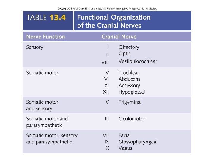
Cranial Nerves Copyright © The Mc. Graw-Hill Companies, Inc. Permission required for reproduction or display. Anterior • Indicated by Olfactory bulb (olfactory nerves [I] enter bulb) Optic nerve (II) Olfactory tract Oculomotor nerve (III) Trochlear nerve (IV) Optic chiasm Trigeminal nerve (V) Pituitary gland Abducens nerve (VI) Mammillary body Facial nerve (VII) Pons Vestibulocochlear nerve (VIII ) Olive of medulla oblongata Glossopharyngeal nerve (IX) Vagus nerve (X) Medulla oblongata Hypoglossal nerve (XII) Accessory nerve (XI) Posterior Inferior view – Roman numerals I-XII from anterior to posterior – Names • May have one or more of three functions – Sensory (special or general) – Somatic motor (control of skeletal muscles) – Parasympathetic (regulation of glands, smooth muscles, cardiac muscle)


Spinal Cord • Extends from foramen magnum to second lumbar vertebra • Segmented – Cervical – Thoracic – Lumbar – Sacral • Gives rise to 31 pairs of spinal nerves • Not uniform in diameter throughout length – Cervical enlargement: supplies upper limbs – Lumbar enlargement: supplies lower limbs • Conus medullaris: tapered inferior end. • Cauda equina: origins of spinal nerves extending inferiorly from lumbosacral enlargement and conus medullaris. Copyright © The Mc. Graw-Hill Companies, Inc. Permission required for reproduction or display. Brain Level of foramen magnum Roots of spinal nerves Spinal nerves Cervical enlargement Spinal cord Lumbosacral enlargement Conus medullaris Level of second lumbar vertebra Cauda equina Filum terminale Posterior view

Meninges of the Spinal Cord • Connective tissue membranes surrounding spinal cord and brain – Dura mater: continuous with epineurium of the spinal nerves – Arachnoid mater: thin and wispy – Pia mater: bound tightly to surface of brain and spinal cord. Forms the filum terminale, which anchors spinal cord to coccyx and the denticulate ligaments that attach the spinal cord to the dura mater Copyright © The Mc. Graw-Hill Companies, Inc. Permission required for reproduction or display. Duramater Subdural space Denticulate ligament Arachnoid mater Subarachnoid space Pia mater Epineurium of spinal nerve Dorsal root ganglion • Spaces – Epidural: anesthesia injected. Contains blood vessels, areolar connective tissue and fat. – Subdural: serous fluid – Subarachnoid: CSF and blood vessels within web-like strands of arachnoid tissue Spinal nerve Ventral root (a) Anterolateral view

Meninges of the Spinal Cord Copyright © The Mc. Graw-Hill Companies, Inc. Permission required for reproduction or display. Posterior Spinous process of vertebra Transverse process of vertebra Dorsal root ganglion Spinal cord Denticulate ligament Spinal nerve Ventral root Body of vertebra Pia mater Subarachnoid space Periosteum Arachnoid mater Subdural space Dura mater Anterior (b) Superior view Epidural space filled with adipose tissue

Cross Section of Spinal Cord Copyright © The Mc. Graw-Hill Companies, Inc. Permission required for reproduction or display. Central canal White matter Dorsal (posterior) column Ventral (anterior) column Lateral column Posterior median sulcus Dorsal root ganglion Central canal Ventral root Gray matter Posterior (dorsal) horn Spinal nerve Lateral horn Anterior (ventral) horn Rootlets Ventral root Gray commissure White commissure Anterior median fissure (a) Anterolateral view Ascending nerve tracts Descending nerve tracts (c) Anterolateral view

Cross Section of Spinal Cord • Anterior median fissure and posterior median sulcus: deep clefts partially separating left and right halves • White matter: myelinated axons forming tracts – Three columns (funiculi): ventral, dorsal, lateral • Each of these divided into tracts (fasciculi; pathways) • Gray matter: neuron, cell bodies, dendrites, axons – Horns • Posterior (dorsal) • Anterior (ventral) • Lateral (associated with ANS)

Cross Section of Spinal Cord Copyright © The Mc. Graw-Hill Companies, Inc. Permission required for reproduction or display. • Commissures: connections between left and right halves • Roots: spinal nerves arise as rootlets then combine to form roots – Dorsal (posterior) root has a ganglion – Ventral (anterior) – Two roots merge laterally and form the spinal nerve Central canal White matter Dorsal (posterior) column Ventral (anterior) column Lateral column Posterior median sulcus Dorsal root ganglion Central canal Ventral root Gray matter Posterior (dorsal) horn Spinal nerve Lateral horn Anterior (ventral) horn Rootlets Ventral root Gray commissure White commissure Anterior median fissure (a) Anterolateral view Ascending nerve tracts Descending nerve tracts (c) Anterolateral view

Organization of Neurons in the Spinal Cord and Spinal Nerves • Dorsal root ganglion: collections of cell bodies of unipolar sensory neurons forming dorsal roots. Copyright © The Mc. Graw-Hill Companies, Inc. Permission required for reproduction or display. Posterior horn Dorsal root ganglion Sensory neuron Spinal nerve Interneuron Autonomic neuron Lateral horn Somatic motor neuron Anterior horn Superior view Ventral root

Reflexes • Basic functional unit of nervous system and simplest portion capable of receiving a stimulus and producing a response • Automatic response to a stimulus that occurs without conscious thought. Homeostatic. • Components – Action potentials produced in sensory receptors transmitted to – Sensory neuron. To-Interneurons. To-Motor neuron. To– Effector organ which responds with a reflex

Reflex Arc Copyright © The Mc. Graw-Hill Companies, Inc. Permission required for reproduction or display. 3 Interneuron Dorsal root ganglion 2 Sensory neuron 1 Sensory receptor Spinal cord Skin 4 Motor neuron Spinal nerve Ventral root 1 A sensory receptor detects a stimulus. 5 Effector organ 2 A sensory neuron conducts action potentials through the nerve and dorsal root to the spinal cord. 3 In the spinal cord, the sensory neuron synapses with an interneuron. (An interneuron is not involved in a monosynaptic reflex arc. ) 4 The interneuron synapses with a motor neuron. 5 A motor neuron axon conducts action potentials through the ventral root and spinal nerve to an effector organ. Skeletal muscle

Withdrawal Reflex • Function is to remove a body limb or other part from a painful stimulus. • Reciprocal innervation: causes relaxation of extensor muscle when flexor muscle contracts. – Also involved in stretch reflex. • Crossed extensor reflex: when a withdrawal reflex is initiated in one lower limb, the crossed extensor reflex causes extension of opposite lower limb. 12 -33

Withdrawal Reflex Copyright © The Mc. Graw-Hill Companies, Inc. Permission required for reproduction or display. Stimulation of pain receptors results in: To brain 1 Pain receptors detect a painful stimulus. 2 Sensory neurons conduct action potentials to the spinal cord. 3 Sensory neurons synapse with excitatory Interneurons that synapse with alpha motor neurons. Sensory neuron 2 Quadriceps femoris muscle (extensor) 3 4 Excitation of the alpha motor neurons results in contraction of the flexor muscles and withdrawal of the limb from the painful stimulus. Excitatory interneuron 4 Alpha motor neuron Hamstring muscles (fexor) Sensory neuron 1 Stimulus Pain receptor Withdrawal reflex

Withdrawal Reflex with Reciprocal Innervation Copyright © The Mc. Graw-Hill Companies, Inc. Permission required for reproduction or display. Reciprocal innervation 1 During the withdrawal reflex, sensory neurons conduct action potentials from pain receptors to the spinal cord. Inhibitory interneuron Quadriceps femoris muscle (extensor) 4 2 Sensory neurons synapse with excitatory interneurons that are part of the withdrawal reflex. 3 Collateral branches of the sensory neurons also synapse with inhibitory interneurons that are part of reciprocal innervation. 4 Collateral branch from sensory neuron Reciprocal innervation 3 1 Hamstring muscles (flexor) Sensory neuron To brain 2 The inhibitory interneurons synapse with alpha motor neurons supplying the extensor muscles, causing them to relax and not oppose the flexor muscles of the withdrawal reflex, which are contracting. Withdrawal reflex Alpha motor neuron Excitatory interneuron

Withdrawal Reflex with Crossed Extensor Reflex Copyright © The Mc. Graw-Hill Companies, Inc. Permission required for reproduction or display. Crossed extensor reflex 1 During the withdrawal reflex, sensory neurons from pain receptors conduct action potentials to the spinal cord. 2 Sensory neurons synapse with excitatory interneurons that are part of the withdrawal reflex. 3 The excitatory interneurons that are part of the withdrawal reflex stimulate alpha motor neurons that innervate flexor muscles, causing withdrawal of the limb from the painful stimulus. 4 Collateral branches of the sensory neurons also synapse with excitatory interneurons that cross to the opposite side of the spinal cord as part of the crossed extensor reflex. 5 The excitatory interneurons that cross the spinal cord stimulate alpha motor neurons supplying extensor muscles in the opposite limb, causing them to contract and support body weight during the withdrawal reflex. Quadriceps femoris muscle (extensor) Sensory neuron To brain 1 5 2 4 Alpha motor neuron 3 Hamstring muscles (flexor) Alpha motor neuron Withdrawal reflex Excitatory interneuron Crossed extensor reflex

Spinal Nerves Copyright © The Mc. Graw-Hill Companies, Inc. Permission required for reproduction or display. • Consist of – Axon bundles – Schwann cells – Connective tissue • Endoneurium: surrounds individual neurons • Perineurium: surrounds axon groups to form fascicles • Epineurium: surrounds the entire nerve Adipose tissue Epineurium Perineurium Artery and vein Endoneurium Loose connective tissue Schwann cell Fascicle Axon

Organization of Spinal Nerves • Thirty-one pairs of spinal nerves • First pair exit vertebral column between skull and atlas • Last four pair exit via the sacral foramina • Others exit through intervertebral foramina • Eight pair cervical, twelve pair thoracic, five pair lumbar, five pair sacral, one pair coccygeal Copyright © The Mc. Graw-Hill Companies, Inc. Permission required for reproduction or display. Cervical nerves Thoracic nerves C 1 2 3 4 5 6 7 8 T 1 2 3 4 5 6 7 8 9 Cervical plexus (C 1–C 4) Brachial plexus (C 5–T 1) Duramater 10 11 12 L 1 Lumbar nerves Cauda equina Lumbar plexus (L 1–L 4) 2 3 4 5 S 1 S 2 S 3 S 4 S 5 Sacral nerves Coccygeal nerves Co Posterior view Sacral plexus (L 4–S 4) Lumbosacral plexus (L 1–S 4) Coccygeal plexus (S 5–Co)

Dermatomal Map • Spinal nerves indicated by capital letter and number • Dermatomal map: skin area supplied with sensory innervation by spinal nerves Copyright © The Mc. Graw-Hill Companies, Inc. Permission required for reproduction or display. Functions Cervical nerves C 1 2 3 4 5 6 7 8 T 1 2 3 7 8 9 10 C 4 C 3 T 2 T 3 T 4 T 5 T 6 T 7 C 5 C 6 T 9 C 6 C 7 T 2 T 1 T 8 Rib movement inbreathing, vertebral column movement, and tone in postural back muscles C 7 T 11 T 1 S 2 T 12 C 8 L 1 S 5 Co S 3 C 6 S 3 L 2 S 4 C 8 S 4 11 Lumbar nerves T 9 T 10 T 11 T 12 L 1 T 1 C 6 T 2 T 10 T 1 L 2 S 3 L 2 C 7 L 3 L 3 12 L 1 T 3 T 4 T 5 T 6 T 7 T 8 C 4 T 1 5 Thoracic nerves C 3 C 2 C 5 4 6 C 2 Head movement Diaphragm movement Neck and shoulder movement Upper limb movement S 2 Hip movement L 4 2 L 4 L 4 L 5 3 4 5 Lower limb movement Sacral nerves Coccygeal nerves (a) Posterior view S 1 S 1 L 5 L 5 S 1 C 5 C 7 C 8

Cervical Plexus Copyright © The Mc. Graw-Hill Companies, Inc. Permission required for reproduction or display. C 1 Roots (ventral rami): C 1, C 2, C 3, C 4 Branches Other nerves (not part of cervical plexus) C 1 Hypoglossal nerve (XII) Accessory nerve (XI) C 2 Lesser occipital nerve Greater auricular nerve Nerve to sternocleidomastoid muscle C 3 Superior root of ansa cervicalis Branch to infrahyoid muscles C 4 Transverse cervical nerve Ansa cervicalis Nerve to trapezius muscle Branches to infrahyoid muscles C 5 Inferior root of ansa cervicalis Supraclavicular nerves Phrenic nerve Anterior view • C 1 -C 4 • Innervates superficial neck structures, skin of neck, posterior portion of head • Ansa cervicalis: loop between C 1 and C 3 • Phrenic nerve – From C 3 -C 5 (cervical and brachial plexuses) – Innervate diaphragm

Copyright © The Mc. Graw-Hill Companies, Inc. Permission required for reproduction or display. Lumbar and Sacral Plexuses L 1 L 4 • Lumbar plexus: ventral rami of L 1 -L 4 • Sacral plexus: ventral rami of L 4 -S 4 • Usually considered together because of their close relationship • Four major nerves exit and enter lower limb – – Obturator Femoral Tibial Common fibular (peroneal) S 4 Roots ( ventral rami) Posterior divisions Anterior divisions Nerves L 1 L 2 Iliohypogastric Ilioinguinal L 3 Lateral femoral cutaneous L 4 Genitofemoral Femoral L 5 Obturator Lumbosacral trunk S 1 Superior gluteal Inferior gluteal Common fibular (peroneal) S 2 Tibial S 3 Sciatic S 4 Posterior femoral cutaneous S 5 Pudendal Anterior view
- Slides: 41