Brain Anatomy Chapter 14 By Sedi Heydari The

Brain Anatomy Chapter 14 By: Sedi Heydari

The forebrain: The brain is the most complex part of the human body. Responsible for a variety of functions including receiving and processing sensory information, thinking, perceiving, producing and understanding language, and controlling motor function. There are two major divisions of forebrain: the diencephalon and the telencephalon. The diencephalon contains structures such as the thalamus and hypothalamus which are responsible for such functions as motor control, relaying sensory information, and controlling autonomic functions. The telencephalon contains the largest part of the brain, the cerebral cortex. Most of the actual information processing in the brain takes place in the cerebral cortex. Brain Divisions

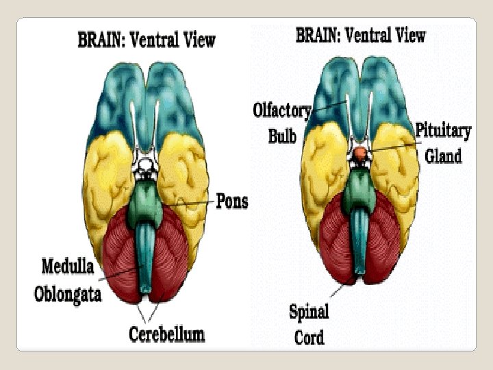
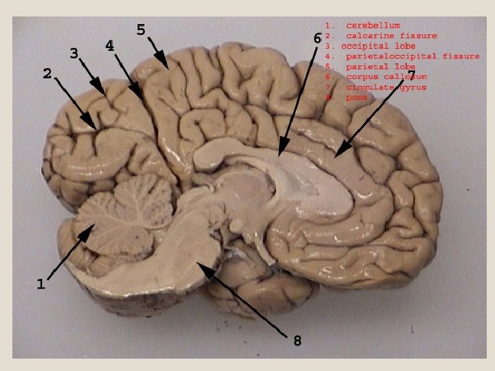
The midbrain and the hindbrain together make up the brainstem. The midbrain is the portion of the brainstem that connects the hindbrain and the forebrain. This region of the brain is involved in auditory and visual responses as well as motor function. The hindbrain extends from the spinal cord and is composed of the metencephalon and myelencephalon. The metencephalon contains structures such as the pons and cerebellum. These regions assists in maintaining balance and equilibrium, movement coordination, and the conduction of sensory information. The myelencephalon is composed of the medulla oblongata which is responsible for controlling such autonomic functions as breathing, heart rate, and digestion.

Brainstem – Most of the cranial nerves come from the brainstem. The brainstem is the pathway for all fiber tracts passing up and down from peripheral nerves and spinal cord to the highest parts of the brain. The lower extension of the brain where it connects to the spinal cord. Neurological functions located in the brainstem include those necessary for survival (breathing, digestion, heart rate, blood pressure) and for arousal (being awake and alert).
,")
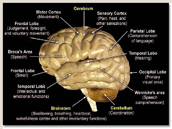
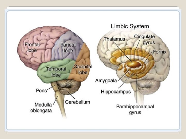
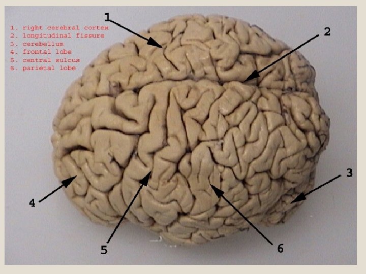
The outside of the brain, showing the major lobes (frontal, parietal, temporal and occipital), The brain stem structures (pons, medulla oblongata and cerebellum). The inside of the brain, The limbic system consists of a number of structures, including the fornix, hippocampus, cingulate gyrus, amygdala, the parahippocampal gyrus and parts of the thalamus. Anatomy of the Brain
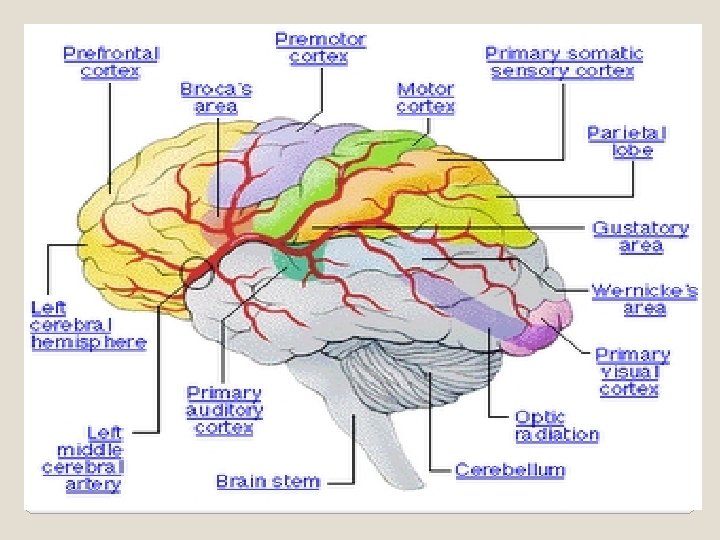

Cerebral Cortex The outermost layer of the cerebral hemisphere which is composed of gray matter. Both hemispheres are able to analyze sensory data, perform memory functions, learn new information, form thoughts and make decisions. Left Hemisphere Sequential Analysis: systematic, logical interpretation of information. Interpretation and production of symbolic information: language, mathematics, abstraction and reasoning. Memory stored in a language format. Right Hemisphere Holistic Functioning: processing multi-sensory input simultaneously to provide "holistic" picture of one's environment. Visual spatial skills. Holistic functions such as dancing and gymnastics are coordinated by the right hemisphere. Memory is stored in auditory, visual and spatial modalities.

Frontal Lobe Function Cognition and memory. Prefrontal area: The ability to concentrate and attend, elaboration of thought. The "Gatekeeper"; (judgment, inhibition). Personality and emotional traits. Movement: Motor Cortex (Brodman's): voluntary motor activity. Premotor Cortex: storage of motor patterns and voluntary activities. Language: motor speech Associated Signs and Symptoms a. Impairment of recent memory, inattentiveness, inability to concentrate, behavior disorders, difficulty in learning new information. Lack of inhibition (inappropriate social and/or sexual behavior). Emotional lability. "Flat" affect. b. Contralateral plegia, paresis. c. Expressive/motor aphasia.

Parietal Lobe Processing of sensory input, sensory discrimination. Body orientation. Primary/ secondary somatic area Associated Signs and Symptoms a. b. c. d. e. Inability to discriminate between sensory stimuli. Inability to locate and recognize parts of the body (Neglect). Severe Injury: Inability to recognize self. Disorientation of environment space. Inability to write

Occipital Lobe Primary visual reception area. Primary visual association area: Allows for visual interpretation. Associated Signs and Symptoms a. Primary Visual Cortex: loss of vision opposite field. b. Visual Association Cortex: loss of ability to recognize object seen in opposite field of vision, "flash of light", "stars".

Temporal Lobe Auditory receptive area and association areas. Expressed behavior. Language: Receptive speech. Memory: Information retrieval. Associated Signs and Symptoms a. Hearing deficits. b. Agitation, irritability, childish behavior. c. Receptive/ sensory aphasia.

Olfactory pathways: Amygdala and their pathways. Hippocampi and their pathways. Limbic lobes: Sex, rage, fear; emotions. Integration of recent memory, biological rhythms. Hypothalamus. Loss of sense of smell. Agitation, loss of control of emotion. Loss of recent memory.

Brain Structure Thalamus Function Processing center of the cerebral cortex. Coordinates and regulates all functional activity of the cortex via the integration of the afferent input to the cortex (except olfaction). Contributes to affectual expression. Associated Signs and Symptoms • Altered level of consciousness. • Loss of perception. • Thalamic syndrome spontaneous pain opposite side of body.
: • Malignant Regulation")
Hypothalamus Integration center of • Hormonal Autonomic Nervous imbalances. System (ANS): • Malignant Regulation of body hypothermia. temperature and • Inability to control endocrine function. temperature. Anterior Hypothalamus: • Diabetes Insipidus parasympathetic activity (DI). (maintenance function). • Inappropriate ADH Posterior (SIADH). Hypothalamus: • Diencephalic sympathetic activity dysfunction: ("Fight" or "Flight", "neurogenic stress response. storms". Behavioral patterns: Physical expression of behavior. Appestat: Feeding center. Pleasure center.

Cerebellum Coordination and control • Tremors. of voluntary movement. • Nystagmus (Involuntary movement of the eye). • Ataxia, lack of coordination.

• Midbrain Nerve pathway of cerebral hemispheres. Auditory and Visual reflex centers. Cranial Nerves: CN III - Oculomotor (Related to eye movement), [motor]. CN IV - Trochlear (Superior oblique muscle of the eye which rotates the eye down and out), [motor]. • Weber's: CN III palsy and ptosis (drooping) ipsalateral (same side of body). • Pupils: • Size: Midposition to dilated. Reactivity: Sluggish to fixed. LOC (Loss of consciousness): Varies • Movement: Abnormal extensor ( muscle that extends a part). • Respiratory: Hyperventilating. • CN (Cranial Nerve) Deficits: CN III, CN IV.

• Pons Respiratory Center. • Pupils: Cranial Nerves: • Size: Pinpoint LOC: CN V - Trigeminal (Skin • Semi-coma "Akinetic of face, tongue, teeth; Mute". "Locked In" muscle of mastication), Syndrome. Movement: [motor and sensory]. CN • Abnormal extensor. VI - Abducens (Lateral Withdrawal. Respiratory: rectus muscle of eye • Apneustic (Abnormal which rotates eye respiration marked by outward), [motor]. CN sustained inhalation). VII - Facial (Muscles of Hyperventilation. CN expression), [motor and Deficits: CN VI, CN VII. sensory]. CN VIII Acoustic (Internal auditory passage), [sensory].

• Medulla Oblongata Crossing of motor tracts. • Movement: Ipsilateral Cardiac Center. (same side) plegia Respiratory Center. (paralysis). Vasomotor (nerves having • Pupils: muscular control of the • Size: Dilated. Reactivity: blood vessel walls) Center Fixed. LOC: Comatose. Centers for cough, gag, • Respiratory: swallow, and vomit. • Abnormal breathing Cranial Nerves: patterns. Ataxic. Clustered. CN IX - Glossopharyneal Hiccups. CN Palsies (Muscles and mucous (Inability to control membranes of pharynx, the movement): constricted openings from Absent Cough. Gag. the mouth and the oral pharynx and the posterior third of tongue. ), [mixed]. CN X - Vagus (Pharynx, larynx, heart, lungs, stomach), [mixed]. CN XI Accessory (Rotation of the head and shoulder), [motor]. CN XII Hypoglossal (Intrinsic muscles of the tongue), [motor].
 The cortex, or cerebrum, is made up")
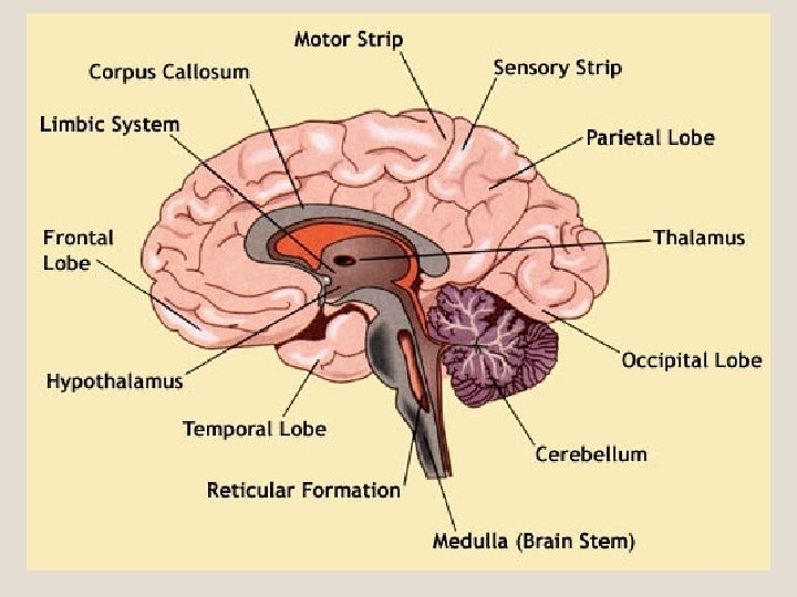
The Parts of the Brain Cortex (Cerebrum) The cortex, or cerebrum, is made up of two hemispheres (or sides) connected by a band of tissue called the corpus callosum. These hemispheres control speech, intelligence, and memory. There are specific centres for specific functions; for example, the speech centre governs the ability to form sounds into meaningful words, phrases, etc. Left Hemisphere The Left hemisphere controls the right side of the body. It controls speech, comprehension, arithmetic, and writing. Right Hemisphere The right hemisphere controls the left side of the body. It is responsible for more abstract skills, such as creativity, spatial ability, and artistic and musical skills.

Each cerebral hemisphere is divided into lobes. (In this illustration, the Frontal Lobe, the Temporal Lobe, the Parietal Lobe, Occipital Lobe, Motor Strip and Sensory Strip are all the different parts that make up the Cortex). Frontal Lobe The frontal lobe is located in front of the cerebrum behind the forehead. It is the centre for judgment, reasoning, personality, motivation, and inhibition of impulses. It also plays a role in controlling emotions, social skills, and expressive language. Parietal Lobe The parietal lobe sits just behind the frontal lobe. It is responsible for receiving and processing the sensations of touch (for example, pain, heat, cold, pressure, size, shape, and texture). It analyzes the combined information coming in from all five senses. It is also closely linked to writing and speech fluency. Temporal Lobe The temporal lobe is located alongside the frontal and parietal lobes, just above the ear. It is the centre for the senses of hearing, taste, and smell. It is also involved in receiving auditory information and in memory. Occipital Lobe The occipital lobe rests in back of the cortex behind the parietal and temporal lobes. Damage to this area may affect sight, such as perceiving or understanding visual information.

Cerebellum The cerebellum is located beneath the cerebral cortex in the back of the skull. It is smaller than the cortex. Its job is to transmit and coordinate the signals from the cortex. It also controls the movement of voluntary muscles, balance, posture, and in coordinating movements. Brain Stem The brain stem is in front of the cerebellum and beneath the cerebral cortex. It connects the spinal cord to the cortex. Its role includes passing messages back and forth between various parts of the body and the cerebral cortex. The brain stem coordinates the body's functions such as breathing, blood pressure and pulse. It also contains the reticular formation which is responsible for consciousness, drowsiness, and attention. Originating in the brain stem are 12 cranial nerves. These nerves control smell, hearing, vision, eye movement, facial sensations, taste, and swallowing. They also control muscle movements in the face, neck, shoulders, and tongue. Damage to one of these areas may affect those areas which it controls. However, no two injuries are alike. It is very important to note that not all areas will be affected; since a brain trauma can be localized, only very specific areas may be affected. The above is intended only to present a brief overview of the different areas of the brain and what they control. It is impossible to generalize which control centres are affected in a survivor of brain injury without extensive medical examinations and long-term observation.

, part
- Slides: 28